Search results
Search for "XAS" in Full Text gives 36 result(s) in Beilstein Journal of Nanotechnology.
Ambient pressure XPS at MAX IV
Beilstein J. Nanotechnol. 2025, 16, 1677–1694, doi:10.3762/bjnano.16.118

- electrochemical (EC) cell; a liquid microjet is also used to a smaller extent. In the EC setup, a three-electrode setup is immersed and retracted from a beaker of liquid electrolyte, forming a thin, electrochemically active meniscus on the working electrode that can be probed using APXPS or XAS, see Figure 1


Insights into the electronic and atomic structures of cerium oxide-based ultrathin films and nanostructures using high-brilliance light sources
Beilstein J. Nanotechnol. 2025, 16, 860–871, doi:10.3762/bjnano.16.65

- with laboratory sources. However, the information it provides primarily pertains to the surface atomic layers of the sample, as it relies on detecting electrons that strongly interact with materials. Studies by X-ray absorption spectroscopy and related techniques X-ray absorption spectroscopy (XAS) has
- provided complementary information to that obtained by synchrotron radiation-based photoemission on cerium oxide-based materials. The depth sensitivity of XAS can be tuned on a much wider range than XPS by choosing the desired absorption edge and by selecting a specific detection mode. Moreover
- , complementary information on the electronic properties and on the local atomic structure can be obtained from the analysis of XAS data. X-ray absorption near-edge spectroscopy (XANES), analyzing the signal within the first few tens of electronvolts above the absorption edge, provides information mainly on the


Retrieval of B1 phase from high-pressure B2 phase for CdO nanoparticles by electronic excitations in CdxZn1−xO composite thin films
Beilstein J. Nanotechnol. 2025, 16, 551–560, doi:10.3762/bjnano.16.43

- film. This observation further supports the presence of the Zn2SiO4 phase in the CZ900_313O thin film, which has not been detected in the XRD pattern due to limitations in detection sensitivity. Thus, X-ray absorption spectroscopy (XAS) has effectively addressed this limitation, providing evidence for


Properties of tin oxide films grown by atomic layer deposition from tin tetraiodide and ozone
Beilstein J. Nanotechnol. 2023, 14, 1085–1092, doi:10.3762/bjnano.14.89

- grazing incidence X-ray diffractometry (GIXRD), using an X-ray diffractometer SmartLab Rigaku with Cu Kα radiation, which corresponds to an X-ray wavelength of 0.15406 nm. X-ray photoelectron emission and X-ray absorption spectroscopy (XPS and XAS, respectively) measurements were made at the FinEstBeAMS
- beamline [18] at a solids research endstation [19]. XPS was carried out using a SPECS Phoibos150 hemispherical photoelectron kinetic energy analyser at an overall spectral resolution of 0.3 eV. XAS was carried out at 0.1 eV spectral resolution in total electron yield (TEY) mode by measuring sample
- eV binding energy, whereas the peak at 5 eV is not as dominant [28][29][35]. X-ray absorption spectra (Figure 11) were additionally recorded to possibly detect any differences between the samples. The Sn 3d XAS band is constituted by transitions from the 3d core orbital to the unoccupied p and f


Upscaling the urea method synthesis of CoAl layered double hydroxides
Beilstein J. Nanotechnol. 2023, 14, 927–938, doi:10.3762/bjnano.14.76

- process and aiming to quantify the amount of this Co-based α-LH impurity, X-ray absorption spectroscopy (XAS) measurements were performed at the CLÆSS BL22 beamline at the ALBA synchrotron. Figure 2A depicts the X-ray absorption near-edge structure (XANES) spectra for the Co K edge. In all samples, the
- -ray absorption spectroscopy (XAS) measurements were performed at the BL-22 (CLÆSS) beamline of the ALBA synchrotron (Barcelona, Spain), proposal: 2022097096. XANES and EXAFS Co K edge spectra were measured at room temperature in transmission mode. Absorbents of as-synthesized fresh samples were
- transmitted X-rays were measured using two ionization chambers. XAS spectra were collected from 7590 to 8550 eV with a reduced step (0.2 eV) in the XANES region (7690 to 7750 eV). The incident photon energy was calibrated using the first inflection point of the Co K edge (7709 eV) from a Co reference foil


Evaluation of electrosynthesized reduced graphene oxide–Ni/Fe/Co-based (oxy)hydroxide catalysts towards the oxygen evolution reaction
Beilstein J. Nanotechnol. 2023, 14, 420–433, doi:10.3762/bjnano.14.34
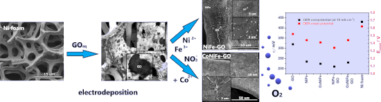
- Figure 3a–d shows the X-ray absorption spectra (XAS) of the L3 edge of nickel (a), iron (b), cobalt (c), and carbon (d) in the studied catalysts. The appearance of a shoulder peak at the L3 edge of the nickel (Figure 3a) at 855 eV indicates the presence of oxides in the structure of the catalysts (Ni in
- a strong crystal field) [28][29]. The shape of the XAS spectra (Ni edge) indicates a similar type of oxides in the structure of the catalysts. The addition of GO to NiFe and CoNiFe intensified both the nickel and iron L3 edge peaks, indicating partial electron transfer from nickel and iron to the
- substitutional GO (carbon) [30]. In the case of the edge of iron (Figure 3b), the XAS spectra indicate the presence of iron atoms in the oxidation state Fe3+ in each of the studied catalysts [28][29]. The iron edge peak observed at 707 eV disappeared after the addition of GO to CoNiFe, indicating a change in the


Theoretical investigations of oxygen vacancy effects in nickel-doped zirconia from ab initio XANES spectroscopy at the oxygen K-edge
Beilstein J. Nanotechnol. 2022, 13, 975–985, doi:10.3762/bjnano.13.85

- and oxidation states of the atom in the sample [30]. In the case of transition metal atoms, L2,3-edge XAS is particularly suitable for probing 3d valence orbitals via the dipole-allowed 2p–3d transitions. Several factors may affect the L2,3-edge spectrum, including the structure of the metal complex


A review on the green and sustainable synthesis of silver nanoparticles and one-dimensional silver nanostructures
Beilstein J. Nanotechnol. 2021, 12, 102–136, doi:10.3762/bjnano.12.9


Green and scalable synthesis of nanocrystalline kuramite
Beilstein J. Nanotechnol. 2019, 10, 2073–2083, doi:10.3762/bjnano.10.202

- microscopy (SEM), principal component analysis (PCA) of the wavelength dispersion spectroscopy (WDS) data, X-ray absorption spectroscopy (XAS) and Raman spectroscopy. Materials and Methods Synthesis The reactants necessary for the three syntheses are: CuCl2·2H2O (Merck), ZnCl2 (Merck), SnCl2·2H2O (Riedel-de
- ][55]. Raman spectroscopy was performed with a He–Ne laser source emitting at 632.8 nm with a laser spot on the sample of about 10 μm2. The main reference for the positions of the Raman peaks is from the RRUFF database [56]. XAS measurements at Cu and Sn K-edge (8978.9 and 29200.1 eV, respectively


The influence of porosity on nanoparticle formation in hierarchical aluminophosphates
Beilstein J. Nanotechnol. 2019, 10, 1952–1957, doi:10.3762/bjnano.10.191

- greater disparity (IW 511 nm, AE 529 nm). This is likely due to the wider range of possible deposition sites and environments, though overall the systems are in good agreement. X-ray adsorption spectroscopy (XAS) was used to probe the gold species, but only subtle variations between the systems was
- the influence of synthesis protocols on active site design. X-ray photoelectron spectroscopy (XPS) data (Figure 4) was in good agreement with the XAS data, as Au/HP-SAPO-5 IW and Au/HP-SAPO-5 AE systems were exclusively fit with Au0 features (Figure 4B). However, the corresponding microporous systems
- the k3-weighted Fourier transform for the XAS data of the Au-deposited microporous MP-SAPO-5 (A) and hierarchical HP-SAPO-5 (B) compared to the Au foil. Associated scattering paths, with a single Au–Au feature are included. Stacked XPS data for Au-doped microporous MP-SAPO-5 (A) and hierarchical HP


Metal–dielectric hybrid nanoantennas for efficient frequency conversion at the anapole mode
Beilstein J. Nanotechnol. 2018, 9, 2306–2314, doi:10.3762/bjnano.9.215

- amplification [22] and enhanced Raman scattering [23] have been recently suggested. In this framework, AlxGa1−xAs, a III–V semiconductor, has become a popular material for nonlinear photonics thanks to its non-centrosymmetric structure and other important key assets including: i) a large band gap enabling TPA


Metal-free catalysis based on nitrogen-doped carbon nanomaterials: a photoelectron spectroscopy point of view
Beilstein J. Nanotechnol. 2018, 9, 2015–2031, doi:10.3762/bjnano.9.191

- ]: Niwa et al. [105] described carbon nanostructure alloys (a precursor of graphene), Nagaiah and co-workers studied nitrogen-doped CNTs [106], and Parvez et al. used N-graphene [107]. In the first report, correlating X-ray absorption spectroscopy (XAS) and ORR activity, the study was focused on the
- agreement with previous reports that correlate XAS and XPS [108][109]. By correlating XAS and RRDE measurements, they assumed that the π* profile observed in the absorption spectra can be used to identify the nitrogen configuration, being therefore an indicator of the ORR activity. Nagaiah and co-workers


Mechanistic insights into plasmonic photocatalysts in utilizing visible light
Beilstein J. Nanotechnol. 2018, 9, 628–648, doi:10.3762/bjnano.9.59

- excitation and electron injection into the semiconductor are still unclear. The verification that plasmon-excited electrons in Au NPs possess sufficient energy to overcome the Schottky junction to be injected into TiO2 was confirmed using high-resolution X-ray absorption spectroscopy (HR-XAS) [105]. The
- adopted experimental setup is depicted in Figure 10. The significant spectral variations observed by X-ray absorption spectroscopy (XAS) and resonant inelastic X-ray scattering (RIXS) suggest that electrons injected from Au NPs upon LSPR excitation could survive longer and become trapped at a Ti site near
- polarized irradiation along various axes was combined with theoretical simulations based on the finite element method (FEM). In situ XAS was used to understand the electronic structural changes caused by the electromagnetic field upon the surface of plasmonic materials [108]. Designing the physical


Charge transfer from and to manganese phthalocyanine: bulk materials and interfaces
Beilstein J. Nanotechnol. 2017, 8, 1601–1615, doi:10.3762/bjnano.8.160

- photoemission spectroscopy (IPES), electron energy-loss spectroscopy (EELS), spectroscopic ellipsometry and X-ray absorption spectroscopy (XAS). Here, we only briefly mention the kind of information that is provided by these methods, and we refer the reader to comprehensive literature for detailed information
- moment of open shells is accessible. Spectroscopic ellipsometry [47][48][49] measures the change in the light polarization after reflection on a sample surface. This information allows for the determination of the real and the imaginary part of the dielectric function. XAS [42][50] is equivalent to EELS
- revealed by polarization dependent X-ray absorption spectroscopy (XAS) studies [127]. In Figure 12 we present corresponding N 1s absorption spectra for pure MnPc and two different film thicknesses of an F16CoPc overlayer, deposited on a gold single crystal. Different light polarizations with respect to the


Modeling of the growth of GaAs–AlGaAs core–shell nanowires
Beilstein J. Nanotechnol. 2017, 8, 506–513, doi:10.3762/bjnano.8.54

- surface limit and deposition has been developed for the evolution of the shell morphology and concentration in AlxGa1−xAs alloys. The model includes a completely faceted shell–vapor interface. The objective is to understand the mechanisms of the formation of the radial heterostructures (Al-rich stripes
- Figure 7b). It is obvious that a model assuming pure materials cannot address the mechanisms leading to the non-homogeneous distribution of Al in the shell. A two-dimensional fully faceted model [8] was developed for the growth of the shell with an AxB1−x (AlxGa1−xAs) alloy on a hexagonal core. Surface


Photocurrent generation in carbon nanotube/cubic-phase HfO2 nanoparticle hybrid nanocomposites
Beilstein J. Nanotechnol. 2016, 7, 1075–1085, doi:10.3762/bjnano.7.101

- is manifested as a broad hump starting above 288 eV and extending up to 305 eV, corresponding to the C 1s → σ* transition for disordered carbon–carbon bonds. On a similar note, other noticeable features are usually observed via X-ray absorption spectroscopy (XAS) in the region between π* and σ


Paramagnetism of cobalt-doped ZnO nanoparticles obtained by microwave solvothermal synthesis
Beilstein J. Nanotechnol. 2015, 6, 1957–1969, doi:10.3762/bjnano.6.200

- was calculated to be 350 nm (Table 4). XAS investigations of Zn1−xCoxO before annealing The data shown in Figure 10 presents the Fourier transformations of EXAFS oscillations taken at the Zn K-edge for the as-synthesized nanopowders. These data indicate that the introduction of Co into ZnO does not


Metal hydrides: an innovative and challenging conversion reaction anode for lithium-ion batteries
Beilstein J. Nanotechnol. 2015, 6, 1821–1839, doi:10.3762/bjnano.6.186

- confirmed by XAS and Mössbauer spectroscopy [22]. Ex situ XAS spectroscopy of the Mg2CoH5 and Mg2NiH4 electrodes revealed the formation of disordered MgCo and Mg2Ni intermetallic compounds. The intensity reduction of the XRD lines, which occurs without broadening, involves shifts of the lattice parameters


Magnetic properties of self-organized Co dimer nanolines on Si/Ag(110)
Beilstein J. Nanotechnol. 2015, 6, 777–784, doi:10.3762/bjnano.6.80

- nanolines X-ray absorption spectroscopy (XAS) spectra were recorded at normal incidence in a magnetic field of 6 T for parallel (σ+) and antiparallel (σ−) alignment of the X-ray helicity with respect to the sample magnetization. Magnetic hysteresis measurements at the L3 resonance confirm that the sample
- magnetization is saturated at 6 T. The strong non-magnetic background signal coming from the Ag substrate was subtracted from the Co L2,3 XAS spectra presented in this paper. The spectra are also normalized to the incident beam intensity, which is set to zero at the L3 pre-edge and to one far above the L2 edge
- . Figure 3a,b shows the XAS spectra for both helicities (upper panel) for ≈1 MLCo and ≈2 MLCo, respectively. Two broad absorption resonances are clearly visible at the L3 and L2 edges. A shoulder peak, indicated in the XAS spectra of Figure 3 by a dotted line, is also present at about 4 eV above the L3


Materials and characterization techniques for high-temperature polymer electrolyte membrane fuel cells
Beilstein J. Nanotechnol. 2015, 6, 68–83, doi:10.3762/bjnano.6.8

- in HT-PEM by using in operando X-ray absorption spectroscopy (XAS) incorporating the Δμ technique. The goal of this study was also to investigate phosphoric acid adsorption on platinum in a real fuel cell. The technique is capable to determine adsorbates on the platinum catalyst particles by


Advances in NO2 sensing with individual single-walled carbon nanotube transistors
Beilstein J. Nanotechnol. 2014, 5, 2179–2191, doi:10.3762/bjnano.5.227

- –SWNT system by utilizing metallicity-sorted, ultrapure carbon nanotubes. They used X-ray photoelectron spectroscopy (XPS) and X-ray absorption spectroscopy (XAS) to investigate the nature of bonding and chemical interaction. Their conclusions, supported by theoretical calculations, are that the process


Two-dimensional and tubular structures of misfit compounds: Structural and electronic properties
Beilstein J. Nanotechnol. 2014, 5, 2171–2178, doi:10.3762/bjnano.5.226

- addition to theoretical considerations, the electronic structure is discernible by spectroscopy as Ohno [12][35] presented in 1991. By performing X-ray photoelectron and absorption spectroscopy (XPS, XAS) and reflection electron energy loss spectroscopy (REELS), it was revealed that the electronic
- as a donor and TMX2 as an acceptor of the transferred electron density. This claim was based on studies of valence band XPS and XAS spectra of the misfit compounds, compared to those of the individual layers, and a spectrum of iron intercalated titanium disulfide (Fe1/3TiS2), which can be interpreted
- as a true intercalated system. Figure 5 in [35] shows this XAS spectra of the K absorption edge of sulfur for the systems TiS2, PbS, PbTiS3 (=MLC (PbS)1.18TiS2), and Fe1/3TiS2. The shapes of the PbTiS3 and Fe1/3TiS2 spectra are quite similar. From this, charge transfer was concluded, which should


UHV deposition and characterization of a mononuclear iron(III) β-diketonate complex on Au(111)
Beilstein J. Nanotechnol. 2014, 5, 2139–2148, doi:10.3762/bjnano.5.223

- high coverages compatible with an ex situ prepared sample cannot be achieved by UHV sublimation, a thick film sample of Fe(dpm)3 was prepared by drop-casting. XAS spectra at the Fe L2,3 edge, acquired at the BACH beamline of the Elettra synchrotron for both circular left (σ+) and circular right (σ


Cathode lens spectromicroscopy: methodology and applications
Beilstein J. Nanotechnol. 2014, 5, 1873–1886, doi:10.3762/bjnano.5.198

- , synchrotron sources greatly extend the application field of XPEEM instruments, which can achieve chemical, magnetic and electronic structure contrast through the implementation of the most popular photoelectron spectroscopies such as X-ray absorption spectroscopy (XAS), photoelectron spectroscopy (XPS), and
- angle-resolved photoemission spectroscopy (ARPES) [5]. XAS based methods. XAS is the only method readily available when using the basic XPEEM instrument installed at a synchrotron beamline with a monochromator in place. Among the variety of detection methods to measure X-ray absorption [18], the
- secondary photoelectrons are collected in XPEEM as a close approximation to the total photoelectron yield measurement. The local XAS spectra are obtained by acquiring image sequences as a function of the photon energy, which can then be processed in order to extract the intensity variation within any region


CoPc and CoPcF16 on gold: Site-specific charge-transfer processes
Beilstein J. Nanotechnol. 2014, 5, 524–531, doi:10.3762/bjnano.5.61

- spectroscopy (UPS) as well as X-ray absorption spectroscopy (XAS). Combined XPS and XAES measurements can be employed as a tool to study the contribution of the polarization energy to chemical shifts at interfaces. XAS gives valuable information about the unoccupied electronic structure and the hybridization
- negatively” charged compared to the bulk. For a strongly related system, namely CoPcF16 on Au(100), X-ray absorption spectroscopy measurements at the fluorine K-edge show that the electron density can change at the fluorine site. Since XAS probes transitions from occupied into unoccupied valence states
- linear dichroism of F K-edge XAS spectra compared to N or C K-edge spectra [30]. The presence of angular dependent π* and σ* transitions indicates that fluorine atoms participate in the conjugated π system. Comparing spectra from the thick film in Figure 5a to spectra at a lower coverage of about 0.8 nm


