Guest Editor: H. Ritter Beilstein J. Org. Chem.2012,8, 1644–1651.https://doi.org/10.3762/bjoc.8.188 Received 18 Jun 2012,
Accepted 27 Aug 2012,
Published 28 Sep 2012
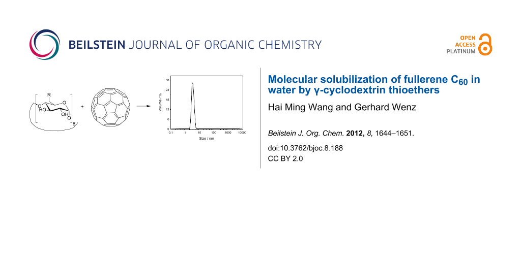
Various hydrophilic γ-cyclodextrin (CD) thioethers, containing neutral or ionic side arms were found to form molecular disperse solutions of C60 in water reaching concentrations of 15 mg/L. Equilibrium state was approached after seven days without the use of organic cosolvents. The 1:2 stoichiometry of the C60/γ-CD thioether complexes was demonstrated by a parabolic phase-solubility diagram. In contrast, native γ-CD forms nanoparticles with C60. Particle sizes of C60 were determined by dynamic light scattering.
Since the first spectroscopic discovery of buckminsterfullerene, C60, by Kroto, Heath, Curl and Smalley in 1985 [1], and its first macroscopic synthesis by Krätschmer et al. in 1990 [2], this third allotropic modification of carbon has been the subject of more than 10,000 publications giving rise to many interesting potential applications both in the biomedical field [3-5] and materials science [6-9].
A good solubility of C60 in water is especially required for biological applications; however, this is not the case at all. Solubility of C60 was estimated to be as low as 10−8 ng/L, equivalent to 10 C60 molecules per millilitre of water [10,11]. Therefore, hydrophilic derivatives of C60 have been synthesized and utilized for the inhibition of therapeutically important enzymes, such as HIV-1 protease [12], for the prevention of bacterial growth [13,14], or for photodynamic therapy of cancer by scission of DNA [3]. Despite these successes, there are still several issues relating to the chemical modification of C60. The regioselectivity of derivatization is difficult to control [15], and derivatization reduces aromaticity, which leads to a change of the distinct electronic and photonic properties of C60.
Dispersions of C60 nanoparticles in water have been discussed as alternatives for molecular solutions. Such C60 dispersions are generally obtained by the so-called solvent-exchange method [16,17], where C60 is dissolved primarily in an organic solvent, such as benzene, THF, or acetone, and afterwards diluted with water. After evaporation of the organic solvent, clusters of C60 in water remain, which are temporarily stable. The biological activities of those dispersions strongly increase with decreasing size of the C60 nanoparticles [13]. Also micellar dispersions of C60 in water stabilized by detergents such as Tween, Triton, or SDS are known [18]. Applications of these C60 dispersions are hampered by the toxicities of the employed organic solvents or surfactants.
The most successful strategy to carry the extremely hydrophobic C60 molecule into water is the use of appropriate water-soluble carriers that can form host–guest complexes, such as calixarenes [19,20] and cyclodextrins (CDs) [21,22]. CDs are cyclic oligosaccharides consisting of six, seven, eight or more glucose subunits connected through α-1,4 glycosidic linkages, called α-, β-, and γ-CD, respectively. CD molecules resemble truncated cones comprising a hydrophilic outer surface and a relatively hydrophobic cavity [23-25]. CDs form water-soluble inclusion complexes with many hydrophobic or amphiphilic guest molecules [26], mainly driven by hydrophobic interactions [27]. Among the commercially available CDs, γ-CD with a clear width of d = 0.74 nm [28] is only large enough to partially accommodate C60, which has a still greater van der Waals diameter of 1.0 nm [2]. Molecular dynamics studies strongly favour a sandwich-like structure of the complex, in which two γ-CD molecules tightly interact through hydrogen bonds between their secondary rims and in which C60 is situated in the middle at the widest sites of both CDs (see Figure 1, [29]).
Figure 1:
Space-filling model of the most stable complex between γ-CD and C60 with 1:2 stoichiometry, calculated in vacuo. Reprinted with permission from reference [29]. Copyright (2010) American Chemical Society.
Figure 1:
Space-filling model of the most stable complex between γ-CD and C60 with 1:2 stoichiometry, calcula...
Andersson et al. reported that C60 formed a water-soluble 1:2 inclusion complex with γ-CD after heating under reflux in water [21]. Mixing of an aqueous solution of γ-CD with a methanolic solution of C60 led to a C60 dispersion with a concentration of ca. 70 mg/L [30]. Even higher concentrations of up to 1 g/L were reached by high-speed vibration milling of C60 in aqueous solutions of γ-CD [31]. The main drawbacks of these aqueous systems are still (a) their lack of stability, leading to crystallization of the C60 complex after some days [31]; and (b) their pronounced tendency to form nanoparticular aggregates [21,32], both of which limit their practical application.
Recently, we developed a new class of highly water-soluble per-6-deoxy-thioethers of β- and γ-CD (>20% w/w), which showed exceptionally high solubilization abilities for several aromatic molecules, such as anthracene and acenaphthylene, in water [33-35]. In this work, we investigated solubilization of C60 by these γ-CD thioethers (compounds 1–7 in Scheme 1) with the hope of achieving high concentrations of solubilized C60. The hydrophilic substituents at the primary face of γ-CD should increase solubility and avoid aggregation, because they cannot form intermolecular hydrogen bonds like the primary hydroxyls of native γ-CD.
Scheme 1:
Chemical structures of γ-CD and γ-CD thioether 1–7 used to solubilize C60 in water.
Scheme 1:
Chemical structures of γ-CD and γ-CD thioether 1–7 used to solubilize C60 in water.
Powdered C60 was stirred at 25 °C in 6 mM aqueous solutions of γ-CD and γ-CD thioethers 1–7 giving rise to clear, dark yellow solutions of C60, which show a narrow absorption band at λmax = 335 nm quite similar to that of a solution of C60 in THF (λmax = 327 nm), shown in Figure 2. Extensive centrifugation caused only a small reduction of signal intensity (less than 8%, Figure 2), which clearly precludes the existence of aggregates, which would otherwise sediment to the bottom. Therefore the molar concentration of C60 could be calculated by using the known molar extinction coefficient of C60 molecularly dissolved in n-hexane at 328 nm, i.e, ε = 52,000 M−1·cm−1 as published previously [36].
Figure 2:
UV–vis spectra of (a) C60 solution in THF, (b) aqueous solutions of C60 with 6 mM γ-CD thioether 5 before centrifugation, and (c) after centrifugation (13,000 rpm) for 60 min. The absorbance intensities of (a) and (b) are shifted by 0.2 and 0.1 AU, respectively.
Figure 2:
UV–vis spectra of (a) C60 solution in THF, (b) aqueous solutions of C60 with 6 mM γ-CD thioether 5 ...
After the dissolution of C60 by γ-CD thioethers had been demonstrated, we were interested in how long it takes to reach equilibrium, within experimental error. Therefore a thinly casted film of C60 was incubated with an aqueous solution of γ-CD thioether 7 at 50 °C and stirred according to Kuroda et al. [37]. The slow increase of C60 concentration was monitored by the increase in absorption intensity at 335 nm. The observed first-order dissolution kinetics of C60 came nearly to an end after 7 d, as shown in Figure 3. The obtained rate constant, k = 0.021 h−1 was somewhat higher than the one already found for native γ-CD, k = 0.011 h−1, which also showed first-order dissolution kinetics [37].
Figure 3:
Isothermal kinetics of the dissolution of C60 in the presence of 10 mM CD 7 in water. Curve: best fit of first-order kinetics k = 0.5 d−1 = 0.021 h−1.
Figure 3:
Isothermal kinetics of the dissolution of C60 in the presence of 10 mM CD 7 in water. Curve: best f...
The observed simple first-order kinetics was puzzling for us, because the concentration of the CD host does not go down significantly (<<1%) over the course of the dissolution of C60. Therefore other reasons for the observed continuous decrease of dissolution rate had to be found.
Dissolution of C60 can be described by a two-step process, originally proposed by Kuroda et al. [37]. Alternatively, a one-step process, in which a C60 molecule is trapped by two CD rings at the same time, appeared reasonable to us. Both models are shown in Scheme 2.
Scheme 2:
Mechanistic description of the two possible mechanisms for the complexation of C60 (G) by CD hosts, in (a) two steps, and (b) one step.
Scheme 2:
Mechanistic description of the two possible mechanisms for the complexation of C60 (G) by CD hosts,...
The two-step model (a) employs a slow, rate-determining complexation of the guest C60 by the first CD molecule and a fast further complexation by the second. The second step is much faster than the first due to the strong stabilization exerted by multiple hydrogen bonds between both CD rings. For the back process, the first dissociation step should be slow and rate determining, because of the necessary cleavage of these intermolecular hydrogen bonds. These assumptions lead to an apparent equilibrium constant K′ in which the CD concentration is only present in first order. The initial rate of complexation is predicted to be proportional to CD concentration, which was already experimentally found for native γ-CD [37].
On the other hand, the one step model (b) leads to the classical binding constant K. The initial complexation rate should be proportional to the square of the host concentration, as shown in Supporting Information File 1.
Both models have in common that the formation of the complex follows pseudo-zero-order kinetics whereas dissociation follows first-order kinetics. Consequently, the integrations of the rate equations for both models (described in Supporting Information File 1) lead to the same final kinetics (Equation 1), a simple first-order equation converging to the equilibrium solubility of the guest [C60·CD2]equ in agreement with the observed experimental data. According to both new models, the obtained rate constants k−2 and k−, respectively, are not due to formation of the complex as proposed previously [37], but due to its dissociation.
(1)
Dependence of the equilibrium concentration of C60 on the host and its concentration
Equilibrium concentrations of C60 in aqueous solutions of 6 mM γ-CD and γ-CD thioethers 1–7, determined from the absorptions (λmax = 335 nm) after being stirred for 7 d, are listed in Table 1. C60 concentrations obtained with γ-CD thioethers were up 35 times higher than the one obtained with native γ-CD, in accordance with previous results for other hydrophobic guests [38]. The improved solubilization potential of the thioethers was attributed to the higher hydrophobicity of sulfur compared to oxygen. The highest concentration of C60 in water, 14.9 μM, was found for CD derivative 5 with attached neutral diol substituents. In general, neutral γ-CD thioethers 1, 5, and 7 performed better than the anionic ones 2, 3, 4. Coulomb repulsion between the anionic groups in between the two CD molecules was held responsible for the reduced binding affinity. Astonishingly, the amino derivative 1 also showed a high solubilization potential, which may originate from the addition of the amine to a double bond of C60, as was already observed by Geckeler for other amino compounds [39].
Table 1:
C60 concentration in 6.0 mM aqueous solutions of γ-CD and γ-CD thioethers 1–7.
host
[C60] (μM)
γ-CD
0.4
1
10.3
2
2.9
3
5.3
4
2.1
5
14.9
6
7.6
7
9.3
The phase-solubility diagram of C60 in the presence of γ-CD thioether 3, according to the method established by Higuchi and Connors [40], was obtained by plotting the concentration of the dissolved C60 versus the concentration of the host, as depicted in Figure 4. The observed parabolic AP-type phase concentration dependence is typical for the formation of complexes with 1:2 stoichiometry [40,41]. The equation for the best fit [C60] = 10−3(0.21 + 0.17[CD] + 0.11[CD]2) indicates that the two-step process (first order in [CD]) as well as the one-step process (second order in [CD]), discussed above, contribute to the dissolution of C60.
Figure 4:
Phase-solubility diagram of C60 in aqueous solution in the presence of CD 3.
Figure 4:
Phase-solubility diagram of C60 in aqueous solution in the presence of CD 3.
Preparation of C60 complexes with the aid of organic solvents
Because equilibration took a long time (7 d) and occupancies of the γ-CD thioethers are still low (<0.3%), organic solvents, such as toluene, DMF or CS2 were added to the aqueous solutions of γ-CD and γ-CD thioether 5, in the hope of accelerating and improving the dissolution of C60 as reported by Murphy et al. [42]. The resulting solutions were characterized by UV–vis spectroscopy. The saturation concentrations of C60 are listed in Table 2.
Table 2:
C60 concentration in 6.0 mM aqueous solutions of γ-CD and γ-CD thioether 5.
Indeed, for native γ-CD much higher C60 concentrations could be achieved in water with the aid of organic solvents. But careful examination of the UV–vis spectra of the C60 solutions produced according to procedure c[42] before filtration, after filtration, and after centrifugation, as shown in Figure 5, revealed significant differences. The unfiltered solution of the C60/γ-CD complex showed an additional absorption maximum 475 nm, which is typical for C60 nanoparticles (nC60) such as those formed by dilution of a solution of C60 in THF with water [16,43]. After centrifugation nearly no C60 was left. Consequently, apparent improvements of the solubilization obtained with the aid of organic solvents were mostly due to the formation of nanoparticulate dispersions. Since these organic solvents are also hazardous for most cells, applications in biomedicine are prohibited. Therefore, organic solvents were avoided for the dissolution of C60 by γ-CD thioethers, because they also did not improve the solubilization process significantly.
Figure 5:
UV–vis spectra of the water solution of C60 produced by stirring C60 in 6mM γ-CD solution in DMF/toluene (v/v 1:1) at rt for 7 d, followed by dissolution of the resulting complex in water after evaporation of the solvents (procedure c): (a) before filtration, (b) after filtration, and (c) after centrifugation; (d) nC60 made from THF [43] before filtration.
Figure 5:
UV–vis spectra of the water solution of C60 produced by stirring C60 in 6mM γ-CD solution in DMF/to...
Investigation of aggregation by dynamic light scattering
Since aggregation of CDs and CD inclusion compounds was already found in previous work [44,45], dynamic light scattering (DLS) investigations were performed to check for any aggregation during solubilization of C60 by γ-CD thioethers. DLS is a relatively fast method for the determination of the particle size distributions of proteins, polymers, micelles, and nanoparticles [46]. In particular, DLS is able to distinguish between a homogenous molecular solution and a dispersion of aggregates [47,48].
The size distribution of the solution of C60 in 6 mM γ-CD thioether 5 before centrifugation, shown in Figure 6, comprised two peaks at particle sizes of 3 and 300 nm. The first peak was attributed to the molecular CD/C60 complex, the second to aggregates of it. Since the intensity of the scattered light increases with the sixth power of the particle size, the content of aggregates is highly overestimated [45]. For getting the right picture, this intensity profile had to be transformed to the volume distribution profile, by using the Mie theory [49]. In the resulting volume distribution profile (Figure 6b) only one peak remains, which corresponds to the molecular complex. Consequently, this solution mainly consists of molecularly dissolved C60. This finding is in accordance with the negligible decrease of C60 absorption caused by centrifugation, shown in Figure 2.
Figure 6:
Size distribution of the molecular solution of C60 with 6 mM CD 5 in water at 25.0 °C: before centrifugation (a) by intensity, and (b) by volume; and after centrifugation (c) by intensity, and (d) by volume.
Figure 6:
Size distribution of the molecular solution of C60 with 6 mM CD 5 in water at 25.0 °C: before centr...
After centrifugation (13,000 rpm) for 60 min, shown in Figure 6c, only a sharp peak at 1 nm was observed for both the intensity and the volume size distributions, which demonstrates the validity of the DLS measurement. For comparison, the corresponding intensity and volume size distributions of a freshly prepared C60 solution in the presence of 6.0 mM native γ-CD according to procedure c[42] (Figure 7) only showed one peak at a diameter of 166 nm, very similar to nC60 prepared from THF. Consequently, the DLS investigations confirmed our previous finding that solubilization of C60 in water with the aid of organic solvents results in dispersions of C60 nanoparticles.
Figure 7:
Size distributions of the aqueous C60 dispersions after filtration, produced by stirring C60 in 6mM γ-CD solution in DMF/toluene (v/v 1:1) according to procedure c) [42]: (a) by intensity and (b) by volume.
Figure 7:
Size distributions of the aqueous C60 dispersions after filtration, produced by stirring C60 in 6mM...
Neutral γ-CD thioethers are especially well suited as solubilizers for C60. Sandwich-like 1:2 complexes are formed at room temperature without the necessity of adding organic cosolvents. These complexes show a much lower aggregation tendency than the corresponding ones of native γ-CD. Molecular solubilization of fullerene C60 in water, reaching concentrations as high as 15 μM, was achieved in the presence of 6.0 mM of a γ-CD thioether. The resulting aqueous molecular solutions of C60 free of toxic organic solvents will hopefully find interesting applications in biomedicine, such as in photodynamic therapy or HIV-protease inhibition.
Experimental
General
Unless otherwise stated, all chemicals were used as received. Powdered fullerene C60 (> 99%) was purchased from Sigma Aldrich. Teflon syringe filters from Roth, Karlsruhe, Germany (0.45 μm) were used to remove insoluble material before UV–vis spectrophotometric analysis. UV–vis spectra of aqueous samples were performed on a Perkin Elmer Lambda 2 spectrometer (λ: 200–600 nm), by using quartz cells with a 1 cm or 1 mm optical path at 298 K.
Synthetic procedures
Hydrophilic thioethers 1–7 at all primary carbon atoms of γ-CD were synthesized from octakis(6-deoxy-6-iodo)-γ-CD by nucleophilic displacement reaction with sulfur nucleophiles by using standard procedures described previously [38].
Phase-solubility investigations
Solubility measurements of C60 in the presence of γ-CD and γ-CD derivatives in water were carried out according to the method proposed by Higuchi and Connors [40]. In glass vials containing excess amounts of C60, aqueous solutions of γ-CD or γ-CD derivatives with different concentrations were added. The vials were sealed, protected from light, and magnetically stirred at room temperature for seven days. The solid residues were removed by filtration with syringe filter. According to the Lambert–Beer law, the concentrations of C60 in pure water and in CDs solutions were determined from UV–vis extinctions at the absorption maxima (log ε = 4.717, λmax = 335 nm) [18].
Procedures for the solubilization of C60
Several procedures were employed for the solubilization of C60. Procedure a: C60 stirred in water in the presence of 6 mM γ-CD thioether at rt for 7 d. Procedure b was modified from a previously described method [21]: heated under reflux in water/toluene (v/v 1:1) for 3 d (γ-CD thioether concentration 6 mM), and then the resulting mixture dissolved in water after evaporation of the solvents. Procedure c was taken from a previous paper [42]: C60 stirred in DMF/toluene (v/v 1:1) at rt for 7 d with 6 mM γ-CD thioether, and then the obtained inclusion complex dissolved in water after evaporation of the solvents. Procedure d: C60 stirred in water/CS2 (v/v 1:1) at rt for 7 d with 6 mM γ-CD thioether, and then the obtained inclusion complex dissolved in water after evaporation of the solvents. nC60 was prepared following a method similar to that reported by Deguchi et al. (procedure e) [16]. nC60: A saturated solution of C60 in THF was prepared by adding an excess amount of solid C60 (>2 mg) into THF (20 mL) and stirring overnight under a nitrogen atmosphere at room temperature. Excess solid was filtered off with a syringe filter. Saturated C60/THF solution (500 mL) was placed in a flask and an equal volume of water was added at a rate of ca. 25 mL/min under vigorous stirring. A rotary evaporator was used to remove THF by using a stepwise evaporation approach. The start temperature was set at 30 °C. When the mixture volume had decreased to 500 mL, the temperature was increased at 1 °C/min to 70 °C and maintained at 70 °C until the volume had decreased to 250 mL, at which time an additional 250 mL volume of water was added. The last step was repeated once. The resulting nC60 solution could be diluted by adding water or concentrated by evaporation as needed.
Isothermal kinetic measurement
The procedure for performing isothermal kinetic investigations was modified from a previously described method [37]: A solution of C60 in chloroform (ca. 0.5 mg/mL, 0.5 mL) was carefully evaporated in a 1 cm quartz cell under nitrogen flow. After the addition of 3 mL of an aqueous solution containing the necessary amount of γ-CD derivative, the cell was sealed, protected from light, and maintained at 50 °C under gentle shaking. The UV–vis spectra of the resultant solution at appropriate time intervals were measured directly.
Dynamic light scattering measurement
Particle size distributions of the aqueous solution or dispersions of C60 were determined by dynamic light scattering (DLS) with a ZetaSizer Nano ZS (Malvern Instruments Ltd., Malvern, United Kingdom). From the diffusion coefficient the radius of the particle was determined via the Stokes–Einstein equation. The samples were filtered through a 0.45 μm syringe filter prior to particle-size measurements. Both intensity and volume size distribution curves were calculated from the scattering data by using the software of the instrument.
Supporting Information
Supporting Information File 1:
Detailed dissolution kinetics.
We gratefully thank the technical support from Annegret Engelke. Financial supports from Saarland University and China Scholarship Council are gratefully acknowledged.
References
Kroto, H. W.; Heath, J. R.; O'Brien, S. C.; Curl, R. F.; Smalley, R. E. Nature1985,318, 162–163. doi:10.1038/318162a0
Return to citation in text:
[1]
Krätschmer, W.; Lamb, L. D.; Fostiropoulos, K.; Huffman, D. R. Nature1990,347, 354–358. doi:10.1038/347354a0
Return to citation in text:
[1]
[2]
Nakamura, E.; Isobe, H. Acc. Chem. Res.2003,36, 807–815. doi:10.1021/ar030027y
Return to citation in text:
[1]
[2]
Da Ros, T.; Prato, M. Chem. Commun.1999, 663–669. doi:10.1039/a809495k
Return to citation in text:
[1]
Jensen, A. W.; Wilson, S. R.; Schuster, D. I. Bioorg. Med. Chem.1996,4, 767–779. doi:10.1016/0968-0896(96)00081-8
Return to citation in text:
[1]
Hirsch, T.; Kettenberger, H.; Wolfbeis, O. S.; Mirsky, V. M. Chem. Commun.2003, 432–433. doi:10.1039/b210554c
Return to citation in text:
[1]
Hebard, A. F.; Rosseinsky, M. J.; Haddon, R. C.; Murphy, D. W.; Glarum, S. H.; Palstra, T. T. M.; Ramirez, A. P.; Kortan, A. R. Nature1991,350, 600–601. doi:10.1038/350600a0
Return to citation in text:
[1]
Halls, J. J. M.; Pichler, K.; Friend, R. H.; Moratti, S. C.; Holmes, A. B. Appl. Phys. Lett.1996,68, 3120–3122. doi:10.1063/1.115797
Return to citation in text:
[1]
Semenov, K. N.; Charykov, N. A.; Keskinov, V. A.; Piartman, A. K.; Blokhin, A. A.; Kopyrin, A. A. J. Chem. Eng. Data2010,55, 13–36. doi:10.1021/je900296s
Return to citation in text:
[1]
Sijbesma, R.; Srdanov, G.; Wudl, F.; Castoro, J. A.; Wilkins, C.; Friedman, S. H.; DeCamp, D. L.; Kenyon, G. L. J. Am. Chem. Soc.1993,115, 6510–6512. doi:10.1021/ja00068a006
Return to citation in text:
[1]
Lyon, D. Y.; Adams, L. K.; Falkner, J. C.; Alvarez, P. J. J. Environ. Sci. Technol.2006,40, 4360–4366. doi:10.1021/es0603655
Return to citation in text:
[1]
[2]
Fortner, J. D.; Lyon, D. Y.; Sayes, C. M.; Boyd, A. M.; Falkner, J. C.; Hotze, E. M.; Alemany, L. B.; Tao, Y. J.; Guo, W.; Ausman, K. D.; Colvin, V. L.; Hughes, J. B. Environ. Sci. Technol.2005,39, 4307–4316. doi:10.1021/es048099n
Return to citation in text:
[1]
Hirsch, A.; Lamparth, I.; Karfunkel, H. R. Angew. Chem., Int. Ed. Engl.1994,33, 437–438. doi:10.1002/anie.199404371
Return to citation in text:
[1]
Deguchi, S.; Alargova, R. G.; Tsujii, K. Langmuir2001,17, 6013–6017. doi:10.1021/la010651o
Return to citation in text:
[1]
[2]
[3]
Scrivens, W. A.; Tour, J. M.; Creek, K. E.; Pirisi, L. J. Am. Chem. Soc.1994,116, 4517–4518. doi:10.1021/ja00089a067
Return to citation in text:
[1]
Torres, V. M.; Posa, M.; Srdjenovic, B.; Simplício, A. L. Colloids Surf., B2011,82, 46–53. doi:10.1016/j.colsurfb.2010.08.012
Return to citation in text:
[1]
[2]
Haino, T.; Yanase, M.; Fukunaga, C.; Fukazawa, Y. Tetrahedron2006,62, 2025–2035. doi:10.1016/j.tet.2005.07.121
Return to citation in text:
[1]
Ikeda, A.; Suzuki, Y.; Yoshimura, M.; Shinkai, S. Tetrahedron1998,54, 2497–2508. doi:10.1016/S0040-4020(98)00012-X
Return to citation in text:
[1]
Andersson, T.; Nilsson, K.; Sundahl, M.; Westman, G.; Wennerström, O. J. Chem. Soc., Chem. Commun.1992, 604–606. doi:10.1039/C39920000604
Return to citation in text:
[1]
[2]
[3]
[4]
Yoshida, Z.-i.; Takekuma, H.; Takekuma, S.-i.; Matsubara, Y. Angew. Chem., Int. Ed. Engl.1994,33, 1597–1599. doi:10.1002/anie.199415971
Return to citation in text:
[1]
Wenz, G. Angew. Chem., Int. Ed. Engl.1994,33, 803–822. doi:10.1002/anie.199408031
Return to citation in text:
[1]
Dodziuk, H. Cyclodextrins and their Complexes: Chemistry, Analytical Methods, Applications; Wiley-VCH: Weinheim, Germany, 2006. doi:10.1002/3527608982
Return to citation in text:
[1]
Rekharsky, M. V.; Inoue, Y. Chem. Rev.1998,98, 1875–1918. doi:10.1021/cr970015o
Return to citation in text:
[1]
Liu, L.; Guo, Q.-X. J. Inclusion Phenom. Macrocyclic Chem.2002,42, 1–14. doi:10.1023/A:1014520830813
Return to citation in text:
[1]
Müller, A.; Wenz, G. Chem.–Eur. J.2007,13, 2218–2223. doi:10.1002/chem.200600764
Return to citation in text:
[1]
Raffaini, G.; Ganazzoli, F. J. Phys. Chem. B2010,114, 7133–7139. doi:10.1021/jp911812j
Return to citation in text:
[1]
[2]
Priyadarsini, K. I.; Mohan, H.; Tyagi, A. K.; Mittal, J. P. J. Phys. Chem.1994,98, 4756–4759. doi:10.1021/j100068a044
Return to citation in text:
[1]
Komatsu, K.; Fujiwara, K.; Murata, Y.; Braun, T. J. Chem. Soc., Perkin Trans. 11999, 2963–2966. doi:10.1039/A904736K
Return to citation in text:
[1]
[2]
Konstantaki, M.; Koudoumas, E.; Couris, S.; Janot, J. M.; Eddaoudi, H.; Deratani, A.; Seta, P.; Leach, S. Chem. Phys. Lett.2000,318, 488–495. doi:10.1016/S0009-2614(00)00037-3
Return to citation in text:
[1]
Wenz, G.; Strassnig, C.; Thiele, C.; Engelke, A.; Morgenstern, B.; Hegetschweiler, K. Chem.–Eur. J.2008,14, 7202–7211. doi:10.1002/chem.200800295
Return to citation in text:
[1]
Steffen, A.; Thiele, C.; Tietze, S.; Strassnig, C.; Kämper, A.; Lengauer, T.; Wenz, G.; Apostolakis, J. Chem.–Eur. J.2007,13, 6801–6809. doi:10.1002/chem.200700661
Return to citation in text:
[1]
Wang, H. M.; Soica, C. M.; Wenz, G. Nat. Prod. Commun.2012,7, 289–291.
Return to citation in text:
[1]
Bensasson, R. V.; Bienvenue, E.; Dellinger, M.; Leach, S.; Seta, P. J. Phys. Chem.1994,98, 3492–3500. doi:10.1021/j100064a035
Return to citation in text:
[1]
Kuroda, Y.; Nozawa, H.; Ogoshi, H. Chem. Lett.1995,24, 47–48. doi:10.1246/cl.1995.47
Return to citation in text:
[1]
[2]
[3]
[4]
[5]
[6]
Wang, H. M.; Wenz, G. Chem.–Asian J.2011,6, 2390–2399. doi:10.1002/asia.201100217
Return to citation in text:
[1]
[2]
Geckeler, K. E.; Hirsch, A. J. Am. Chem. Soc.1993,115, 3850–3851. doi:10.1021/ja00062a091
Return to citation in text:
[1]
Higuchi, T.; Connors, K. Adv. Anal. Chem. Instrum.1965,4, 117–212.
Return to citation in text:
[1]
[2]
[3]
Singh, R.; Tonnesen, H. H.; Vogensen, S. B.; Loftsson, T.; Másson, M. J. Inclusion Phenom. Macrocyclic Chem.2010,66, 335–348. doi:10.1007/s10847-009-9651-5
Return to citation in text:
[1]
Murthy, C. N.; Geckeler, K. E. Chem. Commun.2001, 1194–1195. doi:10.1039/b102142g
Return to citation in text:
[1]
[2]
[3]
[4]
[5]
[6]
Hou, W.-C.; Jafvert, C. T. Environ. Sci. Technol.2009,43, 362–367. doi:10.1021/es802465z
Return to citation in text:
[1]
[2]
Jansook, P.; Moya-Ortega, M. D.; Loftsson, T. J. Inclusion Phenom. Macrocyclic Chem.2010,68, 229–236. doi:10.1007/s10847-010-9779-3
Return to citation in text:
[1]
Puskás, I.; Schrott, M.; Malanga, M.; Szente, L. J. Inclusion Phenom. Macrocyclic Chem.2012, 1–8. doi:10.1007/s10847-012-0127-7
Return to citation in text:
[1]
[2]
Higuchi, T.; Connors, K. Adv. Anal. Chem. Instrum.1965,4, 117–212.
41.
Singh, R.; Tonnesen, H. H.; Vogensen, S. B.; Loftsson, T.; Másson, M. J. Inclusion Phenom. Macrocyclic Chem.2010,66, 335–348. doi:10.1007/s10847-009-9651-5
Semenov, K. N.; Charykov, N. A.; Keskinov, V. A.; Piartman, A. K.; Blokhin, A. A.; Kopyrin, A. A. J. Chem. Eng. Data2010,55, 13–36. doi:10.1021/je900296s
Hirsch, T.; Kettenberger, H.; Wolfbeis, O. S.; Mirsky, V. M. Chem. Commun.2003, 432–433. doi:10.1039/b210554c
7.
Hebard, A. F.; Rosseinsky, M. J.; Haddon, R. C.; Murphy, D. W.; Glarum, S. H.; Palstra, T. T. M.; Ramirez, A. P.; Kortan, A. R. Nature1991,350, 600–601. doi:10.1038/350600a0
Lyon, D. Y.; Adams, L. K.; Falkner, J. C.; Alvarez, P. J. J. Environ. Sci. Technol.2006,40, 4360–4366. doi:10.1021/es0603655
14.
Fortner, J. D.; Lyon, D. Y.; Sayes, C. M.; Boyd, A. M.; Falkner, J. C.; Hotze, E. M.; Alemany, L. B.; Tao, Y. J.; Guo, W.; Ausman, K. D.; Colvin, V. L.; Hughes, J. B. Environ. Sci. Technol.2005,39, 4307–4316. doi:10.1021/es048099n
Sijbesma, R.; Srdanov, G.; Wudl, F.; Castoro, J. A.; Wilkins, C.; Friedman, S. H.; DeCamp, D. L.; Kenyon, G. L. J. Am. Chem. Soc.1993,115, 6510–6512. doi:10.1021/ja00068a006


![[1860-5397-8-188-1]](/bjoc/content/figures/1860-5397-8-188-1.png?scale=2.0&max-width=1024&background=FFFFFF)
![[1860-5397-8-188-i1]](/bjoc/content/inline/1860-5397-8-188-i1.svg?scale=2.0&max-width=1024&background=FFFFFF)
![[1860-5397-8-188-2]](/bjoc/content/figures/1860-5397-8-188-2.png?scale=2.0&max-width=1024&background=FFFFFF)
![[1860-5397-8-188-3]](/bjoc/content/figures/1860-5397-8-188-3.png?scale=2.0&max-width=1024&background=FFFFFF)
![[1860-5397-8-188-i2]](/bjoc/content/inline/1860-5397-8-188-i2.svg?scale=2.0&max-width=1024&background=FFFFFF)
![[1860-5397-8-188-i3]](/bjoc/content/inline/1860-5397-8-188-i3.svg?max-width=590&scale=1.18182)
![[1860-5397-8-188-4]](/bjoc/content/figures/1860-5397-8-188-4.png?scale=2.0&max-width=1024&background=FFFFFF)
![[1860-5397-8-188-5]](/bjoc/content/figures/1860-5397-8-188-5.png?scale=2.0&max-width=1024&background=FFFFFF)
![[1860-5397-8-188-6]](/bjoc/content/figures/1860-5397-8-188-6.png?scale=2.0&max-width=1024&background=FFFFFF)
![[1860-5397-8-188-7]](/bjoc/content/figures/1860-5397-8-188-7.png?scale=2.0&max-width=1024&background=FFFFFF)