Abstract
Surface photovoltage (SPV) measurements are a crucial way of investigating optoelectronic and photocatalytic semiconductors. The local SPV is generally measured consecutively by Kelvin probe force microscopy (KPFM) in darkness and under illumination, in which thermal drift degrades spatial and energy resolutions. In this study, we propose the method of AC bias Kelvin probe force microscopy (AC-KPFM), which controls the AC bias to nullify the modulated signal. We succeeded in directly measuring the local SPV by AC-KPFM with higher resolution, thanks to the exclusion of the thermal drift. We found that AC-KPFM can achieve a SPV response faster by about one to eight orders of magnitude than classical KPFM. Moreover, AC-KPFM is applicable in both amplitude modulation and frequency modulation mode. Thus, it contributes to advancing SPV measurements in various environments, such as vacuum, air, and liquids. This method can be utilized for direct measurements of changes in surface potential induced by modulated external disturbances.
Introduction
Surface photovoltage (SPV) is the change in surface potential caused by light illumination [1,2] and is measured to determine such features as band bending [3,4], the lifetimes of excited carriers [5-7], the minority carrier diffusion length [8,9], and the plasmonic effect [10-12]. The local SPV is usually measured by Kelvin probe force microscopy (KPFM) [13-21], which is based on atomic force microscopy (AFM) [22]. KPFM measures the contact potential difference (CPD), which corresponds to the difference in work function between the tip and the sample, consecutively in darkness and under illumination, to determine the SPV values: SPV = CPDlight − CPDdark. In this method, the thermal drift between darkness and illumination degrades the spatial and energy resolutions, reducing the accuracy of SPV measurements on the nanometer scale [23].
To overcome the above problem, direct SPV measurements by means of laser power modulation and a lock-in technique have been proposed. Streicher et al. used two tandem lock-in amplifiers, tandem SPV-KPFM [24], which can measure only slow SPV responses on the subsecond time scale because it uses closed-loop DC bias feedback on the millisecond-to-second time scale. Sugawara et al. used two parallel lock-in amplifiers, parallel SPV-KPFM [25], which also detects relatively slow SPV responses because it is based on the frequency modulation (FM) method in which bandwidth is limited to below a few kilohertz. Recently, a different type of methodology of directly measuring the SPV that is not based on a standard lock-in technique has been demonstrated and implemented to perform time-resolved measurements of SPV [23,26,27].
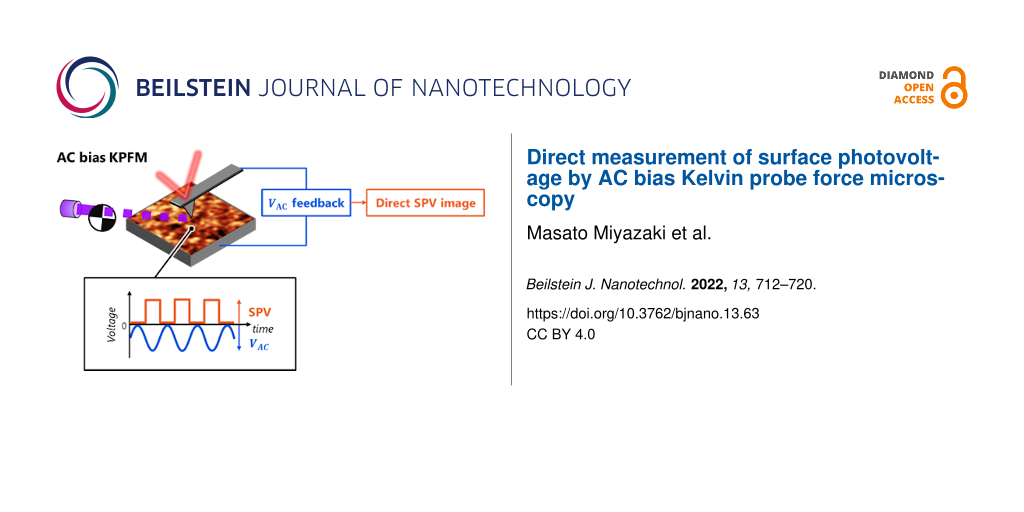
In this paper, we propose a novel method of directly measuring the local SPV, namely AC bias Kelvin probe force microscopy (AC-KPFM), which is based on a lock-in technique (widely used in standard KPFM setups) and controls the AC bias to nullify a modulated signal, referring to the AC bias null method presented by Kohl and co-workers [28]. AC-KPFM avoids the problem of thermal drift and achieves a higher resolution. We provide the theory for both the amplitude modulation (AM) mode [29] and the frequency modulation (FM) mode [30] and demonstrate experiments using AC-KPFM in the FM mode.
Theory of AC-KPFM for SPV Measurements
General concept
KPFM measures the CPD by compensating the electrostatic forces between the tip and the sample. When an AC bias VAC·cos(ωmt) with modulation frequency ωm between the tip and the sample is applied, the electrostatic force Fele in darkness is described as
![[2190-4286-13-63-i1]](/bjnano/content/inline/2190-4286-13-63-i1.svg?max-width=590&scale=1.18182)
where ∂C/∂z is the capacitance gradient of the tip–sample system and VCPD is the CPD in darkness. Applying a modulated laser power with a sinusoidal waveform of frequency ωm, which is synchronized with the AC bias (Figure 1b), induces the SPV with the peak-to-peak amplitude VSPV:
![[2190-4286-13-63-i2]](/bjnano/content/inline/2190-4286-13-63-i2.svg?max-width=590&scale=1.18182)
Therefore, the electrostatic force ![[Graphic 1]](/bjnano/content/inline/2190-4286-13-63-i22.svg?max-width=637&scale=1.18182) under modulated laser irradiation is described as
under modulated laser irradiation is described as
![[2190-4286-13-63-i3]](/bjnano/content/inline/2190-4286-13-63-i3.svg?max-width=590&scale=1.18182)
This equation can be divided into three parts:
![[2190-4286-13-63-i4]](/bjnano/content/inline/2190-4286-13-63-i4.svg?max-width=590&scale=1.18182)
![[2190-4286-13-63-i5]](/bjnano/content/inline/2190-4286-13-63-i5.svg?max-width=590&scale=1.18182)
![[2190-4286-13-63-i6]](/bjnano/content/inline/2190-4286-13-63-i6.svg?max-width=590&scale=1.18182)
![[Graphic 2]](/bjnano/content/inline/2190-4286-13-63-i23.svg?max-width=637&scale=1.18182) (Equation 5) is measured to determine the SPV by controlling VAC and nullifying the modulated force
(Equation 5) is measured to determine the SPV by controlling VAC and nullifying the modulated force ![[Graphic 3]](/bjnano/content/inline/2190-4286-13-63-i24.svg?max-width=637&scale=1.18182) where the SPV is derived as
where the SPV is derived as
![[2190-4286-13-63-i7]](/bjnano/content/inline/2190-4286-13-63-i7.svg?max-width=590&scale=1.18182)
Thus, AC-KPFM controls the AC bias VAC to directly measure the SPV, unlike classical KPFM, in which the DC bias VDC is controlled to determine the CPD or SPV. It is noted that when the SPV is negative, VAC yields a negative amplitude, where the phase of the AC bias is in phase opposition. It is also noted that it would be useful to measure the signal of the second harmonic component ![[Graphic 4]](/bjnano/content/inline/2190-4286-13-63-i25.svg?max-width=637&scale=1.18182) (Equation 6) since zero amplitude of
(Equation 6) since zero amplitude of ![[Graphic 5]](/bjnano/content/inline/2190-4286-13-63-i26.svg?max-width=637&scale=1.18182) indicates that the SPV is correctly compensated by the VAC control.
indicates that the SPV is correctly compensated by the VAC control.
![[2190-4286-13-63-1]](/bjnano/content/figures/2190-4286-13-63-1.png?scale=2.0&max-width=1024&background=FFFFFF)
Figure 1: Schematics of AC-KPFM for direct SPV measurements. (a) Block diagram of AC-KPFM in FM mode. FG is a function generator. (b, c) Scheme of the AC bias nullifying method by laser power modulation with (b) sinusoidal and (c) square waveforms, which are synchronized to VAC.
Figure 1: Schematics of AC-KPFM for direct SPV measurements. (a) Block diagram of AC-KPFM in FM mode. FG is a...
Next, when the laser power is modulated with a square waveform of frequency ωm using, for example, a chopper synchronized to the AC bias (Figure 1c), the SPV with a peak-to-peak amplitude VSPV is expressed as a Fourier series,
![[2190-4286-13-63-i8]](/bjnano/content/inline/2190-4286-13-63-i8.svg?max-width=590&scale=1.18182)
Therefore, the electrostatic force ![[Graphic 6]](/bjnano/content/inline/2190-4286-13-63-i27.svg?max-width=637&scale=1.18182) under square-waveform illumination is described as
under square-waveform illumination is described as
![[2190-4286-13-63-i9]](/bjnano/content/inline/2190-4286-13-63-i9.svg?max-width=590&scale=1.18182)
The modulated force with frequency ωm is described as
![[2190-4286-13-63-i10]](/bjnano/content/inline/2190-4286-13-63-i10.svg?max-width=590&scale=1.18182)
In the same manner as before, the SPV is determined by controlling VAC and nullifying the modulated force ![[Graphic 7]](/bjnano/content/inline/2190-4286-13-63-i28.svg?max-width=637&scale=1.18182)
![[2190-4286-13-63-i11]](/bjnano/content/inline/2190-4286-13-63-i11.svg?max-width=590&scale=1.18182)
Thus, AC-KPFM can directly and quantitatively measure the SPV by laser power modulation with either sinusoidal or square waveforms.
AC-KPFM in AM mode
In the AM mode, AC-KPFM measures the oscillation amplitude with frequency ωm, which is driven by the modulated electrostatic force ![[Graphic 8]](/bjnano/content/inline/2190-4286-13-63-i29.svg?max-width=637&scale=1.18182) This signal is measured with a lock-in amplifier and compensated by VAC control, yielding the SPV value. To improve the sensitivity, ωm is usually tuned to the second (first) resonance frequency of the cantilever, while the first (second) resonance frequency is assigned to the AFM measurement [29]. Since these resonance frequencies are commonly in the kilohertz to megahertz range, the time scale of the measured SPV is from microseconds to milliseconds, which is much faster than that measured by classical KPFM, which measures the slow SPV response of the order of seconds to hours because of the long image acquisition time [31] and the need for consecutive experiments in darkness and under illumination. Here, ωm should be set slower than the intrinsic SPV response, which we aim to observe, otherwise the SPV response cannot follow the modulated laser and yields zero amplitude. The spatial and energy resolutions and the image acquisition time of AC-KPFM in the AM mode are comparable to those of the classical KPFM in the AM mode, because both methods detect the electrostatic force,
This signal is measured with a lock-in amplifier and compensated by VAC control, yielding the SPV value. To improve the sensitivity, ωm is usually tuned to the second (first) resonance frequency of the cantilever, while the first (second) resonance frequency is assigned to the AFM measurement [29]. Since these resonance frequencies are commonly in the kilohertz to megahertz range, the time scale of the measured SPV is from microseconds to milliseconds, which is much faster than that measured by classical KPFM, which measures the slow SPV response of the order of seconds to hours because of the long image acquisition time [31] and the need for consecutive experiments in darkness and under illumination. Here, ωm should be set slower than the intrinsic SPV response, which we aim to observe, otherwise the SPV response cannot follow the modulated laser and yields zero amplitude. The spatial and energy resolutions and the image acquisition time of AC-KPFM in the AM mode are comparable to those of the classical KPFM in the AM mode, because both methods detect the electrostatic force, ![[Graphic 9]](/bjnano/content/inline/2190-4286-13-63-i30.svg?max-width=637&scale=1.18182) and the response time of the bias feedback τ limits the image acquisition time. To reach sufficient sensitivity, the
and the response time of the bias feedback τ limits the image acquisition time. To reach sufficient sensitivity, the ![[Graphic 10]](/bjnano/content/inline/2190-4286-13-63-i31.svg?max-width=637&scale=1.18182) value should typically be larger than 100 mV.
value should typically be larger than 100 mV.
AC-KPFM in FM mode
In the FM mode, AC-KPFM measures the modulated frequency shift ![[Graphic 11]](/bjnano/content/inline/2190-4286-13-63-i32.svg?max-width=637&scale=1.18182) with frequency ωm, which is driven by the modulated electrostatic force
with frequency ωm, which is driven by the modulated electrostatic force ![[Graphic 12]](/bjnano/content/inline/2190-4286-13-63-i33.svg?max-width=637&scale=1.18182) For a small oscillation amplitude,
For a small oscillation amplitude, ![[Graphic 13]](/bjnano/content/inline/2190-4286-13-63-i34.svg?max-width=637&scale=1.18182) under a modulated laser can be approximately expressed as
under a modulated laser can be approximately expressed as
![[2190-4286-13-63-i12]](/bjnano/content/inline/2190-4286-13-63-i12.svg?max-width=590&scale=1.18182)
where
![[2190-4286-13-63-i13]](/bjnano/content/inline/2190-4286-13-63-i13.svg?max-width=590&scale=1.18182)
This signal is measured by a lock-in amplifier and compensated by controlling VAC, yielding the SPV value:
![[2190-4286-13-63-i14]](/bjnano/content/inline/2190-4286-13-63-i14.svg?max-width=590&scale=1.18182)
Since ωm is usually set in a range from a few tens of hertz to several kilohertz, the time scale of the measured SPV is of the order of milliseconds, which is faster than that measured by classical KPFM. The spatial and energy resolutions and the image acquisition time of AC-KPFM in the FM mode are comparable to those of the classical KPFM in the FM mode, because both methods detect the electrostatic force gradient ![[Graphic 14]](/bjnano/content/inline/2190-4286-13-63-i35.svg?max-width=637&scale=1.18182) and the response time of the bias feedback τ limits their image acquisition time [31]. To reach sufficient sensitivity, the
and the response time of the bias feedback τ limits their image acquisition time [31]. To reach sufficient sensitivity, the ![[Graphic 15]](/bjnano/content/inline/2190-4286-13-63-i36.svg?max-width=637&scale=1.18182) value should typically be larger than 1 V.
value should typically be larger than 1 V.
Experimental
The experiments were performed by customized ultrahigh-vacuum (UHV) noncontact atomic force microscopy (NC-AFM, UNISOKU) at a temperature T of 78 K with a base pressure below 5 × 10⁻¹¹ Torr. The NC-AFM was operated in the FM mode [32] with a constant oscillation amplitude A of 500 pm. The cantilever deflection was measured by an optical beam deflection (OBD) method [33].
AC-KPFM was carried out in the FM mode, in which the topography and SPV were measured simultaneously. An AC bias VAC with frequency ωm and a DC bias VDC were applied to the sample. The laser power was modulated to a square waveform by a chopper at frequency ωm, synchronized to the AC bias. VAC is controlled to nullify the modulated frequency shift (Equation 12), yielding the SPV value. We used a digital lock-in amplifier (HF2LI with PID option, Zurich Instruments) to generate and control the AC bias. The typical sensitivity of our measurements was estimated to be δV = 1 mV (see Appendix). We simultaneously measured the tunneling current through the I/V converter as scanning tunneling microscopy (STM) [34] to consider the influence of the photocurrent [35-37].
The ultraviolet (UV) light source was a He–Cd laser (Kimmon Koha) with a wavelength of 325 nm and a laser power of 2 mW. A lens was equipped on a xyz-scanner in the UHV chamber to focus the laser onto the sample with a beam diameter of 500 µm. A band-pass filter was arranged in front of a photodetector of the OBD system to suppress the influence of the UV light on the deflection sensor.
We used a commercial Ir-coated Si cantilever (NANOSENSORS, SD-T7L100) with a resonant frequency f0 of 913 kHz, a spring constant k of 650 N/m, and a quality factor Q of 7748. The tip was cleaned by Ar+ sputtering (0.8 keV, 5 × 10⁻⁷ Torr, 5 min) to remove the contaminants and the native oxide layer. We used a rutile TiO2(110) sample to demonstrate the AC-KPFM. TiO2 is one of the promising photocatalytic materials [38-40] and has been widely studied using AFM and KPFM [41-44]. Rutile TiO2 has a bandgap of 3.0 eV [45] and shows the SPV under UV illumination [46-48]. A clean rutile TiO2(110) surface (Crystal Base) was prepared by several cycles of Ar+ sputtering (1 keV, 1 × 10⁻⁶ Torr, 15 min) and annealing (993 K, less than 2 × 10⁻¹⁰ Torr, 30 min).
Results and Discussion
First, we detected the SPV signal with AC-KPFM in the FM mode. Figure 2a shows the spectrum of the frequency shift Δf under modulated UV laser illumination with frequency fm = 100 Hz. The peak appeared at 100 Hz only when the tip approached the sample. Here, VDC was set to −300 mV to ensure sufficient sensitivity (CPD ≈ 600 mV). Then we applied VAC, which was synchronized with the modulated laser, and measured the amplitude of that peak at 100 Hz depending on VAC (Figure 2c). The amplitude of the peak showed linear behavior as a function of VAC under UV illumination, which is consistent with Equation 12. While the x-intercept was 0 V in darkness, the x-intercept was 22.1 mV under UV illumination, at which VAC nullified the modulated frequency shift and the SPV value |VSPV| of 34.7 mV was determined from Equation 14. Therefore, when VAC was set to 22.1 mV, the peak at 100 Hz disappeared in the spectrum of Δf (Figure 2b). Here, we cannot determine the polarity of the SPV because the phase for the lock-in amplifier was adjusted to maximize the absolute value of the demodulated output. We note that the response time of SPV on the rutile TiO2(110) surface is intrinsically sufficiently fast (of the order of nanoseconds) [46,49] compared with the modulation frequency of 100 Hz. Thus, we confirmed that the SPV signal of AC-KPFM agreed with the present theory described above.
![[2190-4286-13-63-2]](/bjnano/content/figures/2190-4286-13-63-2.png?scale=2.0&max-width=1024&background=FFFFFF)
Figure 2:
PSD of Δf under modulated UV laser illumination (fm = 100 Hz) with (a) VAC = 0 and (b) VAC = 2/π VSPV, where VAC nullifies the SPV. (c) Dependence of the amplitude of ![[Graphic 16]](/bjnano/content/inline/2190-4286-13-63-i37.svg?max-width=637&scale=1.18182) (the peak at 100 Hz in (a)) on VAC in darkness (black) and under UV illumination (blue) measured using a lock-in amplifier. Circles and solid lines are experimental data and fit results, respectively.
(the peak at 100 Hz in (a)) on VAC in darkness (black) and under UV illumination (blue) measured using a lock-in amplifier. Circles and solid lines are experimental data and fit results, respectively.
Figure 2: PSD of Δf under modulated UV laser illumination (fm = 100 Hz) with (a) VAC = 0 and (b) VAC = 2/π VS...
Next, we performed AC-KPFM imaging to directly obtain the SPV distribution. Figure 3a shows the AFM image of the rutile TiO2(110) surface. Terrace and step structures were observed, and the surface was flat within a single step height of 325 pm [50]. Figure 3b shows the SPV image obtained simultaneously with the AFM image. No tunneling current was detected in the AC-KPFM measurement. The SPV profile is shown in Figure 3c. The AC-KPFM successfully resolved the inhomogeneous SPV distribution with fluctuations on scales of 10–50 nm and a few millivolts, whereas classical KPFM observed a homogeneous SPV distribution over the TiO2 surface with sub-micrometer resolution [12,51] because of the influence of thermal drift between darkness and illumination. In the case of semiconductors, an electric field is screened on the scale of the Debye length LD [3],
![[2190-4286-13-63-i15]](/bjnano/content/inline/2190-4286-13-63-i15.svg?max-width=590&scale=1.18182)
where kB is the Boltzmann constant, T is the temperature, ε0 is the vacuum permittivity, εr is the relative permittivity of the semiconductor, e is the elementary charge, and n is the carrier density. The carrier density of TiO2 can be estimated from the crystal color and was 1016–1018 cm−3 for the slightly reduced TiO2 (light-blue color) [52-54]. At a temperature of 78 K, LD is calculated to be 8–80 nm, while a εr value of rutile TiO2 of 170 is used [55,56]. The calculated LD is consistent with the spatial extent of the local SPV observed by AC-KPFM. We could not find any correlation between the topographic and SPV images. The origin of the inhomogeneous SPV would involve the local distribution of the defect concentration, trapping sites, and an intrinsic electric field [57-59]; however, this is beyond the scope of this paper.
![[2190-4286-13-63-3]](/bjnano/content/figures/2190-4286-13-63-3.png?scale=2.0&max-width=1024&background=FFFFFF)
Figure 3: AC-KPFM imaging for SPV measurement. (a) Topographic and (b) SPV images of TiO2(110) surface. (c) SPV profile along the blue line in (b). The acquisition parameters are fm = 100 Hz, Δf = −80 Hz, VDC = −0.3 V, and an imaging size of 200 × 200 nm2.
Figure 3: AC-KPFM imaging for SPV measurement. (a) Topographic and (b) SPV images of TiO2(110) surface. (c) S...
We note that the time scale of SPV measured with AC-KPFM is determined by the modulation frequency of the laser power and is faster (microseconds to milliseconds) than that in the case of classical KPFM (seconds to hours) because of the need for consecutive measurements in darkness and under illumination. Thus, AC-KPFM and classical KPFM measure the SPV derived from different origins, such as charge recombination (nanoseconds to milliseconds) [60], ion transport (milliseconds to seconds) [61], and surface chemical reactions (hours) [62-64]. Particularly for photocatalytic semiconductors, AC-KPFM would be an indispensable tool for detecting the fast SPV distribution related to charge redistribution (microseconds to milliseconds) because SPV measured with classical KFPM is attributed to both charge redistribution and surface modifications by photocatalytic reactions. Intriguingly, this fact implies that performing the AC-KPFM with changing the modulation frequency of the excitation laser between low and high frequencies is useful to determine the time constant of the SPV response and the origin of the SPV. Indeed, there remains an issue that the modulation frequency has the constraint of a transfer function of cantilever dynamics and the bandwidth of the PLL. We believe that this issue will be solved by future work such as using a heterodyne detection scheme [65-67]. It is noted that AC-KPFM observes not an instantaneous photo-response of the system but a photo-response in equilibrium states, which is synchronized to the modulation of the excitation laser. We are convinced that this SPV response also provides crucial and attractive information.
Conclusion
We have proposed the method of AC bias KPFM (AC-KPFM), which nullifies the modulated SPV by controlling the AC bias, to directly measure the SPV distribution. It improves the spatial and energy resolutions thanks to the exclusion of the thermal drift between darkness and illumination, compared with the classical KPFM method. Moreover, we found that AC-KPFM can detect faster SPV responses (microseconds to milliseconds) depending on the modulation frequency of the laser power. AC-KPFM is applicable to both AM and FM modes, so it contributes to advancing SPV measurements in various environments such as vacuum, air, and liquids. Note that it would be useful to operate AC-KPFM with a heterodyne detection scheme [65-67] in order to reduce a photothermal effect on the cantilever dynamics [68,69] and measure the fast SPV phenomena. The AC-KPFM method is utilized not only for SPV measurements, but also for direct measurements of changes in surface potential induced by modulated external disturbances such as electric fields, magnetic fields [70,71], and stress fields [72,73].
Appendix
Sensitivity of AC-KPFM in the FM mode
The sensitivity of AC-KPFM in the FM mode is comparable to the sensitivity of FM-KPFM. The frequency noise density nFM is described as [74]
![[2190-4286-13-63-i16]](/bjnano/content/inline/2190-4286-13-63-i16.svg?max-width=590&scale=1.18182)
where ntherm is the thermal noise density, ndetect is the detector noise density, nosc is the oscillator noise density, and nds is the deflection sensor noise density. The frequency noise δf measured with a bandwidth of B at a center of fm is described as
![[2190-4286-13-63-i17]](/bjnano/content/inline/2190-4286-13-63-i17.svg?max-width=590&scale=1.18182)
When fm ≫ B, δf is described as
![[2190-4286-13-63-i18]](/bjnano/content/inline/2190-4286-13-63-i18.svg?max-width=590&scale=1.18182)
Here, we note that the frequency noise derived from ndetect is proportional to fm and small fm yields a low noise level:
![[2190-4286-13-63-i19]](/bjnano/content/inline/2190-4286-13-63-i19.svg?max-width=590&scale=1.18182)
For a small oscillation amplitude, the measured signal strength ![[Graphic 17]](/bjnano/content/inline/2190-4286-13-63-i38.svg?max-width=637&scale=1.18182) in AC-KPFM is approximately expressed as
in AC-KPFM is approximately expressed as
![[2190-4286-13-63-i20]](/bjnano/content/inline/2190-4286-13-63-i20.svg?max-width=590&scale=1.18182)
Therefore, the minimum detectable voltage δV is described as
![[2190-4286-13-63-i21]](/bjnano/content/inline/2190-4286-13-63-i21.svg?max-width=590&scale=1.18182)
The typical value of δf and δV in our measurements were estimated to be 0.1 Hz and 1 mV, respectively, with T = 80 K, Q = 10,000, k = 650 N/m, A = 500 pm, f0 = 910 kHz, fm = 100 Hz, B = 10 Hz, nds = 100 fm/![[Graphic 18]](/bjnano/content/inline/2190-4286-13-63-i39.svg?max-width=637&scale=1.18182) ,
, ![[Graphic 19]](/bjnano/content/inline/2190-4286-13-63-i40.svg?max-width=637&scale=1.18182) = 111.25 Hz/V2, and
= 111.25 Hz/V2, and ![[Graphic 20]](/bjnano/content/inline/2190-4286-13-63-i41.svg?max-width=637&scale=1.18182) = 1 V. The value of
= 1 V. The value of ![[Graphic 21]](/bjnano/content/inline/2190-4286-13-63-i42.svg?max-width=637&scale=1.18182) was calculated from Kelvin probe force spectroscopy (KPFS) [75,76], as shown in Figure 4.
was calculated from Kelvin probe force spectroscopy (KPFS) [75,76], as shown in Figure 4.
![[2190-4286-13-63-4]](/bjnano/content/figures/2190-4286-13-63-4.png?scale=2.0&max-width=1024&background=FFFFFF)
Figure 4:
KPFS measurement on a TiO2(110) surface. KPFS data (blue dots) was fitted by Δf(V) = a1V2 + b1 (red solid line), where 2a1 provides the value of ![[Graphic 22]](/bjnano/content/inline/2190-4286-13-63-i43.svg?max-width=637&scale=1.18182) .
.
Figure 4: KPFS measurement on a TiO2(110) surface. KPFS data (blue dots) was fitted by Δf(V) = a1V2 + b1 (red...
Funding
This work was supported by a Grant-in-Aid for Scientific Research from Japan Society for the Promotion of Science (JSPS) from the Ministry of Education, Culture, Sports, Science, and Technology of Japan (JP16H06327, JP16H06504, JP17H01061 and JP20J20223), the International Joint Research Promotion Program of Osaka University (J171013014, J171013007, J181013004, J181013006, Ja1999001, and Ja19990011), JSPS, and the National Natural Science Foundation of China (NSFC) (JSPSNSFC J191053055).
References
-
Kronik, L.; Shapira, Y. Surf. Sci. Rep. 1999, 37, 1–206. doi:10.1016/s0167-5729(99)00002-3
Return to citation in text: [1] -
Schroder, D. K. Meas. Sci. Technol. 2001, 12, R16–R31. doi:10.1088/0957-0233/12/3/202
Return to citation in text: [1] -
Zhang, Z.; Yates, J. T., Jr. Chem. Rev. 2012, 112, 5520–5551. doi:10.1021/cr3000626
Return to citation in text: [1] [2] -
Gleason-Rohrer, D. C.; Brunschwig, B. S.; Lewis, N. S. J. Phys. Chem. C 2013, 117, 18031–18042. doi:10.1021/jp401585s
Return to citation in text: [1] -
Takihara, M.; Takahashi, T.; Ujihara, T. Appl. Phys. Lett. 2008, 93, 021902. doi:10.1063/1.2957468
Return to citation in text: [1] -
Borowik, Ł.; Lepage, H.; Chevalier, N.; Mariolle, D.; Renault, O. Nanotechnology 2014, 25, 265703. doi:10.1088/0957-4484/25/26/265703
Return to citation in text: [1] -
Ozawa, K.; Yamamoto, S.; Mase, K.; Matsuda, I. e-J. Surf. Sci. Nanotechnol. 2019, 17, 130–147. doi:10.1380/ejssnt.2019.130
Return to citation in text: [1] -
Johnson, E. O. J. Appl. Phys. 1957, 28, 1349–1353. doi:10.1063/1.1722650
Return to citation in text: [1] -
Takihara, M.; Takahashi, T.; Ujihara, T. Appl. Phys. Lett. 2009, 95, 191908. doi:10.1063/1.3264081
Return to citation in text: [1] -
Gwon, M.; Sohn, A.; Cho, Y.; Phark, S.-H.; Ko, J.; Sang Kim, Y.; Kim, D.-W. Sci. Rep. 2015, 5, 16727. doi:10.1038/srep16727
Return to citation in text: [1] -
Gao, Y.; Nie, W.; Zhu, Q.; Wang, X.; Wang, S.; Fan, F.; Li, C. Angew. Chem., Int. Ed. 2020, 59, 18218–18223. doi:10.1002/anie.202007706
Return to citation in text: [1] -
Kazuma, E.; Tatsuma, T. Adv. Mater. Interfaces 2014, 1, 1400066. doi:10.1002/admi.201400066
Return to citation in text: [1] [2] -
Nonnenmacher, M.; O’Boyle, M. P.; Wickramasinghe, H. K. Appl. Phys. Lett. 1991, 58, 2921–2923. doi:10.1063/1.105227
Return to citation in text: [1] -
Zhu, J.; Fan, F.; Chen, R.; An, H.; Feng, Z.; Li, C. Angew. Chem., Int. Ed. 2015, 54, 9111–9114. doi:10.1002/anie.201504135
Return to citation in text: [1] -
Shearer, M. J.; Li, M.-Y.; Li, L.-J.; Jin, S.; Hamers, R. J. J. Phys. Chem. C 2018, 122, 13564–13571. doi:10.1021/acs.jpcc.7b12579
Return to citation in text: [1] -
Grévin, B.; Schwartz, P.-O.; Biniek, L.; Brinkmann, M.; Leclerc, N.; Zaborova, E.; Méry, S. Beilstein J. Nanotechnol. 2016, 7, 799–808. doi:10.3762/bjnano.7.71
Return to citation in text: [1] -
Glatzel, T.; Rusu, M.; Sadewasser, S.; Lux-Steiner, M. C. Nanotechnology 2008, 19, 145705. doi:10.1088/0957-4484/19/14/145705
Return to citation in text: [1] -
Marchat, C.; Dai, L.; Alvarez, J.; Le Gall, S.; Kleider, J.-P.; Misra, S.; Roca i Cabarrocas, P. Nanoscale Res. Lett. 2019, 14, 398. doi:10.1186/s11671-019-3230-5
Return to citation in text: [1] -
Deeb, M. A.; Ledig, J.; Wei, J.; Wang, X.; Wehmann, H.-H.; Waag, A. J. Appl. Phys. 2017, 122, 085307. doi:10.1063/1.5000137
Return to citation in text: [1] -
Minj, A.; Cros, A.; Auzelle, T.; Pernot, J.; Daudin, B. Nanotechnology 2016, 27, 385202. doi:10.1088/0957-4484/27/38/385202
Return to citation in text: [1] -
Qian, Y.; Wang, P.; Rao, L.; Song, C.; Yin, H.; Wang, X.; Zhou, G.; Nötzel, R. Sci. Rep. 2020, 10, 5930. doi:10.1038/s41598-020-62820-3
Return to citation in text: [1] -
Binnig, G.; Quate, C. F.; Gerber, C. Phys. Rev. Lett. 1986, 56, 930–933. doi:10.1103/physrevlett.56.930
Return to citation in text: [1] -
Schumacher, Z.; Miyahara, Y.; Spielhofer, A.; Grutter, P. Phys. Rev. Appl. 2016, 5, 044018. doi:10.1103/physrevapplied.5.044018
Return to citation in text: [1] [2] -
Streicher, F.; Sadewasser, S.; Lux-Steiner, M. C. Rev. Sci. Instrum. 2009, 80, 013907. doi:10.1063/1.3072661
Return to citation in text: [1] -
Sugawara, Y.; Yamanishi, J.; Tokuyama, T.; Naitoh, Y.; Li, Y. J. Phys. Rev. Appl. 2015, 3, 044020. doi:10.1103/physrevapplied.3.044020
Return to citation in text: [1] -
Collins, L.; Ahmadi, M.; Qin, J.; Liu, Y.; Ovchinnikova, O. S.; Hu, B.; Jesse, S.; Kalinin, S. V. Nanotechnology 2018, 29, 445703. doi:10.1088/1361-6528/aad873
Return to citation in text: [1] -
Grévin, B.; Bardagot, O.; Demadrille, R. Beilstein J. Nanotechnol. 2020, 11, 323–337. doi:10.3762/bjnano.11.24
Return to citation in text: [1] -
Kohl, D.; Mesquida, P.; Schitter, G. Microelectron. Eng. 2017, 176, 28–32. doi:10.1016/j.mee.2017.01.005
Return to citation in text: [1] -
Kikukawa, A.; Hosaka, S.; Imura, R. Appl. Phys. Lett. 1995, 66, 3510–3512. doi:10.1063/1.113780
Return to citation in text: [1] [2] -
Kitamura, S.; Iwatsuki, M. Appl. Phys. Lett. 1998, 72, 3154–3156. doi:10.1063/1.121577
Return to citation in text: [1] -
Melitz, W.; Shen, J.; Kummel, A. C.; Lee, S. Surf. Sci. Rep. 2011, 66, 1–27. doi:10.1016/j.surfrep.2010.10.001
Return to citation in text: [1] [2] -
Albrecht, T. R.; Grütter, P.; Horne, D.; Rugar, D. J. Appl. Phys. 1991, 69, 668–673. doi:10.1063/1.347347
Return to citation in text: [1] -
Meyer, G.; Amer, N. M. Appl. Phys. Lett. 1988, 53, 1045–1047. doi:10.1063/1.100061
Return to citation in text: [1] -
Binnig, G.; Rohrer, H.; Gerber, C.; Weibel, E. Phys. Rev. Lett. 1982, 49, 57–61. doi:10.1103/physrevlett.49.57
Return to citation in text: [1] -
Kuk, Y.; Becker, R. S.; Silverman, P. J.; Kochanski, G. P. Phys. Rev. Lett. 1990, 65, 456–459. doi:10.1103/physrevlett.65.456
Return to citation in text: [1] -
Terada, Y.; Yoshida, S.; Takeuchi, O.; Shigekawa, H. Nat. Photonics 2010, 4, 869–874. doi:10.1038/nphoton.2010.235
Return to citation in text: [1] -
Weymouth, A. J.; Wutscher, T.; Welker, J.; Hofmann, T.; Giessibl, F. J. Phys. Rev. Lett. 2011, 106, 226801. doi:10.1103/physrevlett.106.226801
Return to citation in text: [1] -
Fujishima, A.; Zhang, X.; Tryk, D. A. Surf. Sci. Rep. 2008, 63, 515–582. doi:10.1016/j.surfrep.2008.10.001
Return to citation in text: [1] -
Diebold, U. Surf. Sci. Rep. 2003, 48, 53–229. doi:10.1016/s0167-5729(02)00100-0
Return to citation in text: [1] -
Chen, R.; Fan, F.; Dittrich, T.; Li, C. Chem. Soc. Rev. 2018, 47, 8238–8262. doi:10.1039/c8cs00320c
Return to citation in text: [1] -
Zhang, Q.; Brndiar, J.; Konôpka, M.; Wen, H. F.; Adachi, Y.; Miyazaki, M.; Turanský, R.; Xu, R.; Cheng, Z. H.; Sugawara, Y.; Štich, I.; Li, Y. J. J. Phys. Chem. C 2021, 125, 27607–27614. doi:10.1021/acs.jpcc.1c07997
Return to citation in text: [1] -
Adachi, Y.; Wen, H. F.; Zhang, Q.; Miyazaki, M.; Sugawara, Y.; Li, Y. J. Nanoscale Adv. 2020, 2, 2371–2375. doi:10.1039/c9na00776h
Return to citation in text: [1] -
Miyazaki, M.; Sugawara, Y.; Li, Y. J. Langmuir 2021, 37, 10588–10593. doi:10.1021/acs.langmuir.1c01845
Return to citation in text: [1] -
Wen, H. F.; Sugawara, Y.; Li, Y. J. Nanomaterials 2020, 10, 1506. doi:10.3390/nano10081506
Return to citation in text: [1] -
Kavan, L.; Grätzel, M.; Gilbert, S. E.; Klemenz, C.; Scheel, H. J. J. Am. Chem. Soc. 1996, 118, 6716–6723. doi:10.1021/ja954172l
Return to citation in text: [1] -
Ozawa, K.; Emori, M.; Yamamoto, S.; Yukawa, R.; Yamamoto, S.; Hobara, R.; Fujikawa, K.; Sakama, H.; Matsuda, I. J. Phys. Chem. Lett. 2014, 5, 1953–1957. doi:10.1021/jz500770c
Return to citation in text: [1] [2] -
Henning, A.; Günzburger, G.; Jöhr, R.; Rosenwaks, Y.; Bozic-Weber, B.; Housecroft, C. E.; Constable, E. C.; Meyer, E.; Glatzel, T. Beilstein J. Nanotechnol. 2013, 4, 418–428. doi:10.3762/bjnano.4.49
Return to citation in text: [1] -
Sheppard, L. R.; Dittrich, T.; Nowotny, J.; Bak, T. Appl. Phys. Lett. 2010, 96, 072104. doi:10.1063/1.3318465
Return to citation in text: [1] -
Schneider, J.; Matsuoka, M.; Takeuchi, M.; Zhang, J.; Horiuchi, Y.; Anpo, M.; Bahnemann, D. W. Chem. Rev. 2014, 114, 9919–9986. doi:10.1021/cr5001892
Return to citation in text: [1] -
Onishi, H.; Iwasawa, Y. Surf. Sci. 1994, 313, L783–L789. doi:10.1016/0039-6028(94)91146-0
Return to citation in text: [1] -
Yoo, H.; Bae, C.; Yang, Y.; Lee, S.; Kim, M.; Kim, H.; Kim, Y.; Shin, H. Nano Lett. 2014, 14, 4413–4417. doi:10.1021/nl501381a
Return to citation in text: [1] -
Li, M.; Hebenstreit, W.; Diebold, U.; Tyryshkin, A. M.; Bowman, M. K.; Dunham, G. G.; Henderson, M. A. J. Phys. Chem. B 2000, 104, 4944–4950. doi:10.1021/jp9943272
Return to citation in text: [1] -
Iguchi, E.; Yajima, K.; Asahina, T.; Kanamori, Y. J. Phys. Chem. Solids 1974, 35, 597–599. doi:10.1016/s0022-3697(74)80016-8
Return to citation in text: [1] -
Nilius, N.; Ernst, N.; Freund, H.-J. Chem. Phys. Lett. 2001, 349, 351–357. doi:10.1016/s0009-2614(01)01232-5
Return to citation in text: [1] -
Parker, R. A. Phys. Rev. 1961, 124, 1719–1722. doi:10.1103/physrev.124.1719
Return to citation in text: [1] -
Bonkerud, J.; Zimmermann, C.; Weiser, P. M.; Vines, L.; Monakhov, E. V. Sci. Rep. 2021, 11, 12443. doi:10.1038/s41598-021-92021-5
Return to citation in text: [1] -
Qian, R.; Zong, H.; Schneider, J.; Zhou, G.; Zhao, T.; Li, Y.; Yang, J.; Bahnemann, D. W.; Pan, J. H. Catal. Today 2019, 335, 78–90. doi:10.1016/j.cattod.2018.10.053
Return to citation in text: [1] -
Liu, B.; Zhao, X.; Yu, J.; Parkin, I. P.; Fujishima, A.; Nakata, K. J. Photochem. Photobiol., C 2019, 39, 1–57. doi:10.1016/j.jphotochemrev.2019.02.001
Return to citation in text: [1] -
Onoda, J.; Pang, C. L.; Yurtsever, A.; Sugimoto, Y. J. Phys. Chem. C 2014, 118, 13674–13679. doi:10.1021/jp503402w
Return to citation in text: [1] -
Gao, Y.; Nie, W.; Wang, X.; Fan, F.; Li, C. Chem. Commun. 2020, 56, 1007–1021. doi:10.1039/c9cc07128h
Return to citation in text: [1] -
Shi, J.; Li, Y.; Li, Y.; Li, D.; Luo, Y.; Wu, H.; Meng, Q. Joule 2018, 2, 879–901. doi:10.1016/j.joule.2018.04.010
Return to citation in text: [1] -
Dagdeviren, O. E.; Glass, D.; Sapienza, R.; Cortés, E.; Maier, S. A.; Parkin, I. P.; Grütter, P.; Quesada-Cabrera, R. Nano Lett. 2021, 21, 8348–8354. doi:10.1021/acs.nanolett.1c02853
Return to citation in text: [1] -
Fischer, S.; Bokareva, O. S.; Barsch, E.; Bokarev, S. I.; Kühn, O.; Ludwig, R. ChemCatChem 2016, 8, 404–411. doi:10.1002/cctc.201500872
Return to citation in text: [1] -
Zhan, X.; Yan, C.; Zhang, Y.; Rinke, G.; Rabsch, G.; Klumpp, M.; Schäfer, A. I.; Dittmeyer, R. React. Chem. Eng. 2020, 5, 1658–1670. doi:10.1039/d0re00238k
Return to citation in text: [1] -
Sugawara, Y.; Kou, L.; Ma, Z.; Kamijo, T.; Naitoh, Y.; Li, Y. J. Appl. Phys. Lett. 2012, 100, 223104. doi:10.1063/1.4723697
Return to citation in text: [1] [2] -
Yamanishi, J.; Naitoh, Y.; Li, Y. J.; Sugawara, Y. Appl. Phys. Lett. 2017, 110, 123102. doi:10.1063/1.4978755
Return to citation in text: [1] [2] -
Sugawara, Y.; Miyazaki, M.; Li, Y. J. J. Phys. Commun. 2020, 4, 075015. doi:10.1088/2399-6528/aba477
Return to citation in text: [1] [2] -
Umeda, N.; Ishizaki, S.; Uwai, H. J. Vac. Sci. Technol., B: Microelectron. Nanometer Struct.–Process., Meas., Phenom. 1991, 9, 1318. doi:10.1116/1.585187
Return to citation in text: [1] -
Sadewasser, S.; Nicoara, N.; Solares, S. D. Beilstein J. Nanotechnol. 2018, 9, 1272–1281. doi:10.3762/bjnano.9.119
Return to citation in text: [1] -
Dremov, V. V.; Grebenchuk, S. Y.; Shishkin, A. G.; Baranov, D. S.; Hovhannisyan, R. A.; Skryabina, O. V.; Lebedev, N.; Golovchanskiy, I. A.; Chichkov, V. I.; Brun, C.; Cren, T.; Krasnov, V. M.; Golubov, A. A.; Roditchev, D.; Stolyarov, V. S. Nat. Commun. 2019, 10, 4009. doi:10.1038/s41467-019-11924-0
Return to citation in text: [1] -
Chu, Z.; Dong, C.; Tu, C.; Liang, X.; Chen, H.; Sun, C.; Yu, Z.; Dong, S.; Sun, N.-X. Appl. Phys. Lett. 2019, 115, 162901. doi:10.1063/1.5122774
Return to citation in text: [1] -
Hu, Y.; Pan, Y.; Wang, Z.; Lin, T.; Gao, Y.; Luo, B.; Hu, H.; Fan, F.; Liu, G.; Wang, L. Nat. Commun. 2020, 11, 2129. doi:10.1038/s41467-020-15993-4
Return to citation in text: [1] -
Yang, M.-M.; Kim, D. J.; Alexe, M. Science 2018, 360, 904–907. doi:10.1126/science.aan3256
Return to citation in text: [1] -
Kobayashi, K.; Yamada, H.; Matsushige, K. Rev. Sci. Instrum. 2009, 80, 043708. doi:10.1063/1.3120913
Return to citation in text: [1] -
Gross, L.; Mohn, F.; Liljeroth, P.; Repp, J.; Giessibl, F. J.; Meyer, G. Science 2009, 324, 1428–1431. doi:10.1126/science.1172273
Return to citation in text: [1] -
Adachi, Y.; Brndiar, J.; Wen, H. F.; Zhang, Q.; Miyazaki, M.; Thakur, S.; Sugawara, Y.; Sang, H.; Li, Y.; Štich, I.; Kantorovich, L. Commun. Mater. 2021, 2, 71. doi:10.1038/s43246-021-00176-5
Return to citation in text: [1]
| 12. | Kazuma, E.; Tatsuma, T. Adv. Mater. Interfaces 2014, 1, 1400066. doi:10.1002/admi.201400066 |
| 51. | Yoo, H.; Bae, C.; Yang, Y.; Lee, S.; Kim, M.; Kim, H.; Kim, Y.; Shin, H. Nano Lett. 2014, 14, 4413–4417. doi:10.1021/nl501381a |
| 3. | Zhang, Z.; Yates, J. T., Jr. Chem. Rev. 2012, 112, 5520–5551. doi:10.1021/cr3000626 |
| 52. | Li, M.; Hebenstreit, W.; Diebold, U.; Tyryshkin, A. M.; Bowman, M. K.; Dunham, G. G.; Henderson, M. A. J. Phys. Chem. B 2000, 104, 4944–4950. doi:10.1021/jp9943272 |
| 53. | Iguchi, E.; Yajima, K.; Asahina, T.; Kanamori, Y. J. Phys. Chem. Solids 1974, 35, 597–599. doi:10.1016/s0022-3697(74)80016-8 |
| 54. | Nilius, N.; Ernst, N.; Freund, H.-J. Chem. Phys. Lett. 2001, 349, 351–357. doi:10.1016/s0009-2614(01)01232-5 |
| 1. | Kronik, L.; Shapira, Y. Surf. Sci. Rep. 1999, 37, 1–206. doi:10.1016/s0167-5729(99)00002-3 |
| 2. | Schroder, D. K. Meas. Sci. Technol. 2001, 12, R16–R31. doi:10.1088/0957-0233/12/3/202 |
| 10. | Gwon, M.; Sohn, A.; Cho, Y.; Phark, S.-H.; Ko, J.; Sang Kim, Y.; Kim, D.-W. Sci. Rep. 2015, 5, 16727. doi:10.1038/srep16727 |
| 11. | Gao, Y.; Nie, W.; Zhu, Q.; Wang, X.; Wang, S.; Fan, F.; Li, C. Angew. Chem., Int. Ed. 2020, 59, 18218–18223. doi:10.1002/anie.202007706 |
| 12. | Kazuma, E.; Tatsuma, T. Adv. Mater. Interfaces 2014, 1, 1400066. doi:10.1002/admi.201400066 |
| 29. | Kikukawa, A.; Hosaka, S.; Imura, R. Appl. Phys. Lett. 1995, 66, 3510–3512. doi:10.1063/1.113780 |
| 65. | Sugawara, Y.; Kou, L.; Ma, Z.; Kamijo, T.; Naitoh, Y.; Li, Y. J. Appl. Phys. Lett. 2012, 100, 223104. doi:10.1063/1.4723697 |
| 66. | Yamanishi, J.; Naitoh, Y.; Li, Y. J.; Sugawara, Y. Appl. Phys. Lett. 2017, 110, 123102. doi:10.1063/1.4978755 |
| 67. | Sugawara, Y.; Miyazaki, M.; Li, Y. J. J. Phys. Commun. 2020, 4, 075015. doi:10.1088/2399-6528/aba477 |
| 8. | Johnson, E. O. J. Appl. Phys. 1957, 28, 1349–1353. doi:10.1063/1.1722650 |
| 9. | Takihara, M.; Takahashi, T.; Ujihara, T. Appl. Phys. Lett. 2009, 95, 191908. doi:10.1063/1.3264081 |
| 31. | Melitz, W.; Shen, J.; Kummel, A. C.; Lee, S. Surf. Sci. Rep. 2011, 66, 1–27. doi:10.1016/j.surfrep.2010.10.001 |
| 68. | Umeda, N.; Ishizaki, S.; Uwai, H. J. Vac. Sci. Technol., B: Microelectron. Nanometer Struct.–Process., Meas., Phenom. 1991, 9, 1318. doi:10.1116/1.585187 |
| 69. | Sadewasser, S.; Nicoara, N.; Solares, S. D. Beilstein J. Nanotechnol. 2018, 9, 1272–1281. doi:10.3762/bjnano.9.119 |
| 5. | Takihara, M.; Takahashi, T.; Ujihara, T. Appl. Phys. Lett. 2008, 93, 021902. doi:10.1063/1.2957468 |
| 6. | Borowik, Ł.; Lepage, H.; Chevalier, N.; Mariolle, D.; Renault, O. Nanotechnology 2014, 25, 265703. doi:10.1088/0957-4484/25/26/265703 |
| 7. | Ozawa, K.; Yamamoto, S.; Mase, K.; Matsuda, I. e-J. Surf. Sci. Nanotechnol. 2019, 17, 130–147. doi:10.1380/ejssnt.2019.130 |
| 29. | Kikukawa, A.; Hosaka, S.; Imura, R. Appl. Phys. Lett. 1995, 66, 3510–3512. doi:10.1063/1.113780 |
| 62. | Dagdeviren, O. E.; Glass, D.; Sapienza, R.; Cortés, E.; Maier, S. A.; Parkin, I. P.; Grütter, P.; Quesada-Cabrera, R. Nano Lett. 2021, 21, 8348–8354. doi:10.1021/acs.nanolett.1c02853 |
| 63. | Fischer, S.; Bokareva, O. S.; Barsch, E.; Bokarev, S. I.; Kühn, O.; Ludwig, R. ChemCatChem 2016, 8, 404–411. doi:10.1002/cctc.201500872 |
| 64. | Zhan, X.; Yan, C.; Zhang, Y.; Rinke, G.; Rabsch, G.; Klumpp, M.; Schäfer, A. I.; Dittmeyer, R. React. Chem. Eng. 2020, 5, 1658–1670. doi:10.1039/d0re00238k |
| 3. | Zhang, Z.; Yates, J. T., Jr. Chem. Rev. 2012, 112, 5520–5551. doi:10.1021/cr3000626 |
| 4. | Gleason-Rohrer, D. C.; Brunschwig, B. S.; Lewis, N. S. J. Phys. Chem. C 2013, 117, 18031–18042. doi:10.1021/jp401585s |
| 30. | Kitamura, S.; Iwatsuki, M. Appl. Phys. Lett. 1998, 72, 3154–3156. doi:10.1063/1.121577 |
| 65. | Sugawara, Y.; Kou, L.; Ma, Z.; Kamijo, T.; Naitoh, Y.; Li, Y. J. Appl. Phys. Lett. 2012, 100, 223104. doi:10.1063/1.4723697 |
| 66. | Yamanishi, J.; Naitoh, Y.; Li, Y. J.; Sugawara, Y. Appl. Phys. Lett. 2017, 110, 123102. doi:10.1063/1.4978755 |
| 67. | Sugawara, Y.; Miyazaki, M.; Li, Y. J. J. Phys. Commun. 2020, 4, 075015. doi:10.1088/2399-6528/aba477 |
| 24. | Streicher, F.; Sadewasser, S.; Lux-Steiner, M. C. Rev. Sci. Instrum. 2009, 80, 013907. doi:10.1063/1.3072661 |
| 23. | Schumacher, Z.; Miyahara, Y.; Spielhofer, A.; Grutter, P. Phys. Rev. Appl. 2016, 5, 044018. doi:10.1103/physrevapplied.5.044018 |
| 26. | Collins, L.; Ahmadi, M.; Qin, J.; Liu, Y.; Ovchinnikova, O. S.; Hu, B.; Jesse, S.; Kalinin, S. V. Nanotechnology 2018, 29, 445703. doi:10.1088/1361-6528/aad873 |
| 27. | Grévin, B.; Bardagot, O.; Demadrille, R. Beilstein J. Nanotechnol. 2020, 11, 323–337. doi:10.3762/bjnano.11.24 |
| 60. | Gao, Y.; Nie, W.; Wang, X.; Fan, F.; Li, C. Chem. Commun. 2020, 56, 1007–1021. doi:10.1039/c9cc07128h |
| 23. | Schumacher, Z.; Miyahara, Y.; Spielhofer, A.; Grutter, P. Phys. Rev. Appl. 2016, 5, 044018. doi:10.1103/physrevapplied.5.044018 |
| 28. | Kohl, D.; Mesquida, P.; Schitter, G. Microelectron. Eng. 2017, 176, 28–32. doi:10.1016/j.mee.2017.01.005 |
| 61. | Shi, J.; Li, Y.; Li, Y.; Li, D.; Luo, Y.; Wu, H.; Meng, Q. Joule 2018, 2, 879–901. doi:10.1016/j.joule.2018.04.010 |
| 22. | Binnig, G.; Quate, C. F.; Gerber, C. Phys. Rev. Lett. 1986, 56, 930–933. doi:10.1103/physrevlett.56.930 |
| 55. | Parker, R. A. Phys. Rev. 1961, 124, 1719–1722. doi:10.1103/physrev.124.1719 |
| 56. | Bonkerud, J.; Zimmermann, C.; Weiser, P. M.; Vines, L.; Monakhov, E. V. Sci. Rep. 2021, 11, 12443. doi:10.1038/s41598-021-92021-5 |
| 13. | Nonnenmacher, M.; O’Boyle, M. P.; Wickramasinghe, H. K. Appl. Phys. Lett. 1991, 58, 2921–2923. doi:10.1063/1.105227 |
| 14. | Zhu, J.; Fan, F.; Chen, R.; An, H.; Feng, Z.; Li, C. Angew. Chem., Int. Ed. 2015, 54, 9111–9114. doi:10.1002/anie.201504135 |
| 15. | Shearer, M. J.; Li, M.-Y.; Li, L.-J.; Jin, S.; Hamers, R. J. J. Phys. Chem. C 2018, 122, 13564–13571. doi:10.1021/acs.jpcc.7b12579 |
| 16. | Grévin, B.; Schwartz, P.-O.; Biniek, L.; Brinkmann, M.; Leclerc, N.; Zaborova, E.; Méry, S. Beilstein J. Nanotechnol. 2016, 7, 799–808. doi:10.3762/bjnano.7.71 |
| 17. | Glatzel, T.; Rusu, M.; Sadewasser, S.; Lux-Steiner, M. C. Nanotechnology 2008, 19, 145705. doi:10.1088/0957-4484/19/14/145705 |
| 18. | Marchat, C.; Dai, L.; Alvarez, J.; Le Gall, S.; Kleider, J.-P.; Misra, S.; Roca i Cabarrocas, P. Nanoscale Res. Lett. 2019, 14, 398. doi:10.1186/s11671-019-3230-5 |
| 19. | Deeb, M. A.; Ledig, J.; Wei, J.; Wang, X.; Wehmann, H.-H.; Waag, A. J. Appl. Phys. 2017, 122, 085307. doi:10.1063/1.5000137 |
| 20. | Minj, A.; Cros, A.; Auzelle, T.; Pernot, J.; Daudin, B. Nanotechnology 2016, 27, 385202. doi:10.1088/0957-4484/27/38/385202 |
| 21. | Qian, Y.; Wang, P.; Rao, L.; Song, C.; Yin, H.; Wang, X.; Zhou, G.; Nötzel, R. Sci. Rep. 2020, 10, 5930. doi:10.1038/s41598-020-62820-3 |
| 25. | Sugawara, Y.; Yamanishi, J.; Tokuyama, T.; Naitoh, Y.; Li, Y. J. Phys. Rev. Appl. 2015, 3, 044020. doi:10.1103/physrevapplied.3.044020 |
| 57. | Qian, R.; Zong, H.; Schneider, J.; Zhou, G.; Zhao, T.; Li, Y.; Yang, J.; Bahnemann, D. W.; Pan, J. H. Catal. Today 2019, 335, 78–90. doi:10.1016/j.cattod.2018.10.053 |
| 58. | Liu, B.; Zhao, X.; Yu, J.; Parkin, I. P.; Fujishima, A.; Nakata, K. J. Photochem. Photobiol., C 2019, 39, 1–57. doi:10.1016/j.jphotochemrev.2019.02.001 |
| 59. | Onoda, J.; Pang, C. L.; Yurtsever, A.; Sugimoto, Y. J. Phys. Chem. C 2014, 118, 13674–13679. doi:10.1021/jp503402w |
| 33. | Meyer, G.; Amer, N. M. Appl. Phys. Lett. 1988, 53, 1045–1047. doi:10.1063/1.100061 |
| 31. | Melitz, W.; Shen, J.; Kummel, A. C.; Lee, S. Surf. Sci. Rep. 2011, 66, 1–27. doi:10.1016/j.surfrep.2010.10.001 |
| 70. | Dremov, V. V.; Grebenchuk, S. Y.; Shishkin, A. G.; Baranov, D. S.; Hovhannisyan, R. A.; Skryabina, O. V.; Lebedev, N.; Golovchanskiy, I. A.; Chichkov, V. I.; Brun, C.; Cren, T.; Krasnov, V. M.; Golubov, A. A.; Roditchev, D.; Stolyarov, V. S. Nat. Commun. 2019, 10, 4009. doi:10.1038/s41467-019-11924-0 |
| 71. | Chu, Z.; Dong, C.; Tu, C.; Liang, X.; Chen, H.; Sun, C.; Yu, Z.; Dong, S.; Sun, N.-X. Appl. Phys. Lett. 2019, 115, 162901. doi:10.1063/1.5122774 |
| 32. | Albrecht, T. R.; Grütter, P.; Horne, D.; Rugar, D. J. Appl. Phys. 1991, 69, 668–673. doi:10.1063/1.347347 |
| 72. | Hu, Y.; Pan, Y.; Wang, Z.; Lin, T.; Gao, Y.; Luo, B.; Hu, H.; Fan, F.; Liu, G.; Wang, L. Nat. Commun. 2020, 11, 2129. doi:10.1038/s41467-020-15993-4 |
| 73. | Yang, M.-M.; Kim, D. J.; Alexe, M. Science 2018, 360, 904–907. doi:10.1126/science.aan3256 |
| 74. | Kobayashi, K.; Yamada, H.; Matsushige, K. Rev. Sci. Instrum. 2009, 80, 043708. doi:10.1063/1.3120913 |
| 46. | Ozawa, K.; Emori, M.; Yamamoto, S.; Yukawa, R.; Yamamoto, S.; Hobara, R.; Fujikawa, K.; Sakama, H.; Matsuda, I. J. Phys. Chem. Lett. 2014, 5, 1953–1957. doi:10.1021/jz500770c |
| 49. | Schneider, J.; Matsuoka, M.; Takeuchi, M.; Zhang, J.; Horiuchi, Y.; Anpo, M.; Bahnemann, D. W. Chem. Rev. 2014, 114, 9919–9986. doi:10.1021/cr5001892 |
| 50. | Onishi, H.; Iwasawa, Y. Surf. Sci. 1994, 313, L783–L789. doi:10.1016/0039-6028(94)91146-0 |
| 45. | Kavan, L.; Grätzel, M.; Gilbert, S. E.; Klemenz, C.; Scheel, H. J. J. Am. Chem. Soc. 1996, 118, 6716–6723. doi:10.1021/ja954172l |
| 46. | Ozawa, K.; Emori, M.; Yamamoto, S.; Yukawa, R.; Yamamoto, S.; Hobara, R.; Fujikawa, K.; Sakama, H.; Matsuda, I. J. Phys. Chem. Lett. 2014, 5, 1953–1957. doi:10.1021/jz500770c |
| 47. | Henning, A.; Günzburger, G.; Jöhr, R.; Rosenwaks, Y.; Bozic-Weber, B.; Housecroft, C. E.; Constable, E. C.; Meyer, E.; Glatzel, T. Beilstein J. Nanotechnol. 2013, 4, 418–428. doi:10.3762/bjnano.4.49 |
| 48. | Sheppard, L. R.; Dittrich, T.; Nowotny, J.; Bak, T. Appl. Phys. Lett. 2010, 96, 072104. doi:10.1063/1.3318465 |
| 38. | Fujishima, A.; Zhang, X.; Tryk, D. A. Surf. Sci. Rep. 2008, 63, 515–582. doi:10.1016/j.surfrep.2008.10.001 |
| 39. | Diebold, U. Surf. Sci. Rep. 2003, 48, 53–229. doi:10.1016/s0167-5729(02)00100-0 |
| 40. | Chen, R.; Fan, F.; Dittrich, T.; Li, C. Chem. Soc. Rev. 2018, 47, 8238–8262. doi:10.1039/c8cs00320c |
| 41. | Zhang, Q.; Brndiar, J.; Konôpka, M.; Wen, H. F.; Adachi, Y.; Miyazaki, M.; Turanský, R.; Xu, R.; Cheng, Z. H.; Sugawara, Y.; Štich, I.; Li, Y. J. J. Phys. Chem. C 2021, 125, 27607–27614. doi:10.1021/acs.jpcc.1c07997 |
| 42. | Adachi, Y.; Wen, H. F.; Zhang, Q.; Miyazaki, M.; Sugawara, Y.; Li, Y. J. Nanoscale Adv. 2020, 2, 2371–2375. doi:10.1039/c9na00776h |
| 43. | Miyazaki, M.; Sugawara, Y.; Li, Y. J. Langmuir 2021, 37, 10588–10593. doi:10.1021/acs.langmuir.1c01845 |
| 44. | Wen, H. F.; Sugawara, Y.; Li, Y. J. Nanomaterials 2020, 10, 1506. doi:10.3390/nano10081506 |
| 34. | Binnig, G.; Rohrer, H.; Gerber, C.; Weibel, E. Phys. Rev. Lett. 1982, 49, 57–61. doi:10.1103/physrevlett.49.57 |
| 75. | Gross, L.; Mohn, F.; Liljeroth, P.; Repp, J.; Giessibl, F. J.; Meyer, G. Science 2009, 324, 1428–1431. doi:10.1126/science.1172273 |
| 76. | Adachi, Y.; Brndiar, J.; Wen, H. F.; Zhang, Q.; Miyazaki, M.; Thakur, S.; Sugawara, Y.; Sang, H.; Li, Y.; Štich, I.; Kantorovich, L. Commun. Mater. 2021, 2, 71. doi:10.1038/s43246-021-00176-5 |
| 35. | Kuk, Y.; Becker, R. S.; Silverman, P. J.; Kochanski, G. P. Phys. Rev. Lett. 1990, 65, 456–459. doi:10.1103/physrevlett.65.456 |
| 36. | Terada, Y.; Yoshida, S.; Takeuchi, O.; Shigekawa, H. Nat. Photonics 2010, 4, 869–874. doi:10.1038/nphoton.2010.235 |
| 37. | Weymouth, A. J.; Wutscher, T.; Welker, J.; Hofmann, T.; Giessibl, F. J. Phys. Rev. Lett. 2011, 106, 226801. doi:10.1103/physrevlett.106.226801 |
© 2022 Miyazaki et al.; licensee Beilstein-Institut.
This is an open access article licensed under the terms of the Beilstein-Institut Open Access License Agreement (https://www.beilstein-journals.org/bjnano/terms), which is identical to the Creative Commons Attribution 4.0 International License (https://creativecommons.org/licenses/by/4.0). The reuse of material under this license requires that the author(s), source and license are credited. Third-party material in this article could be subject to other licenses (typically indicated in the credit line), and in this case, users are required to obtain permission from the license holder to reuse the material.