Abstract
Novel N-α-haloacylated homoserine lactones, in which a halogen atom was introduced at the α-position of the carbonyl function of the N-acyl chain, have been studied as quorum sensing (QS) modulators and compared with a library of natural N-acylated homoserine lactones (AHLs). The series of novel analogues consists of α-chloro, α-bromo and α-iodo AHL analogues. Furthermore, the biological QS activity of the synthetic AHL analogues compared to the natural AHLs was evaluated. Halogenated analogues demonstrated a reduced activity in the Escherichia coli JB523 bioassay, with the α-iodo lactones being the less active ones and the α-chloro AHLs the most potent QS agonists. Most of the α-haloacylated analogues did not exhibit a significant reduction when tested in the QS inhibition test. Therefore, these novel analogues could be utilized as chemical probes for QS structure–activity studies.

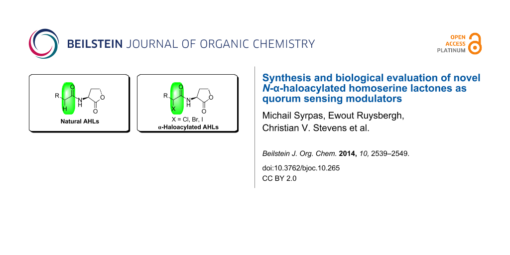
Graphical Abstract
Introduction
Quorum sensing (QS) is the communication system used by bacteria allowing them to adapt to their environment [1]. Bacteria relying on QS secrete signal molecules, called autoinducers, in order to control the expression of specific target genes important for phenotype expression (e.g., biofilm formation, bioluminescence, virulence expression, etc.) in a population density dependent manner [2]. The importance of QS for virulence development in pathogenic bacteria nowadays is obvious [3]. Therefore, quorum sensing modulation is seen as a new anti-infective strategy [4]. Although the described QS systems can regulate virulence, it is believed that they do not affect the viability of bacteria and it could be expected that disruption of QS will not likely create a selective pressure towards resistant organisms [5]. Despite recent reports supporting this theory, it has to be noted that this view point has been challenged and is still under debate among scientists [6,7]. Most Gram-negative bacteria use N-acylated homoserine lactones (AHLs) as signal molecules [8]. The general structure of these compounds consist of a lactone ring with an acyl side chain [9]. The length of the acyl chain is usually between 4–18 carbons and the functionality at the β-carbon position can vary. The QS mechanism in these bacteria relies generally upon two components: a LuxI like protein which is a synthase that produces the autoinducer and a transcriptional activator, i.e, a LuxR like protein which binds to the autoinducer [10]. Different bacterial strains were shown to respond to and produce different but specific autoinducers [11]. Furthermore, recent studies reveal that AHLs facilitate both intra-species communication and inter-kingdom interactions [12].
The strong impact of AHLs on bacterial behaviour has gained the attention of scientists towards designing AHL mimics that can modulate QS [13,14]. However, the high specificity and affinity that most of the receptors show towards their natural ligands does not allow the design of analogues with high structural deviation from the parent autoinducer [15]. In biological processes, covalent binding and non-covalent interactions are encountered in many protein-ligand interactions as well as in protein secondary structures and are of great significance. In the past years, it was shown that the proximity of carbonyl groups can influence the conformation of molecules by an n→π* interaction [16,17]. Newberry and Raines, very recently proposed that tailored small molecules, that could attenuate this n→π* interaction, in which the lone pair (n) of the N-acyl oxygen overlays with the π* orbital of the lactone carbonyl group, could increase the affinity of AHLs to their cognate receptors [18]. It was shown that introduction of halogens as electron-withdrawing groups in the N-acyl chain reduced the nucleophilicity of the donor oxygen and led to attenuation of the n→π* interaction (Figure 1). Moreover, inhibition of bacterial QS was recently demonstrated by a set of electrophilic probes (which included halogenated analogues of AHLs) that could covalently bind in the LasR binding pocket of Pseudomonas aeruginosa [15]. Therefore, designing analogues with small reactive moieties, such as halogenated carbon atoms, that could be involved in such interactions seem an interesting strategy to be explored.
![[1860-5397-10-265-1]](/bjoc/content/figures/1860-5397-10-265-1.png?scale=2.0&max-width=1024&background=FFFFFF)
Figure 1: Structure of N-acyl homoserine lactone (AHL) and putative n→π* interaction. Attenuation of n→π* interaction in N-tribromoacetyl homoserine lactone (TBAHL) as suggested by Newberry and Raines [18]. General structure of compounds used in this study (α-halo-AHL).
Figure 1: Structure of N-acyl homoserine lactone (AHL) and putative n→π* interaction. Attenuation of n→π* int...
Surprisingly, most of the studies focus on altering the fatty acid tail or the lactone ring of the AHL molecules [14,19], while modifications at the α-carbon position with respect to the carbonyl function of the N-acyl chain are scarce up to date. It was envisioned that modification of an AHL molecule by introduction of a halogen atom in α-position of the carbonyl group of the N-acyl chain could possibly generate a novel class of QS modulators. Halogenated fatty acids have been reported as minor components of microorganisms, algae, marine invertebrates, and some plants and animals [20,21]. Interest in these naturally occurring products is high as they often exhibit fascinating biological activities [22-24]. Furthermore, due to the reactivity of halogen substituents, this novel class of compounds can be regarded as building blocks for further elaborations towards new analogues of AHLs. Therefore this study aimed towards the synthesis and biological evaluation of a series of novel halogenated analogues, in which a chlorine, bromine or iodine atom was introduced. To further evaluate the influence of a halogen introduction, the bioactivity of these novel N-α-haloacylated homoserine lactones was directly compared with a broad library of naturally occurring AHLs.
Results and Discussion
Synthesis of N-acylated homoserine lactones and halogenated analogues
(S)-Homoserine lactone hydrobromide (1) was prepared as previously described by reaction of (S)-methionine with bromoacetic acid [25]. Reaction of homoserine lactone hydrobromide (1) with the corresponding acid chloride 2 under Schotten–Baumann conditions gave the desired N-acylated homoserine lactones 3a–f (Scheme 1) [26]. These reaction conditions were used for the preparation of AHLs with unfunctionalized acyl chains in a convenient way. The brominated fatty acids 5, except the commercially available α-bromohexanoic acid (5a), were prepared by a Hell–Volhard–Zelinsky bromination of the corresponding fatty acid 4 with molecular bromine and thionyl chloride [27]. These reaction conditions allowed the synthesis of α-brominated fatty acids 5 in a highly selective and efficient manner.
![[1860-5397-10-265-i1]](/bjoc/content/inline/1860-5397-10-265-i1.svg?scale=2.0&max-width=1024&background=FFFFFF)
Scheme 1: Synthesis of natural AHLs 3a–f.
Scheme 1: Synthesis of natural AHLs 3a–f.
Brominated AHL analogues 6a–f (dr 1:1) were prepared in acceptable yields by 1-ethyl-3-(3-dimethylaminopropyl)carbodiimide (EDC)-mediated coupling of the appropriate α-bromo fatty acid 5 with (S)-homoserine lactone 1 (Scheme 2).
![[1860-5397-10-265-i2]](/bjoc/content/inline/1860-5397-10-265-i2.svg?scale=2.0&max-width=1024&background=FFFFFF)
Scheme 2: Synthesis of brominated AHLs 6a-f and iodinated AHLs 8a–f (#commercially available compound).
Scheme 2: Synthesis of brominated AHLs 6a-f and iodinated AHLs 8a–f (#commercially available compound).
For α-iodo fatty acids 7, two different routes were evaluated. Iodinated fatty acids 7e,f were prepared via reaction of the corresponding fatty acids 4e,f with iodine and chlorosulfonic acid [28]. However, this transformation was not quantitative as it resulted in a mixture of both iodinated and non-iodinated fatty acids. Therefore, α-iodo fatty acids 7a–d were prepared via direct substitution of the corresponding α-bromo fatty acids 5a–d with sodium iodide in 2-butanone [27]. The iodinated AHL analogues 8a–f (dr 1:1) were subsequently prepared under the same coupling conditions as for the brominated analogues 6 (Scheme 2). For compounds 8e,f, the above mentioned mixtures of α-iodo fatty acids 7e,f and fatty acids 4e,f were used and the desired α-iodinated lactones 8e,f could successfully be separated from the reaction mixture via column chromatography.
Chlorinated AHL analogues 11a–f were prepared via a one pot procedure (Scheme 3). In this route the acylphosphonates 9a–f were prepared by an Arbuzov reaction and were subsequently chlorinated via reaction with sulfuryl chloride [29,30].
![[1860-5397-10-265-i3]](/bjoc/content/inline/1860-5397-10-265-i3.svg?scale=2.0&max-width=1024&background=FFFFFF)
Scheme 3: Synthesis of chlorinated AHL analogues 11a–f.
Scheme 3: Synthesis of chlorinated AHL analogues 11a–f.
The phosphonate function is used as a strong activating group for the enolization of the carbonyl function facilitating α-monochlorination under mild conditions. α-Chlorinated acylphosphonates can be easily cleaved by a nucleophile with the expulsion of phosphite. Therefore, the α-chloro acylphosphonates 10a–f were transformed to the corresponding chlorinated AHL analogues 11a–f via reaction with S-homoserine lactone hydrobromide (1) and triethylamine (Scheme 3).
Biological evaluation
The above mentioned natural AHLs 3 and their halogenated analogues 6, 8 and 11 were first tested for their ability to induce fluorescence in the Escherichia coli JB523 biosensor (Table 1). Escherichia coli JB523 is a highly sensitive reporter strain that contains plasmid pJBA130 derived from the LuxR-PluxI quorum sensing operon of Vibrio fischeri expressing the production of stable green fluorescent protein (GFP) in response to exogenous AHLs [31]. In addition, the ability of this biosensor to respond to a broad range of chain lengths and functionalization makes it a good choice for the study of structure–activity relationships of synthesized analogues.
Table 1: Quorum sensing-regulated GFP production by Escherichia coli JB523 induced by natural AHLs 3 and α-haloacylated analogues 6, 8 and 11a.
| Compound | Concentration | |||||||
|---|---|---|---|---|---|---|---|---|
![[Graphic 1]](/bjoc/content/inline/1860-5397-10-265-i4.svg?max-width=637&scale=1.18182)
|
R | X | 1000 nM | 750 nM | 500 nM | 250 nM | 100 nM | 10 nM |
| 3a | C4H9 | H | 98 ± 4.3 | 72.9 ± 5.3 | 58.2 ± 3.3 | 40 ± 3.6 | 34.8 ± 2.3 | 5.7 ± 1.4 |
| 11a | C4H9 | Cl | 48.1 ± 4.0 | 46.9 ± 6.7 | 42 ± 3.9 | 37.4 ± 1.5 | 25.7 ± 2.9 | 1.2 ± 0.9 |
| 6a | C4H9 | Br | 19.6 ± 2.1 | 21 ± 2.5 | 14.8 ± 3.4 | 7.3 ± 4.4 | 2.6 ± 1.3 | 0 ± 1.1 |
| 8a | C4H9 | I | 26.8 ± 6.0 | 16.9 ± 5.2 | 12.5 ± 6.9 | 7.5 ± 7.8 | 1.2 ± 2.1 | 0.1 ± 1.4 |
| 3b | C5H11 | H | 74.6 ± 6.1 | 91.6 ± 2.1 | 74.5 ± 3.8 | 81.1 ± 7.4 | 70.6 ± 6.0 | 29.1 ± 11.2 |
| 11b | C5H11 | Cl | 63 ± 3.3 | 69 ± 10.0 | 56.9 ± 3.1 | 54.2 ± 7.9 | 27.5 ± 5.2 | 3.2 ± 10.0 |
| 6b | C5H11 | Br | 24.2 ± 0.5 | 20 ± 2.6 | 14.4 ± 2.7 | 10.5 ± 3.7 | 4.4 ± 3.9 | 0.2 ± 2.9 |
| 8b | C5H11 | I | 4 ± 8.9 | 2.2 ± 3.3 | 1.6 ± 3.1 | 0.8 ± 1.9 | 0.3 ± 2.5 | 0.1 ± 1.3 |
| 3c | C6H13 | H | 57 ± 9.3 | 59.8 ± 6.3 | 45.4 ± 1.5 | 50.8 ± 7.2 | 39.1 ± 3.2 | 12.1 ± 2.9 |
| 11c | C6H13 | Cl | 31.4 ± 3.0 | 28.4 ± 5.3 | 24 ± 2.6 | 16.8 ± 3.6 | 7.7 ± 3.7 | 0.5 ± 1.4 |
| 6c | C6H13 | Br | 30.8 ± 42.4 | 21.8 ± 20.7 | 12.1 ± 33.1 | 13.4 ± 8.4 | 2.5 ± 29.2 | 1.4 ± 5.7 |
| 8c | C6H13 | I | 21.7 ± 37.3 | 30.9 ± 2.8 | 8.5 ± 30.0 | 7.1 ± 11.3 | 1 ± 18.0 | 0.6 ± 5.3 |
| 3d | C7H15 | H | 45 ± 5.7 | 51.5 ± 5.6 | 42.5 ± 8.3 | 41.2 ± 4.8 | 24.2 ± 3.8 | 1.1 ± 1.2 |
| 11d | C7H15 | Cl | 12.4 ± 5.9 | 9.8 ± 2.1 | 5.8 ± 3.1 | 2.9 ± 2.4 | 1.2 ± 2.9 | 0 ± 4.8 |
| 6d | C7H15 | Br | 2.4 ± 2.2 | 2.2 ± 20.2 | 0.6 ± 3.1 | 0.5 ± 15.1 | 0 ± 2.9 | 0 ± 6.7 |
| 8d | C7H15 | I | 0.2 ± 1.6 | 0.2 ± 15.3 | 0 ± 2.0 | 0 ± 10.2 | 0 ± 12.9 | 0 ± 9.3 |
| 3e | C8H17 | H | 61.7 ± 4.9 | 42 ± 4.4 | 27 ± 4.2 | 8.5 ± 3.0 | 1.9 ± 3.7 | 0 ± 3.6 |
| 11e | C8H17 | Cl | 50 ± 6.5 | 54.2 ± 5.0 | 47.9 ± 3.1 | 30.7 ± 2.4 | 10.9 ± 3.8 | 0.5 ± 2.9 |
| 6e | C8H17 | Br | 4.4 ± 13.9 | 2.2 ± 4.7 | 1.3 ± 3.1 | 0 ± 1.9 | 0 ± 2.1 | 0 ± 2.4 |
| 8e | C8H17 | I | 0 ± 8.9 | 0 ± 6.5 | 0 ± 12.1 | 0 ± 1.9 | 0 ± 3.5 | 0 ± 2.0 |
| 3f | C10H21 | H | 18.1 ± 2.7 | 7 ± 5.3 | 2.8 ± 3.4 | 1.4 ± 7.3 | 0.4 ± 2.9 | 0 ± 5.9 |
| 11f | C10H21 | Cl | 0.8 ± 2.0 | 0.5 ± 4.8 | 1.3 ± 36.9 | 0.3 ± 2.8 | 0.1 ± 1.6 | 0 ± 2.4 |
| 6f | C10H21 | Br | 1.3 ± 4.6 | 0.3 ± 2.5 | 0.4 ± 2.1 | 0.1 ± 1.5 | 3.6 ± 2.8 | 0 ± 1.9 |
| 8f | C10H21 | I | 2.9 ± 4.9 | 1.4 ± 2.5 | 0.8 ± 1.5 | 0.4 ± 2.8 | 0.3 ± 2.9 | 0 ± 2.9 |
aGFP production was determined by measuring specific fluorescence. GFP fluorescence was corrected for cell density of the reporter strain (fluorescence/OD600 nm). Phosphate buffered saline was used as control; the specific fluorescence observed for 50 nM of OHHL was set at 100% and the other values were normalized accordingly. Results are expressed as mean value ± standard deviation of six repetitions.
The ability of the natural AHLs 3 and their halogenated analogues 6, 8 and 11 to induce fluorescence was expressed as percentage of relative fluorescence. The activity of these compounds was compared with fluorescence induced by 50 nM of N-3-oxohexanoyl homoserine lactone (OHHL), the most active autoinducer in the Escherichia coli bioassay. Most of the natural AHLs 3a–f showed agonistic activity in the whole range of tested concentrations (1000–10 nM) (Table 1). In correspondence with previous studies this indicates that the 3-oxo moiety of the fatty acid chain is not an absolute requirement for activity in this biosensor [32].
As expected, the activity of the natural AHLs 3 decreased with increasing acyl chain length with N-hexanoyl and N-heptanoyl homoserine lactone 3a and 3b showing the highest activity. Introduction of a chlorine atom at the α-position of the acyl chain lead to a decrease of GFP production as can be seen for analogues 11a,b (Figure 2).
![[1860-5397-10-265-2]](/bjoc/content/figures/1860-5397-10-265-2.png?scale=2.0&max-width=1024&background=FFFFFF)
Figure 2: Normalized fluorescence values for natural AHLs 3a,b and α-haloacylated analogues 6, 8 and 11 tested in various concentrations. A; comparison of AHLs with chain length of 6 carbons. B; comparison of AHLs with chain length of 7 carbons. See Supporting Information File 1 for full experimental data.
Figure 2: Normalized fluorescence values for natural AHLs 3a,b and α-haloacylated analogues 6, 8 and 11 teste...
In general chlorinated lactones 11a–f were the most active among the α-haloacylated analogues. Nevertheless, this modification reduced the activity as compared to the natural AHLs 3a–f with the same N-acyl chain length except for 11e. Brominated analogues 6a–f showed a further reduced activity when compared to their corresponding natural and chlorinated analogues. α-Bromo lactones 6a–b showed a significantly lower activity (less than half of the activity of their corresponding natural AHLs 3a,b) whereas 6d–f showed practically no activity in the whole concentration range (1000–10 nM). Iodinated lactones 8a–f demonstrated the lowest activity, with very low or no activity in the concentration range of 500 nM to 10 nM. Even at the highest concentration tested (1000 nM), introduction of iodine had a tremendous effect and reduced the activity with more than 75% as can be seen for analogues 8a,b (Figure 2). Overall, these results demonstrate that the agonistic activity of the different halogenated analogues (Cl > Br > I) correlated with the electronegativity and possible n→π* attenuation of the α-halogen (Cl > Br > I). The α-halogenated analogues 6, 8 and 11 were then screened in an inhibition test competing with OHHL in the Escherichia coli JB523 bioassay to evaluate their ability to disrupt QS (Table 2). None of the α-chloro lactones 11 showed a significant reduction of QS regulated GFP-production in the tested concentration range (50–0.05 μΜ). Brominated analogues 6 demonstrated a small decrease in the activity. Among these analogues, compounds 6d and 6a inhibited QS the most in the concentration of 50 μΜ and 0.5 μΜ, respectively. α-Iodo lactones 8c,d exhibited a 25% decrease in the 50 μΜ concentration.
Table 2: Inhibition of QS regulated GFP production in the Escherichia coli JB 523 bioassay by α-haloacylated analogues 6, 8 and 11a.
| Compound | Concentration | ||||||||
|---|---|---|---|---|---|---|---|---|---|
![[Graphic 2]](/bjoc/content/inline/1860-5397-10-265-i5.svg?max-width=637&scale=1.18182)
|
R | X | 50 µM | 25 µM | 12.5 µM | 5 µM | 0.5 µM | 0.05 µM | 0 µM |
| 11a | C4H9 | Cl | 77.9 ± 51.5 | 108 ± 2.3 | 97.6 ± 2.5 | 95.1 ± 1.8 | 91.1 ± 1.7 | 95.4 ± 2.5 | 100 ± 2.0 |
| 6a | C4H9 | Br | 97.9 ± 4.7 | 101 ± 5.0 | 90.9 ± 6.9 | 86.2 ± 7.4 | 71.6 ± 3.8 | 84.1 ± 2.0 | 100 ± 3.5 |
| 8a | C4H9 | I | 101 ± 7.1 | 97.5 ± 2.5 | 95.4 ± 3.7 | 91.8 ± 2.0 | 98.2 ± 1.5 | 97.7 ± 2.1 | 100 ± 2.4 |
| 11b | C5H11 | Cl | 105 ± 1.2 | 104 ± 2.3 | 105 ± 1.9 | 103 ± 1.9 | 99.8 ± 2.3 | 94.6 ± 2 | 100 ± 2.8 |
| 6b | C5H11 | Br | 82.5 ± 6.0 | 79 ± 2.0 | 78.9 ± 2.1 | 82.5 ± 2.9 | 88.1 ± 4.1 | 93.8 ± 4.0 | 100 ± 3.1 |
| 8b | C5H11 | I | 73.4 ± 2.9 | 69.4 ± 15.3 | 77.7 ± 3.4 | 83.9 ± 2.6 | 94.3 ± 3.5 | 97 ± 2.7 | 100 ± 2.6 |
| 11c | C6H13 | Cl | 98.6 ± 3.0 | 95.5 ± 2.3 | 91.4 ± 5.9 | 85.9 ± 2.6 | 93.2 ± 3.0 | 96.4 ± 2.9 | 100 ± 4.4 |
| 6c | C6H13 | Br | 85.2 ± 2.5 | 77.9 ± 3.0 | 79.8 ± 4.4 | 74.9 ± 1.4 | 91 ± 2.6 | 97 ± 3.7 | 100 ± 13.1 |
| 8c | C6H13 | I | 75.5 ± 1.7 | 79.3 ± 1.8 | 85.1 ± 4.2 | 88.8 ± 1.6 | 98.2 ± 2.0 | 94.1 ± 2.4 | 100 ± 2.7 |
| 11d | C7H15 | Cl | 84.7 ± 2.7 | 81.5 ± 1.5 | 81.7 ± 2.3 | 84.6 ± 3.0 | 94.2 ± 1.6 | 96.4 ± 0.9 | 100 ± 2.0 |
| 6d | C7H15 | Br | 68.2 ± 2.0 | 73.6 ± 3.5 | 77.7 ± 10.8 | 88.3 ± 2.0 | 95.6 ± 3.3 | 95.3 ± 1.5 | 100 ± 2.1 |
| 8d | C7H15 | I | 75.3 ± 1.5 | 83.1 ± 5.2 | 94.2 ± 3.1 | 93.5 ± 1.3 | 101 ± 2.2 | 98.2 ± 3.7 | 100 ± 2.4 |
| 11e | C8H17 | Cl | 84.3 ± 33.8 | 118 ± 7.4 | 93.2 ± 3.5 | 106 ± 5.0 | 93.9 ± 7.8 | 105 ± 4.0 | 100 ± 1.7 |
| 6e | C8H17 | Br | 86.9 ± 4.6 | 94.8 ± 2.3 | 90.9 ± 8.8 | 104 ± 2.3 | 105 ± 3.2 | 113 ± 9.6 | 100 ± 3.5 |
| 8e | C8H17 | I | 100 ± 6.6 | 108 ± 7.8 | 111 ± 2.5 | 101 ± 4.5 | 119 ± 14.6 | 120 ± 12.6 | 100 ± 16.1 |
| 11f | C10H21 | Cl | 86 ± 5.6 | 93.4 ± 4.4 | 90.6 ± 3.0 | 90.5 ± 3.1 | 90.6 ± 2.2 | 98.5 ± 4.1 | 100 ± 1.0 |
| 6f | C10H21 | Br | 77.9 ± 5.4 | 91.4 ± 11.0 | 81.4 ± 7.5 | 89.6 ± 3.6 | 78 ± 5.2 | 84.9 ± 2.6 | 100 ± 2.3 |
| 8f | C10H21 | I | 90.7 ± 2.0 | 90.1 ± 2.5 | 90.2 ± 1.8 | 93.8 ± 1.6 | 98.8 ± 1.7 | 96.9 ± 0.9 | 100 ± 2.0 |
aInhibition of quorum sensing-regulated GFP production in the Escherichia coli JB523 bioassay by α-haloacylated analogues 6, 8 and 11 in the presence of 50 nM of OHHL. GFP production was determined by measuring specific fluorescence. GFP fluorescence was corrected for cell density of the reporter strain (Fluorescence/OD600 nm). Phosphate buffer saline was used as control; the specific fluorescence observed for 50 nM of OHHL was set at 100% and the other values were normalized accordingly. Results are expressed as mean value ± standard deviation of six repetitions.
It could be expected that, due to attenuation of the n→π* interaction and the subsequent effect on the conformation of the molecules as it was recently suggested, introduction of electron-withdrawing groups in α-position of the carbonyl group could lead to higher affinity of these analogues towards the receptor [18]. However, under the tested conditions the halogenated analogues 6, 8 and 11 did not demonstrate a significantly higher QS activity, although most α-chlorinated analogues 11 retained a significant amount of QS activity. It has been reported that the trans conformation, in which the dihedral angle between the carbonyl oxygen and the α-chloro atom is ~180°, is the most stable conformation of 2-chloroacetamide in solution. In this conformation the C–Cl bond dipole is aligned in parallel with the N–H bond dipole, promoting a favorable electrostatic interaction [33].
The occurrence of these favorable trans conformations would result in opposite face orientations of the alkyl chain of the different diastereoisomers of N-chloroacyl-(S)-homoserine lactones 11. As can be seen from the proposed models (Chem3D Pro) in which N-(2R)- and N-(2S)-chlorohexanoyl-(S)-homoserine lactone 11a (Figure 3) are aligned with the optimized structure of N-tribromoacetyl homoserine lactone [18], for the N-(2R)-chlorohexanoyl-(S)-homoserine lactone 11a the alkyl group is in a position with more steric interactions between the alkyl chain and the lacton ring as compared to the conformation of N-(2S)-chlorohexanoyl-(S)-homoserine lactone 11a. This possible steric hindrance could contribute to attenuation of the n→π* interaction. The influence of these possible conformational effects, combined with the electron-withdrawing effect of the halogens, will be subject of future theoretical calculations.
![[1860-5397-10-265-3]](/bjoc/content/figures/1860-5397-10-265-3.png?scale=2.0&max-width=1024&background=FFFFFF)
Figure 3: Proposed models (Chem3D Pro) of N-(2R)- (left) and N-(2S)-chlorohexanoyl-(S)-homoserine lactone (right) 11a aligned with the optimized structure of N-tribromoacetyl homoserine lactone (middle) [18].
Figure 3: Proposed models (Chem3D Pro) of N-(2R)- (left) and N-(2S)-chlorohexanoyl-(S)-homoserine lactone (ri...
Due to the presence of the halogen as an electrophilic moiety, it could be possible that the reduced activity of the novel halogenated analogues 6, 8 and 11 originates from a covalent binding of the halogenated analogues in the binding site of the target receptor [15]. The absence of antagonistic activity of these molecules in this biosensor however indicates that the reduction in QS activity results from the presence of the larger electron-withdrawing halogen substituents (covalent radius H: 0.37 Å; Cl: 0.99 Å; Br: 1.14 Å; I: 1.33 Å) which causes changes in the steric and electronic properties of the N-acyl chain. These results are in agreement with a previous study where it was demonstrated that the final cleavage of the acyl chain and not the halogenation itself leads to the deactivation of β-keto AHLs [34]. In summary, it can be suggested that some of these novel halogenated analogues 6, 8 and especially 11 could be utilized as tools in QS research, e.g., with wild type bacteria or other reporter strains because of their partially retained activity and the absence of antagonistic effects presently demonstrated.
Conclusion
In conclusion, new AHL analogues were designed by introduction of a halogen atom in α-position of the N-acyl chain and their bioactivity as QS modulators has been evaluated. α-Halogenated AHL analogues exhibited a reduction in activity as compared to the activity of the corresponding natural analogues with the same N-acyl chain in the E. coli JB523 bioassay. These results do not directly support the recent suggestion that modifications, i.e., introduction of halogens that weakens the n→π* interaction should increase the affinity of AHL analogues with the cognate receptor [18]. However, the activity of the novel compounds is modulated by the steric and electronic effects induced by the larger halo substituents. Furthermore, the absence of antagonistic activity of the novel analogues and the retained QS activity of most of the chlorinated analogues in comparison with the natural ligands encourages the utilization of these functionalized compounds as possible tools for further exploration of QS structure–activity studies.
Experimental
Chemistry
Synthesis of homoserine lactone hydrobromide 1
As already described in [34] a mixture of (S)-methionine (15.2 g; 0.10 mol) and bromoacetic acid (1.1 equiv, 15.4 g, 0.11 mol) in 150 mL of a H2O–iPrOH–AcOH mixture (5:5:2 v:v:v) was stirred under reflux overnight. The solvent was then removed under reduced pressure. Subsequently, the orange sticky oil was partly dissolved in 50 mL of a 4:1 mixture (v:v) of iPrOH–HBr (30% in AcOH). The title compound was collected by filtration and the purification procedure was repeated starting by evaporation of the orange filtrate to dryness. Compound 1 was collected as a beige powder, mp 227–229 °C (mp 226–228 °C [25]). All other physical and analytical data were in agreement with those previously reported.
Synthesis of natural AHLs 3 using Schotten–Baumann conditions
The appropriate acid chloride 2 (4 mmol) was added dropwise at room temperature to a vigorously stirred mixture of (S)-homoserine lactone hydrobromide (1, 1.3 equiv, 0.95 g, 5.2 mmol) and Na2CO3 (2.6 equiv, 0.55 g, 10.4 mmol) in water (5 mL per mmol acid chloride) and CH2Cl2 (5 mL per mmol acid chloride). The mixture was stirred vigorously at room temperature for 2 h. CH2Cl2 was added, the aqueous and organic phases were separated and the aqueous phase was extracted with CH2Cl2 (2 × 20 mL). The combined organic extracts were washed with saturated aqueous NaHCO3 (2 × 40 mL), dried (MgSO4), filtered and the solvent removed under reduced pressure to yield the desired product 3. The crude product was purified via column chromatography on silica gel (EtOAc/petroleum ether 4:1).
Synthesis of α-brominated fatty acids 5b–f
To a flask equipped with a sodium hydroxide trap the corresponding fatty acid 4 (0.065 mol) and thionyl chloride (5 equiv, 40 mL, 0.325 mol) were added. The reaction mixture was stirred at 50 °C in a water bath. Then, liquid bromine (3 equiv, 5 mL, 0.195 mol) was added dropwise and the reaction mixture was stirred under reflux for 6 h and then at room temperature overnight. The excess of thionyl chloride was then removed by distillation. To a beaker containing distilled water in an ice bath, the dark red residue was slowly added and manually stirred. Afterwards, the solution was heated and subsequently cooled, resulting in the precipitation of a pale yellow solid for the longer chain fatty acids which was decanted, washed with distilled water, and once again submitted to the crystallization procedure.
Synthesis of α-iodinated fatty acids 7a–d
The appropriate α-bromo fatty acid 5 (3 mmol) and sodium iodide (2 equiv, 0.90 g, 6 mmol) were dissolved in 10 mL of 2-butanone. The reaction mixture was stirred under reflux for 7 h. Subsequently the solvent was removed under reduced pressure and the brown residue was dissolved in distilled water and subsequently decolorized by dropwise addition of 10% aq NaHSO3. The solution was then acidified with 5% aq H2SO4, yielding a black oil which was separated from the aqueous phase by decantation. Distilled water was then added and the suspension was heated to boiling with the dropwise addition of 10% aq NaHSO3 until complete clearing of the oil. The aqueous phase was removed and the yellow oil was dissolved in a saturated aq NaHCO3 solution, the medium was acidified with 5% aq H2SO4 and heated. The dense yellow oil was extracted with ethyl acetate, dried over anhydrous magnesium sulfate, filtered and evaporated under reduced pressure.
Synthesis of α-iodinated fatty acids 7e,f
As described in [28] fatty acid 4e or 4f (1.56 mmol), iodine (0.5 equiv, 0.20 g, 0.78 mmol) and chlorosulfonic acid (1 equiv, 0.1 mL, 1.56 mmol) were dissolved in 1.6 mL of dry 1,2-dichloroethane. The reaction mixture was heated at 80 °C for 5 h, after which it was diluted with 3 mL 1,2-dichloroethane and washed successively with water (2 × 5 mL) and an aqueous 0.1 M Na2S2O3 solution until the color changed from pink to white. The organic phase was dried with MgSO4, filtered and evaporated in vacuo.
Synthesis of α-bromo-acylated homoserine lactones 6a–f and α-iodo acylated homoserine lactones 8a–f
As described in [34] to a stirred solution of (S)-homoserine lactone hydrobromide (1, 0.36 g, 2 mmol) in 5 mL of water, triethylamine (1 equiv, 0.28 mL, 2 mmol) was added followed by the addition of the appropriate brominated or iodinated fatty acid 5 or 7 (1.5 equiv, 3 mmol) and 1-ethyl-3-(3-dimethylaminopropyl)carbodiimide (1.5 equiv, 0.58 g, 3 mmol). The mixture was stirred overnight at room temperature. The aqueous phase was extracted two times with ethyl acetate and the organic phase was washed with saturated aq NaHCO3 solution and brine. Drying with MgSO4, filtration and evaporation of the solvent gave the corresponding acylated homoserine lactones 6 and 8. The crude product was purified via column chromatography on silica gel (EtOAc/petroleum ether 4:1).
Synthesis of α-chloro acylated homoserine lactones 11a–f
In a flask containing the appropriate acid chloride 2 (5 mmol) which was kept under a nitrogen atmosphere at 0 °C, P(OMe)3 (1.16 equiv, 1.45 mL, 5.8 mmol) was added. The mixture was stirred for 4 h at room temperature. Then the excess of P(OMe)3 and MeCl were evaporated under reduced pressure. The flask was covered with aluminium foil and SO2Cl2 (1.3 equiv, 0.52 mL, 6.5 mmol) was added via a syringe at 0 °C. After stirring for 14 h at room temperature, the reaction was quenched by adding 5 mL of dry CH2Cl2 and bubbling nitrogen through the solution to remove excess of SO2Cl2 and HCl. The chlorinated acylphosphonates 10 were added at 0 °C to a bulb containing (S)-homoserine lactone 1 (1.3 equiv, 1.18 g, 6.5 mmol) and Et3N (2.6 equiv, 1.8 mL, 13 mmol) in 10 mL of dry CH2Cl2. This reaction mixture was stirred for 4 h at room temperature under nitrogen. Then the mixture was poured into 20 mL 0.1 M aq HCl and extracted with 20 mL of CH2Cl2. The organic phase was dried with MgSO4, filtered to remove the MgSO4 and the solvent was removed under reduced pressure. The crude product was purified by column chromatography on silica gel (EtOAc/petroleum ether 4:1) to give the desired product.
Characterization of novel compounds
Representative characterization data for novel analogues. Characterization of all compounds can be found in the Supporting Information File 2 of this article.
2-Bromo-N-[(3S)-tetrahydro-2-oxo-3-furanyl]hexanamide) (6a)
1H NMR (300 MHz, CDCl3) δ 0.92 (t, J = 7.0 Hz, 3H, CH3isomer 1 and 2), 1.30–1.53 (m, 4H, (CH2)2CH3isomer 1 and 2), 1.94–2.07 (m, 1H, CHH'CHBrisomer 1 and 2), 2.08–2.16 (m, 1H, CHH'CHBrisomer 1 and 2), 2.16–2.30 (m, 1H, OCH2CHH'isomer 1 and 2), 2.79–2.90 (m, 1H, OCH2CHH'isomer 1 and 2), 4.22–4.36 (m, 2H, CHBrisomer 1 and 2 and OCHH'isomer 1 and 2), 4.50 (t, J = 9.1 Hz, 1H, OCHH'isomer 1 and 2), 4.52–4.61 (m, 1H, CHNisomer 1 and 2), 7.01 (d, J = 5.5 Hz, 1H, NH,isomer 1 and 2); 13C NMR (75 MHz, CDCl3) δ 13.9 (CH3isomer 1 and 2), 22.0 (CH2CH3isomer 1 and 2), 29.35 (CH2CH2CHBrisomer 1), 29.39 (CH2CH2CHBrisomer 2), 29.9 (CH2CHNisomer 1 and 2), 35.36 (CH2CHBrisomer 1), 35.41 (CH2CHBrisomer 2), 49.8 (CHNisomer 1 and 2), 50.3 (CHBrisomer 1), 50.4 (CHBrisomer 2), 66.17 (CH2Oisomer 1), 66.20 (CH2Oisomer 2), 169.8 (NC=Oisomer 1), 169.9 (NC=Oisomer 2), 174.9 (OC=Oisomer 1), 175.0 (OC=Oisomer 2); MS (ESI) m/z (%): 278/280 (M + H+, 100); HRMS calcd for C10H16BrNO3H+, 278.0386; found, 278.0378; IR (cm−1) νmax: 1008, 1178 (C-O), 1554 (HN-C=O), 1655 (HN-C=O), 1769 (C=Olactone), 2860 (CH), 2924 (CH), 2957 (CH), 3298 (NH); chromatography: EtOAc/PE 4:1 Rf 0.48; melting point: 148 °C; white powder; yield: 54%.
2-Iodo-N-[(3S)-tetrahydro-2-oxo-3-furanyl]hexanamide) (8a)
1H NMR (300 MHz, CDCl3) δ 0.91 (t, J = 7.2 Hz, 3H, CH3, isomer 1 and 2), 1.21–1.50 (m, 4H, (CH2)2CH3, isomer 1 and 2), 1.99 (q, J =7.2 Hz, 2H, CH2CHIisomer 1 and 2), 2.11–2.29 (m, 1H, OCH2CHH'isomer 1 and 2), 2.82–2.94 (m, 1H, OCH2CHH'isomer 1 and 2), 4.26–4.36 (m, 1H, OCHH'isomer 1 and 2), 4.30 (t, J = 7.2 Hz, 1H, CHIisomer 1 and 2), 4.46–4.53 (m, 0.5H, CHNisomer 1), 4.51 (t, J = 8.8 Hz, 1H, OCHH'isomer 1 and 2), 4.60 (ddd, J = 11.8 Hz, 8.5 Hz, 6.1 Hz, 0.5H, CHNisomer 2), 6.43 (d, J = 6.1 Hz, 0.5H, NHisomer 1), 6.52 (d, J = 6.1 Hz, 0.5H, NHisomer 2); 13C NMR (75 MHz, CDCl3) δ 13.9 (CH3, isomer 1 and 2), 22.0 and 31.6 ((CH2)2CH3, isomer 1 and 2), 24.9 (CHIisomer 1 and 2), 29.6 (CH2CHNisomer 1), 29.7 (CH2CHNisomer 2), 36.2 (CH2CHIisomer 1), 36.3 (CH2CHIisomer 2), 49.6 (CHNisomer 1), 49.8 (CHNisomer 2), 66.3 (CH2Oisomer 1 and 2), 171.4 (NC=Oisomer 1 ), 171.5 (NC=Oisomer 2), 175.3 (OC=Oisomer 1), 175.4 (OC=Oisomer 2); MS (ESI) m/z (%): 326 (M + H+, 100); HRMS calcd for C10H16INO3H+, 326.0253; found, 326.0244; IR (cm−1) νmax: 1016, 1176 (C-O), 1546 (HN-C=O), 1642 (HN-C=O), 1770 (C=Olactone), 2858 (CH), 2929 (CH), 2956 (CH), 3296 (NH); chromatography: EtOAc/PE 4:1 Rf 0.62; melting point: 152 °C; yellowish powder; yield: 49%.
2-Chloro-N-[(3S)-tetrahydro-2-oxo-3-furanyl]hexanamide) (11a)
1H NMR (300 MHz, CDCl3) δ 0.92 (t, J = 7.2 Hz, 3H, CH3, isomer 1 and 2), 1.26–1.55 (m, 4H, (CH2)2CH3isomer 1 and 2), 1.87–2.01 (m, 1H, CHH'CHClisomer 1 and 2), 2.05–2.18 (m, 1H, CHH'CHClisomer 1 and 2), 2.26 (dddd, J = 11.8 Hz, 11.8 Hz, 11.7 Hz, 8.8 Hz, 1H, OCH2CHH'isomer 1 and 2), 2.74–2.84 (m, 1H, OCH2CHH’isomer 1 and 2), 4.27–4.40 (m, 2H, CHClisomer 1 and 2 and OCHH'isomer 1 and 2), 4.50 (t, J = 8.8 Hz, 1H, OCHH'isomer 1 and 2), 4.55–4.64 (m, 1H, CHNisomer 1 and 2), 7.22 (d, J = 6.1 Hz, 0.5H, NHisomer 1), 7.25 (d, J = 6.1 Hz, 0.5H, NHisomer 2); 13C NMR (75 MHz, CDCl3) δ 13.9 (CH3, isomer 1 and 2), 22.0 (CH2CH3, isomer 1 and 2), 27.9 (CH2CH2CHClisomer 1), 28.0 (CH2CH2CHClisomer 2), 29.7 (CH2CHNisomer 1 and 2), 35.2 (CH2CHClisomer 1 and 2), 49.4 (CHNisomer 1), 49.5 (CHNisomer 2), 60.47 (CHClisomer 1), 60.50 (CHClisomer 2), 66.07 (CH2Oisomer 1), 66.12 (CH2Oisomer 2), 169.96 (NC=Oisomer 1), 170.00 (NC=Oisomer 2), 174.8 (OC=Oisomer 1), 174.9 (OC=Oisomer 2); MS (ESI) m/z (%): 234/236 (M + H+, 100); HRMS calcd for C10H16ClNO3H+, 234.0897; found, 234.0889; IR (cm−1) νmax: 1014, 1172 (C-O), 1553 (HN-C=O), 1656 (HN-C=O), 1771 (C=Olactone), 2860 (CH), 2930 (CH), 2958 (CH), 3298 (NH); chromatography: EtOAc/PE 4:1 Rf 0.48; melting point: 144 °C; white powder; yield: 42%.
Biological evaluation
Escherichia coli JB523 green fluorescent protein (GFP) microplate assay
Strain JB523 was grown overnight at 28 °C in Luria–Bertani (LB) medium supplemented with 20 mg/L tetracycline until optical density (OD) reached approximately 1 at 550 nm. The bacteria were then diluted to an OD600 of 0.1. Then 100 μL of the diluted culture was mixed with 100 μL of the appropriate concentration of the tested compound. Phosphate buffered saline (PBS) was used to prepare the stock solutions for all concentrations. PBS was used as a negative control for activation tests. For the inhibition tests the procedure was the same with the exception of the addition of 50 nM of OHHL to the culture medium. The plate was incubated at 28 °C for 24 h. QS-regulated GFP production was then assessed by fluorescence measurements (excitation at 475 nm and emission at 515 nm) using a PerkinElmer VICTOR X multilabel plate reader. The fluorescence was normalised for cell density of the reporter strain.
Supporting Information
| Supporting Information File 1: Graphical representation of QS activity of natural and α-haloacylated analogues. | ||
| Format: PDF | Size: 285.3 KB | Download |
| Supporting Information File 2: 1H and 13C NMR data and spectra, IR, HRMS, optical rotation and melting point data, chromatographic separation and yields of synthesized compounds. | ||
| Format: PDF | Size: 2.9 MB | Download |
Acknowledgments
The authors are indebted to the “Research Foundation Flanders (FWO)” (G:0374.11N) and Ghent University (BOF) for financial support of this research. The authors would also like to thank Prof. Peter Bossier (A.R.C., Ghent University) for providing the reporter strain for the bioassay.
References
-
Bassler, B. L. Curr. Opin. Microbiol. 1999, 2, 582–587. doi:10.1016/S1369-5274(99)00025-9
Return to citation in text: [1] -
de Kievit, T. R. Environ. Microbiol. 2009, 11, 279–288. doi:10.1111/j.1462-2920.2008.01792.x
Return to citation in text: [1] -
Rutherford, S. T.; Bassler, B. L. Cold Spring Harbor Perspect. Med. 2012, 2, a012427. doi:10.1101/cshperspect.a012427
Return to citation in text: [1] -
Defoirdt, T.; Boon, N.; Bossier, P.; Verstraete, W. Aquaculture 2004, 240, 69–88. doi:10.1016/j.aquaculture.2004.06.031
Return to citation in text: [1] -
Bjarnsholt, T.; Givskov, M. Anal. Bioanal. Chem. 2007, 387, 409–414. doi:10.1007/s00216-006-0774-x
Return to citation in text: [1] -
Gerdt, J. P.; Blackwell, H. E. ACS Chem. Biol. 2014, 9, 2291–2299. doi:10.1021/cb5004288
Return to citation in text: [1] -
Maeda, T.; Garcia-Contreras, R.; Pu, M.; Sheng, L.; Garcia, L. R.; Tomás, M.; Wood, T. K. ISME J. 2012, 6, 493–501. doi:10.1038/ismej.2011.122
Return to citation in text: [1] -
Whitehead, N. A.; Barnard, A. M. L.; Slater, H.; Simpson, N. J. L.; Salmond, G. P. C. FEMS Microbiol. Rev. 2001, 25, 365–404. doi:10.1111/j.1574-6976.2001.tb00583.x
Return to citation in text: [1] -
Decho, A. W.; Frey, R. L.; Ferry, J. L. Chem. Rev. 2011, 111, 86–99. doi:10.1021/cr100311q
Return to citation in text: [1] -
Fuqua, W. C.; Winans, S. C.; Greenberg, E. P. J. Bacteriol. 1994, 176, 269–275.
Return to citation in text: [1] -
Taga, M. E.; Bassler, B. L. Proc. Natl. Acad. Sci. U. S. A. 2003, 100, 14549–14554. doi:10.1073/pnas.1934514100
Return to citation in text: [1] -
Williams, P. Microbiology 2007, 153, 3923–3938. doi:10.1099/mic.0.2007/012856-0
Return to citation in text: [1] -
Geske, G. D.; O'Neill, J. C.; Blackwell, H. E. Chem. Soc. Rev. 2008, 37, 1432–1447. doi:10.1039/b703021p
Return to citation in text: [1] -
Galloway, W. R. J. D.; Hodgkinson, J. T.; Bowden, S. D.; Welch, M.; Spring, D. R. Chem. Rev. 2011, 111, 28–67. doi:10.1021/cr100109t
Return to citation in text: [1] [2] -
Amara, N.; Mashiach, R.; Amar, D.; Krief, P.; Spieser, S. A. H.; Bottomley, M. J.; Aharoni, A.; Meijler, M. M. J. Am. Chem. Soc. 2009, 131, 10610–10619. doi:10.1021/ja903292v
Return to citation in text: [1] [2] [3] -
Bartlett, G. J.; Choudhary, A.; Raines, R. T.; Woolfson, D. N. Nat. Chem. Biol. 2010, 6, 615–620. doi:10.1038/nchembio.406
Return to citation in text: [1] -
Choudhary, A.; Fry, C. G.; Raines, R. T. ARKIVOC 2010, No. viii, 251–262. doi:10.3998/ark.5550190.0011.817
Return to citation in text: [1] -
Newberry, R. W.; Raines, R. T. ACS Chem. Biol. 2014, 9, 880–883. doi:10.1021/cb500022u
Return to citation in text: [1] [2] [3] [4] [5] [6] -
Geske, G. D.; O'Neill, J. C.; Miller, D. M.; Mattmann, M. E.; Blackwell, H. E. J. Am. Chem. Soc. 2007, 129, 13613–13625. doi:10.1021/ja074135h
Return to citation in text: [1] -
Dembitsky, V. M.; Srebnik, M. Prog. Lipid Res. 2002, 41, 315–367. doi:10.1016/S0163-7827(02)00003-6
Return to citation in text: [1] -
Mu, H.; Wesén, C.; Sundin, P. TrAC, Trends Anal. Chem. 1997, 16, 266–274. doi:10.1016/S0165-9936(97)00030-7
Return to citation in text: [1] -
Parang, K.; Knaus, E. E.; Wiebe, L. I.; Sardari, S.; Daneshtalab, M.; Csizmadia, F. Arch. Pharm. 1996, 329, 475–482. doi:10.1002/ardp.19963291102
Return to citation in text: [1] -
Anbukumar, D. S.; Shornick, L. P.; Albert, C. J.; Steward, M. M.; Zoeller, R. A.; Neumann, W. L.; Ford, D. A. J. Lipid Res. 2010, 51, 1085–1092. doi:10.1194/jlr.M003673
Return to citation in text: [1] -
Hernanz, D.; Fabrias, G.; Camps, F. J. Lipid Res. 1997, 38, 1988–1994.
Return to citation in text: [1] -
Persson, T.; Hansen, T. H.; Rasmussen, T. B.; Skindersø, M. E.; Givskov, M.; Nielsen, J. Org. Biomol. Chem. 2005, 3, 253–262. doi:10.1039/b415761c
Return to citation in text: [1] [2] -
Hodgkinson, J. T.; Galloway, W. R. J. D.; Casoli, M.; Keane, H.; Su, X.; Salmond, G. P. C.; Welch, M.; Spring, D. R. Tetrahedron Lett. 2011, 52, 3291–3294. doi:10.1016/j.tetlet.2011.04.059
Return to citation in text: [1] -
Pomini, A. M.; Cruz, P. L. R.; Gai, C.; Araújo, W. L.; Marsaioli, A. J. J. Nat. Prod. 2009, 72, 2125–2129. doi:10.1021/np900043j
Return to citation in text: [1] [2] -
Van den Bergen, H.; Daloze, D.; Braekman, J.-C. J. Braz. Chem. Soc. 1999, 10, 1–12. doi:10.1590/S0103-50531999000100002
Return to citation in text: [1] [2] -
Stevens, C. V.; Vanderhoydonck, B. Tetrahedron 2001, 57, 4793–4800. doi:10.1016/S0040-4020(01)00407-0
Return to citation in text: [1] -
Stevens, C.; De Buyck, L.; De Kimpe, N. Tetrahedron Lett. 1998, 39, 8739–8742. doi:10.1016/S0040-4039(98)01939-X
Return to citation in text: [1] -
Andersen, J. B.; Heydorn, A.; Hentzer, M.; Eberl, L.; Geisenberger, O.; Christensen, B. B.; Molin, S.; Givskov, M. Appl. Environ. Microbiol. 2001, 67, 575–585. doi:10.1128/AEM.67.2.575-585.2001
Return to citation in text: [1] -
Olsen, J. A.; Severinsen, R.; Rasmussen, T. B.; Hentzer, M.; Givskov, M.; Nielsen, J. Bioorg. Med. Chem. Lett. 2002, 12, 325–328. doi:10.1016/S0960-894X(01)00756-9
Return to citation in text: [1] -
Pedersoli, S.; Tormena, C. F.; Rittner, R. J. Mol. Struct. 2008, 875, 235–243. doi:10.1016/j.molstruc.2007.04.037
Return to citation in text: [1] -
Syrpas, M.; Ruysbergh, E.; Blommaert, L.; Vanelslander, B.; Sabbe, K.; Vyverman, W.; De Kimpe, N.; Mangelinckx, S. Mar. Drugs 2014, 12, 352–367. doi:10.3390/md12010352
Return to citation in text: [1] [2] [3]
| 32. | Olsen, J. A.; Severinsen, R.; Rasmussen, T. B.; Hentzer, M.; Givskov, M.; Nielsen, J. Bioorg. Med. Chem. Lett. 2002, 12, 325–328. doi:10.1016/S0960-894X(01)00756-9 |
| 18. | Newberry, R. W.; Raines, R. T. ACS Chem. Biol. 2014, 9, 880–883. doi:10.1021/cb500022u |
| 33. | Pedersoli, S.; Tormena, C. F.; Rittner, R. J. Mol. Struct. 2008, 875, 235–243. doi:10.1016/j.molstruc.2007.04.037 |
| 1. | Bassler, B. L. Curr. Opin. Microbiol. 1999, 2, 582–587. doi:10.1016/S1369-5274(99)00025-9 |
| 5. | Bjarnsholt, T.; Givskov, M. Anal. Bioanal. Chem. 2007, 387, 409–414. doi:10.1007/s00216-006-0774-x |
| 18. | Newberry, R. W.; Raines, R. T. ACS Chem. Biol. 2014, 9, 880–883. doi:10.1021/cb500022u |
| 25. | Persson, T.; Hansen, T. H.; Rasmussen, T. B.; Skindersø, M. E.; Givskov, M.; Nielsen, J. Org. Biomol. Chem. 2005, 3, 253–262. doi:10.1039/b415761c |
| 4. | Defoirdt, T.; Boon, N.; Bossier, P.; Verstraete, W. Aquaculture 2004, 240, 69–88. doi:10.1016/j.aquaculture.2004.06.031 |
| 15. | Amara, N.; Mashiach, R.; Amar, D.; Krief, P.; Spieser, S. A. H.; Bottomley, M. J.; Aharoni, A.; Meijler, M. M. J. Am. Chem. Soc. 2009, 131, 10610–10619. doi:10.1021/ja903292v |
| 28. | Van den Bergen, H.; Daloze, D.; Braekman, J.-C. J. Braz. Chem. Soc. 1999, 10, 1–12. doi:10.1590/S0103-50531999000100002 |
| 3. | Rutherford, S. T.; Bassler, B. L. Cold Spring Harbor Perspect. Med. 2012, 2, a012427. doi:10.1101/cshperspect.a012427 |
| 15. | Amara, N.; Mashiach, R.; Amar, D.; Krief, P.; Spieser, S. A. H.; Bottomley, M. J.; Aharoni, A.; Meijler, M. M. J. Am. Chem. Soc. 2009, 131, 10610–10619. doi:10.1021/ja903292v |
| 18. | Newberry, R. W.; Raines, R. T. ACS Chem. Biol. 2014, 9, 880–883. doi:10.1021/cb500022u |
| 2. | de Kievit, T. R. Environ. Microbiol. 2009, 11, 279–288. doi:10.1111/j.1462-2920.2008.01792.x |
| 16. | Bartlett, G. J.; Choudhary, A.; Raines, R. T.; Woolfson, D. N. Nat. Chem. Biol. 2010, 6, 615–620. doi:10.1038/nchembio.406 |
| 17. | Choudhary, A.; Fry, C. G.; Raines, R. T. ARKIVOC 2010, No. viii, 251–262. doi:10.3998/ark.5550190.0011.817 |
| 34. | Syrpas, M.; Ruysbergh, E.; Blommaert, L.; Vanelslander, B.; Sabbe, K.; Vyverman, W.; De Kimpe, N.; Mangelinckx, S. Mar. Drugs 2014, 12, 352–367. doi:10.3390/md12010352 |
| 10. | Fuqua, W. C.; Winans, S. C.; Greenberg, E. P. J. Bacteriol. 1994, 176, 269–275. |
| 12. | Williams, P. Microbiology 2007, 153, 3923–3938. doi:10.1099/mic.0.2007/012856-0 |
| 15. | Amara, N.; Mashiach, R.; Amar, D.; Krief, P.; Spieser, S. A. H.; Bottomley, M. J.; Aharoni, A.; Meijler, M. M. J. Am. Chem. Soc. 2009, 131, 10610–10619. doi:10.1021/ja903292v |
| 9. | Decho, A. W.; Frey, R. L.; Ferry, J. L. Chem. Rev. 2011, 111, 86–99. doi:10.1021/cr100311q |
| 13. | Geske, G. D.; O'Neill, J. C.; Blackwell, H. E. Chem. Soc. Rev. 2008, 37, 1432–1447. doi:10.1039/b703021p |
| 14. | Galloway, W. R. J. D.; Hodgkinson, J. T.; Bowden, S. D.; Welch, M.; Spring, D. R. Chem. Rev. 2011, 111, 28–67. doi:10.1021/cr100109t |
| 34. | Syrpas, M.; Ruysbergh, E.; Blommaert, L.; Vanelslander, B.; Sabbe, K.; Vyverman, W.; De Kimpe, N.; Mangelinckx, S. Mar. Drugs 2014, 12, 352–367. doi:10.3390/md12010352 |
| 8. | Whitehead, N. A.; Barnard, A. M. L.; Slater, H.; Simpson, N. J. L.; Salmond, G. P. C. FEMS Microbiol. Rev. 2001, 25, 365–404. doi:10.1111/j.1574-6976.2001.tb00583.x |
| 18. | Newberry, R. W.; Raines, R. T. ACS Chem. Biol. 2014, 9, 880–883. doi:10.1021/cb500022u |
| 6. | Gerdt, J. P.; Blackwell, H. E. ACS Chem. Biol. 2014, 9, 2291–2299. doi:10.1021/cb5004288 |
| 7. | Maeda, T.; Garcia-Contreras, R.; Pu, M.; Sheng, L.; Garcia, L. R.; Tomás, M.; Wood, T. K. ISME J. 2012, 6, 493–501. doi:10.1038/ismej.2011.122 |
| 11. | Taga, M. E.; Bassler, B. L. Proc. Natl. Acad. Sci. U. S. A. 2003, 100, 14549–14554. doi:10.1073/pnas.1934514100 |
| 18. | Newberry, R. W.; Raines, R. T. ACS Chem. Biol. 2014, 9, 880–883. doi:10.1021/cb500022u |
| 20. | Dembitsky, V. M.; Srebnik, M. Prog. Lipid Res. 2002, 41, 315–367. doi:10.1016/S0163-7827(02)00003-6 |
| 21. | Mu, H.; Wesén, C.; Sundin, P. TrAC, Trends Anal. Chem. 1997, 16, 266–274. doi:10.1016/S0165-9936(97)00030-7 |
| 18. | Newberry, R. W.; Raines, R. T. ACS Chem. Biol. 2014, 9, 880–883. doi:10.1021/cb500022u |
| 34. | Syrpas, M.; Ruysbergh, E.; Blommaert, L.; Vanelslander, B.; Sabbe, K.; Vyverman, W.; De Kimpe, N.; Mangelinckx, S. Mar. Drugs 2014, 12, 352–367. doi:10.3390/md12010352 |
| 14. | Galloway, W. R. J. D.; Hodgkinson, J. T.; Bowden, S. D.; Welch, M.; Spring, D. R. Chem. Rev. 2011, 111, 28–67. doi:10.1021/cr100109t |
| 19. | Geske, G. D.; O'Neill, J. C.; Miller, D. M.; Mattmann, M. E.; Blackwell, H. E. J. Am. Chem. Soc. 2007, 129, 13613–13625. doi:10.1021/ja074135h |
| 29. | Stevens, C. V.; Vanderhoydonck, B. Tetrahedron 2001, 57, 4793–4800. doi:10.1016/S0040-4020(01)00407-0 |
| 30. | Stevens, C.; De Buyck, L.; De Kimpe, N. Tetrahedron Lett. 1998, 39, 8739–8742. doi:10.1016/S0040-4039(98)01939-X |
| 31. | Andersen, J. B.; Heydorn, A.; Hentzer, M.; Eberl, L.; Geisenberger, O.; Christensen, B. B.; Molin, S.; Givskov, M. Appl. Environ. Microbiol. 2001, 67, 575–585. doi:10.1128/AEM.67.2.575-585.2001 |
| 28. | Van den Bergen, H.; Daloze, D.; Braekman, J.-C. J. Braz. Chem. Soc. 1999, 10, 1–12. doi:10.1590/S0103-50531999000100002 |
| 27. | Pomini, A. M.; Cruz, P. L. R.; Gai, C.; Araújo, W. L.; Marsaioli, A. J. J. Nat. Prod. 2009, 72, 2125–2129. doi:10.1021/np900043j |
| 26. | Hodgkinson, J. T.; Galloway, W. R. J. D.; Casoli, M.; Keane, H.; Su, X.; Salmond, G. P. C.; Welch, M.; Spring, D. R. Tetrahedron Lett. 2011, 52, 3291–3294. doi:10.1016/j.tetlet.2011.04.059 |
| 27. | Pomini, A. M.; Cruz, P. L. R.; Gai, C.; Araújo, W. L.; Marsaioli, A. J. J. Nat. Prod. 2009, 72, 2125–2129. doi:10.1021/np900043j |
| 22. | Parang, K.; Knaus, E. E.; Wiebe, L. I.; Sardari, S.; Daneshtalab, M.; Csizmadia, F. Arch. Pharm. 1996, 329, 475–482. doi:10.1002/ardp.19963291102 |
| 23. | Anbukumar, D. S.; Shornick, L. P.; Albert, C. J.; Steward, M. M.; Zoeller, R. A.; Neumann, W. L.; Ford, D. A. J. Lipid Res. 2010, 51, 1085–1092. doi:10.1194/jlr.M003673 |
| 24. | Hernanz, D.; Fabrias, G.; Camps, F. J. Lipid Res. 1997, 38, 1988–1994. |
| 25. | Persson, T.; Hansen, T. H.; Rasmussen, T. B.; Skindersø, M. E.; Givskov, M.; Nielsen, J. Org. Biomol. Chem. 2005, 3, 253–262. doi:10.1039/b415761c |
© 2014 Syrpas et al; licensee Beilstein-Institut.
This is an Open Access article under the terms of the Creative Commons Attribution License (http://creativecommons.org/licenses/by/2.0), which permits unrestricted use, distribution, and reproduction in any medium, provided the original work is properly cited.
The license is subject to the Beilstein Journal of Organic Chemistry terms and conditions: (http://www.beilstein-journals.org/bjoc)