This search combines search strings from the content search (i.e. "Full Text", "Author", "Title", "Abstract", or "Keywords") with "Article Type" and "Publication Date Range" using the AND operator.
Beilstein J. Nanotechnol. 2026, 17, 1142–1158, doi:10.3762/bjnano.17.78
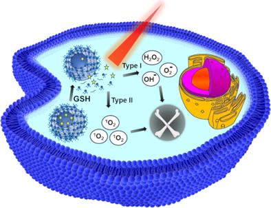
Figure 1: Chemical structures of clinically approved porphyrin-based photosensitizers used in PDT.
Scheme 1: Synthesis of THPP@p(MVCA-BAC) nanoparticles and their GSH-triggered disassembly for ROS generation ...
Figure 2: Optimization of nanoparticle formulation parameters showing hydrodynamic diameter distributions fro...
Figure 3: Data for THPP@p(MVCA-BAC): (A) Hydrodynamic size distribution determined by DLS; three measurements...
Figure 4: (A) Optical characterization of THPP and THPP@p(MVCA-BAC) in PB pH 7.4: UV–vis absorption (black), ...
Figure 5: 1O2 generation monitored via DPBF degradation: (A, B) UV–vis spectral changes of DPBF upon 650 nm i...
Figure 6: (A, B) Cell viability after treatment with p(MVCA-BAC) and THPP@p(MVCA-BAC): (A) in the dark and (B...
Figure 7: Real-time monitoring of the PC-3 cell response to THPP@p(MVCA-BAC) using impedance-based proliferat...
Figure 8: Induction of apoptosis in PC-3 cells incubated with p(MVCA-BAC) and THPP@p(MVCA-BAC), C(THPP) = 2 µ...
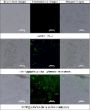
Figure 9: Cellular uptake and localization of THPP@p(MVCA-BAC) in PC-3 cells, bright-field, fluorescence, and...
Figure 10: Light-triggered intracellular ROS generation in PC-3 cells, H2DCFDA fluorescence (green) after trea...