This search combines search strings from the content search (i.e. "Full Text", "Author", "Title", "Abstract", or "Keywords") with "Article Type" and "Publication Date Range" using the AND operator.
Beilstein J. Nanotechnol. 2026, 17, 1087–1102, doi:10.3762/bjnano.17.74
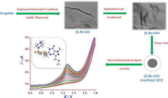
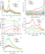
Figure 1: (a) XRD patterns, (b) nitrogen adsorption/desorption isotherms, (c) FTIR spectra, (d) Raman spectra...
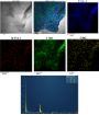
Figure 2: HRTEM observation of (S,N)-GO (a) and (S,N)-rGO (b).
Figure 3: EDX mapping of (S,N)-rGO.
Figure 4: (a) Cyclic voltammograms (CVs) recorded at different electrodes (GCE, GO/GCE, (S,N)-GO/GCE, rGO/GCE...
Figure 5: (a) CVs of (S,N)-rGO/GCE in 0.1 M Britton–Robinson buffer at different pH values (2.0–8.0) containi...
Figure 6: (a) CVs of (S,N)-rGO/GCE in 0.1 M Britton–Robinson buffer (pH 4.0) containing 20 µM SDN at differen...
Figure 7: (a) Molecular structures, (b) CVs, and (c) DPVs of SDN, ciprofloxacin, and ofloxacin with concentra...
Figure 8: (a) DPV curves of (S,N)-rGO/GCE in 0.1 M Britton–Robinson buffer (pH 4.0) for different concentrati...
Figure 9: (a) Variation of peak current (Ip) for SDN at four concentrations (1.0, 2.0, 13.8, and 19.6 µM) rec...
Beilstein J. Nanotechnol. 2026, 17, 541–554, doi:10.3762/bjnano.17.35
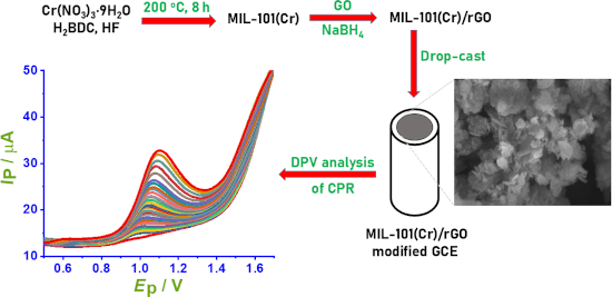
Figure 1: (a) X-ray diffraction (XRD) patterns of graphene oxide (GO), reduced graphene oxide (rGO), and MIL-...
Figure 2: EDX elemental mapping and elemental composition of MIL-101/rGO (3.3), (a) EDX spectrum, (b) electro...
Figure 3: XPS spectra of the MIL-101/rGO (3.3): (a) survey spectrum, (b) high-resolution Cr 2p spectrum, (c) ...
Figure 4: SEM observation of (a) rGO, (b) MIL-101, and (c) MIL-101(Cr)/rGO (3.3).
Figure 5: (a) Cyclic voltammograms (CVs) recorded at different electrodes in 0.1 M BRS buffer (pH 4) containi...
Figure 6: (a) CVs of MIL-101/rGO(3.3)/GCE recorded in 0.1 M BR buffer over the pH range of 2–9 containing 15 ...
Figure 7: (a) Cyclic voltammograms of MIL-101/rGO(3.3)/GCE recorded in 0.1 M BR buffer (pH 4) containing 15 µ...
Scheme 1: The oxidation mechanism of CPR at MIL-101/rGO/CGE.
Figure 8: (a) DPV curves of MIL-101/rGO(3.3)/GCE recorded in 0.1 M BRS buffer (pH 4) at CPR concentrations ra...
Figure 9: (a) Relative standard deviation of the anodic peak currents obtained from DPV measurements recorded...
Figure 10: Relative error (RE%) caused by the interference of selected inorganic (a) and organic (b) species a...