This search combines search strings from the content search (i.e. "Full Text", "Author", "Title", "Abstract", or "Keywords") with "Article Type" and "Publication Date Range" using the AND operator.
Beilstein J. Nanotechnol. 2026, 17, 1087–1102, doi:10.3762/bjnano.17.74
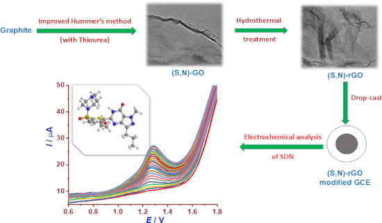
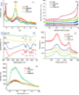
Figure 1: (a) XRD patterns, (b) nitrogen adsorption/desorption isotherms, (c) FTIR spectra, (d) Raman spectra...
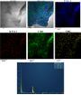
Figure 2: HRTEM observation of (S,N)-GO (a) and (S,N)-rGO (b).
Figure 3: EDX mapping of (S,N)-rGO.
Figure 4: (a) Cyclic voltammograms (CVs) recorded at different electrodes (GCE, GO/GCE, (S,N)-GO/GCE, rGO/GCE...
Figure 5: (a) CVs of (S,N)-rGO/GCE in 0.1 M Britton–Robinson buffer at different pH values (2.0–8.0) containi...
Figure 6: (a) CVs of (S,N)-rGO/GCE in 0.1 M Britton–Robinson buffer (pH 4.0) containing 20 µM SDN at differen...
Figure 7: (a) Molecular structures, (b) CVs, and (c) DPVs of SDN, ciprofloxacin, and ofloxacin with concentra...
Figure 8: (a) DPV curves of (S,N)-rGO/GCE in 0.1 M Britton–Robinson buffer (pH 4.0) for different concentrati...
Figure 9: (a) Variation of peak current (Ip) for SDN at four concentrations (1.0, 2.0, 13.8, and 19.6 µM) rec...
Beilstein J. Nanotechnol. 2024, 15, 719–732, doi:10.3762/bjnano.15.60
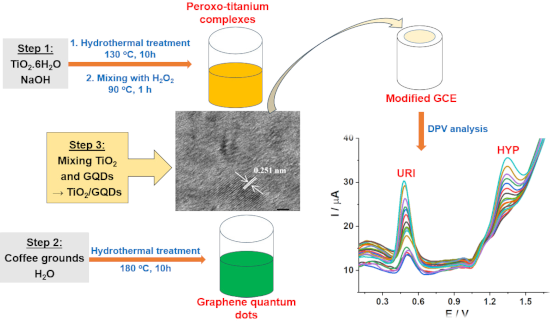
Figure 1: Images of aqueous suspension of TiO2/GQDs samples (a) under white light and (b) under UV light. (c)...
Figure 2: (a) XRD patterns of TiO2, GQDs, and (1:4)TiO2/GQDs in suspension form. (b) XRD pattern of (1:4)TiO2...
Figure 3: HRTEM images of (a) TiO2, (b) GQDs, (c) (1:4)TiO2/GQDs, and (d) of (1:4)TiO2/GQDs with high magnifi...
Figure 4: EDX mapping of (1:4)TiO2/GQDs.
Figure 5: (a) CV curves at different electrodes in a solution (0.05 M BR buffer, pH 4) containing CURI = CHYP...
Figure 6: (a) CV curves of URI and HYP at equal concentrations of 25 µM in 0.05 M BR pH 2–6 at the (1:4)TiO2/...
Figure 7: (a) CV curves of (1:4)TiO2/GQDs-GCE in 0.05 M BR buffer (pH 3) with CURI = CHYP = 25 µM; scanning r...
Scheme 1: Proposed oxidation mechanisms of URI and HYP at the (1:4)TiO2/GQDs-GCE.
Figure 8: DPV curves at the (1:4)TiO2/GQDs-GCE. (a) 0.05 M BR pH 3 with CURI = CHYP = 1.00–15.26 µM. (b) Line...
Figure 9: The RSD of ten replicate measurements of (a) URI and (b) HYP at the (1:4)TiO2/GQDs-GCE in 0.05 M BR...
Figure 10: Effect of possible interferents. (a) Inorganic compounds and (b) organic compounds.
Beilstein J. Nanotechnol. 2024, 15, 475–489, doi:10.3762/bjnano.15.43
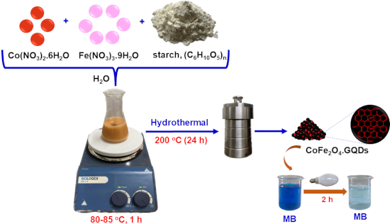
Figure 1: XRD patterns of (a) CF and (b) CF/GQDs.
Figure 2: (a) UV–vis absorption spectra of the GQD solution; inset: GQD solution under white light (left) and...
Figure 3: (a) FTIR spectra of CF/GQDs and CoFe2O4; (b, c) Raman spectra; (d) TG/dTG curves of CF/GQDs-200.
Figure 4: SEM images of (a) CF/GQD-140, (b) CF/GQD-180, and (c) CF/GQD-200; TEM observations and correspondin...
Figure 5: EDX-mapping of CF/GQDs-200. (a) Electron microscopy image, (b) EDX spectrum, (c) carbon mapping, (d...
Figure 6: (a) PL spectra, (b) UV–vis diffuse reflectance spectra with Tauc’s plots in the inset, (c) magnetiz...
Figure 7: (a) MB decolourisation over different catalysts (V = 100 mL, C0(MB) = 10 ppm, m = 0.05 g, time of d...
Figure 8: (a) Effects of addition of KI, IPA, KBrO3, and BQ on the visible-light-driven photocatalytic degrad...
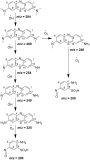
Scheme 1: Proposed intermediates in MB degradation monitored by liquid chromatography coupled with tandem mas...
Figure 9: (a) Cyclic photocatalytic degradation experiments of MB with CF/GQDs-200 photocatalyst; (b) XRD pat...
Scheme 2: Mechanism of MB degradation over the CF/GQDs catalyst.