


Search results
Search for "AgNPs" in Full Text gives 59 result(s) in Beilstein Journal of Nanotechnology.
Controlled graphene oxide assembly on silver nanocube monolayers for SERS detection: dependence on nanocube packing procedure
Beilstein J. Nanotechnol. 2016, 7, 9–21, doi:10.3762/bjnano.7.2
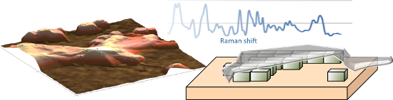
- in two orthogonal electron oscillations with respect to the surface plane when a strong near-field interaction between AgNPs occurs in the 2D array. The cubic geometry of the nanoparticles provides a large nanoparticle–substrate contact area, leading to efficient hybridization of dipolar (D) and
- of a broad intense band due to strong interparticle dipole–dipole coupling centred at 642 nm and 665 nm for LB films transferred at 15 mN/m and 20 mN/m, respectively. Experimental and theoretical studies [45] on two dimensional (2D) arrays of AgNPs with the different edge-to-edge distances showed
- quadrupolar bands, our results show that regular arrangement of close-packed AgNPs contributes to the efficient coupling of dipole modes and that such coupling is less efficient for samples transferred at 20 mN/m, likely due to crystalline fusion in the collapsed 3D microdomains found in this samples. These


Sonochemical co-deposition of antibacterial nanoparticles and dyes on textiles
Beilstein J. Nanotechnol. 2016, 7, 1–8, doi:10.3762/bjnano.7.1
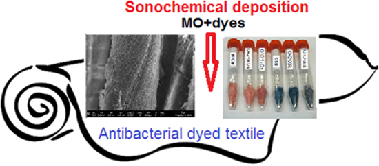
- , however, find only one report in which sonochemistry was used for the co-deposition of AgNPs and AgCl on TiO2 NPs to form Ag@TiO2 and Ag/AgCl@TiO2 [18]. The synthetic process revealed that in fact it was a one-step process combining Ag+, TiO2 NPs, and NaCl in ethylene glycol solution. The sonochemical




Ultrastructural changes in methicillin-resistant Staphylococcus aureus induced by positively charged silver nanoparticles
Beilstein J. Nanotechnol. 2015, 6, 2396–2405, doi:10.3762/bjnano.6.246
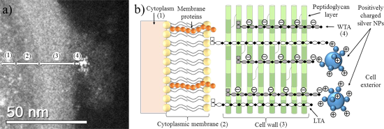
- encouraging path of research in nanobiotechnology involves silver nanoparticles (AgNPs) [24]. AgNPs are potent bactericidal agents with broad-spectrum activity [25]. In fact, the number of papers on this topic has exploded during the last decade [26][27][28][29][30][31][32][33]. AgNPs also have antifungal [34
- ] and antiviral [35][36][37] activities. Due to their small-scale diameters and enhanced surface area to volume ratios, metallic nanoparticles have large contact areas available to interreact with pathogens [24]. AgNPs can disturb the physiology of bacterial cell membranes by affecting their
- permeability [38]. After penetrating the cell membrane, AgNPs can also alter sulfur-containing amino acids and phosphorus (a main constituent of DNA), inhibiting replication via attaching to the bacterial ribosome [39][40]. The proteomic signatures of AgNP-treated E. coli demonstrated an accumulation of


In situ SU-8 silver nanocomposites
Beilstein J. Nanotechnol. 2015, 6, 1661–1665, doi:10.3762/bjnano.6.168
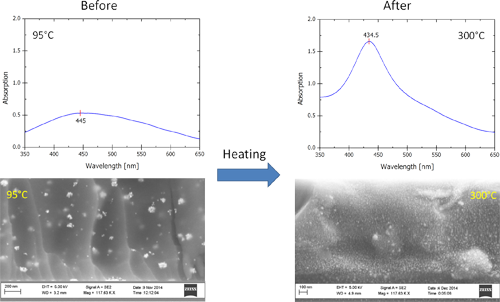
- used for depositing thin films in the range of 1–1.8 µm as documented by the spin curve shown in Figure 1. The resulting film thickness is smaller than with unmodified SU-8, but the thickness can be increased by using more viscous SU-8 formulations if desired. Mostly, AgNPs are formed during the heat
- treatments before and after UV exposure. The exact process is not known as acetonitrile and the many constituents of SU-8 play a role in the NP formation. The AgNPs formed in the SU-8 polymer matrix show a clear plasmonic absorption [16] in the visible region as seen in Figure 2. The high temperature post
- nm is a typical value for the absorption band of AgNPs [17]. Also; this peak resembles the plasmonic peak obtained for AgNPs in cyclopentanone. The shoulder appearing at higher wavelengths for the nanocomposite indicates the retention of the AgNP clusters, formed during the lower temperature


Formation of substrate-based gold nanocage chains through dealloying with nitric acid
Beilstein J. Nanotechnol. 2015, 6, 1362–1368, doi:10.3762/bjnano.6.140
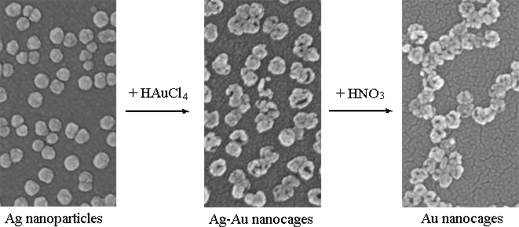
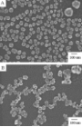
- of nanocages In our previous reports [23][24][25], we have electro-deposited template silver nanoparticles (AgNPs) on ITO substrates and carried out the galvanic replacement reactions. Figure 1 shows top-view and tilted-view SEM images of unreacted AgNP templates and those exposed to aqueous 0.1 mM
- to 83 ± 10 nm. This is attributed to the gold deposited on the surface of AgNP templates. Since AgNPs, AuNPs and nanocages often exhibit different LSPR bands in the UV–vis–NIR wavelength region, the galvanic replacement reactions between the AgNPs and the aqueous HAuCl4 solution can be monitored
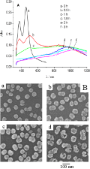
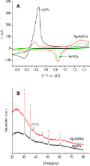
- using absorption spectroscopy. Figure 2A shows the UV–vis absorption spectra recorded from ITO-supported AgNPs after immersion in 0.1 mM HAuCl4 solution for various times. The spectra correspond to the excitation of dipolar and quadrupolar oscillations. The AgNPs exhibited typical LSPR peaks at 455 and

Influence of size, shape and core–shell interface on surface plasmon resonance in Ag and Ag@MgO nanoparticle films deposited on Si/SiOx
Beilstein J. Nanotechnol. 2015, 6, 404–413, doi:10.3762/bjnano.6.40
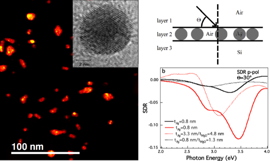
- physical effect: since the reflectivity spectrum for this grazing angle approaches zero in both cases, the ratio (Rsub+AgNPs − Rsub) / Rsub defining the SDR can give rise to numerical instability. Finally, the effect of the MgO layer as a protective ultrathin coating for the optical properties of Ag NPs



Synthesis of boron nitride nanotubes and their applications
Beilstein J. Nanotechnol. 2015, 6, 84–102, doi:10.3762/bjnano.6.9
- combined with the properties of other nanomaterials to construct novel sensor devices for humidity, carbon dioxide detection, and clinical diagnostics. A highly sensitive humidity sensor using BNNTs and silver nanoparticles (AgNPs) for the rapid detection of humidity was fabricated [90]. Figure 8 shows the




Interaction of dermatologically relevant nanoparticles with skin cells and skin
Beilstein J. Nanotechnol. 2014, 5, 2363–2373, doi:10.3762/bjnano.5.245
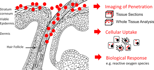
- ]. Results obtained from SERS indicate that single AgNPs can penetrate deeply into the stratum corneum. The Raman and SERS spectra of porcine skin pre-treated with AgNP are shown in Figure 2a. The results illustrate that for AgNP, the SERS effect can be used to monitor the skin penetration depth of single
- the skin surface down to a depth of 50 µm, in 2 µm steps. The measurement time for one spectrum was 5 s. The surface enhanced Raman scattering (SERS) signal as a result of interaction between AgNPs and the porcine skin was generated by using the same excitation conditions. The utilized Raman


Protein-coated pH-responsive gold nanoparticles: Microwave-assisted synthesis and surface charge-dependent anticancer activity
Beilstein J. Nanotechnol. 2014, 5, 1452–1462, doi:10.3762/bjnano.5.158
- different proteins by adding only Ag ions to study the formation of AgNPs during microwave irradiation. Surprisingly, no AgNPs were formed with any of the protein samples based on the absence of UV absorbance at approximately 400 nm, which is characteristic of AgNPs. Although no AgNPs were observed at an Ag
- ion concentration of 0.1 mM, AgNPs were formed at higher concentrations. A yellow coloration and an UV absorbance near 400 nm suggested that AgNPs were formed for all of the proteins samples except for TRY and GOX. Hence, the presence of Ag ions in the reaction mixture might result in a very low
- concentration of AgNPs, which are consumed in a galvanic exchange reaction converting Au ions to AuNPs at a faster rate [33][34]. From the UV–vis spectra (Figure 1B) for the AuNPs formed in the absence of Ag ions, the peaks were broader because the reaction is slow, which results in larger AuNPs. However, when


Other Beilstein-Institut Open Science Activities



