


Search results
Search for "endocytosis" in Full Text gives 105 result(s) in Beilstein Journal of Nanotechnology.
Protein-coated pH-responsive gold nanoparticles: Microwave-assisted synthesis and surface charge-dependent anticancer activity
Beilstein J. Nanotechnol. 2014, 5, 1452–1462, doi:10.3762/bjnano.5.158
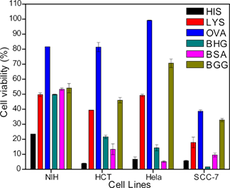
- the attached charged particles through endocytosis or other methods [55]. Because of their higher positive surface charge, HIS-AuNPs are easily internalized by cells, after which they cause cell death. A recent report suggests that the interior of lysosomes possesses an acidic pH (pH 4–5), and it
- charged nanoparticles are formed that binds randomly to the cationic sites, which then undergoes endocytosis [59]. In this study, exposure to OVA-AuNPs resulted in a high cell viability due to the poor ability of the nanoparticles to internalize into the cells because of their low level of cellular


The cell-type specific uptake of polymer-coated or micelle-embedded QDs and SPIOs does not provoke an acute pro-inflammatory response in the liver
Beilstein J. Nanotechnol. 2014, 5, 1432–1440, doi:10.3762/bjnano.5.155
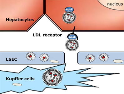
- canonical lipoprotein receptor-mediated apolipoprotein E-dependent endocytosis into hepatocytes. Future studies by using radiolabelled QDs are needed to quantify the precise contribution of different hepatic cell types for QDs-micelles uptake. However, only 5–10% of liver cells are Kupffer cells [25][33


Nanodiamond-DGEA peptide conjugates for enhanced delivery of doxorubicin to prostate cancer
Beilstein J. Nanotechnol. 2014, 5, 937–945, doi:10.3762/bjnano.5.107
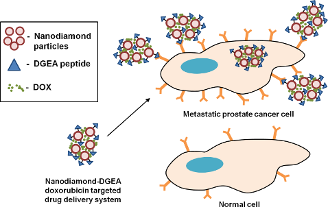
- size and zeta potential are particularly important for drug delivery applications as they give representations of size limitations and potential colloidal stability issues of a system. NDs permeate the cell membrane by endocytosis [20][36][37]. Through this pathway, NDs can effectively deliver drugs


Manipulation of isolated brain nerve terminals by an external magnetic field using D-mannose-coated γ-Fe2O3 nano-sized particles and assessment of their effects on glutamate transport
Beilstein J. Nanotechnol. 2014, 5, 778–788, doi:10.3762/bjnano.5.90
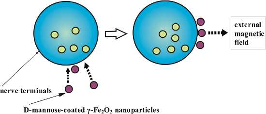
- level, nanoparticles can pass through the plasma membrane of the cells by means of endocytosis [27]. Opposite point of view argues that uptake of nanoparticles into the cells does not occur by endocytic processes, but rather by diffusion or adhesive interactions [28]. In this study, we have presented a



Near-infrared dye loaded polymeric nanoparticles for cancer imaging and therapy and cellular response after laser-induced heating
Beilstein J. Nanotechnol. 2014, 5, 313–322, doi:10.3762/bjnano.5.35
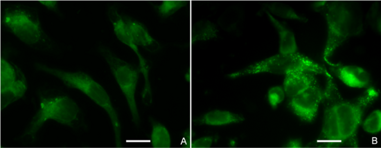
- the cells through an endocytosis pathway (Supporting Information File 1, Figure S5). Calculated image ratio values, R, from the fluorescence microscope images show that the NP formulation produces a higher intracellular fluorescence intensity (R = 3.75 ± 0.54) (mean ± SD) than the free dye (R = 2.89


Other Beilstein-Institut Open Science Activities



