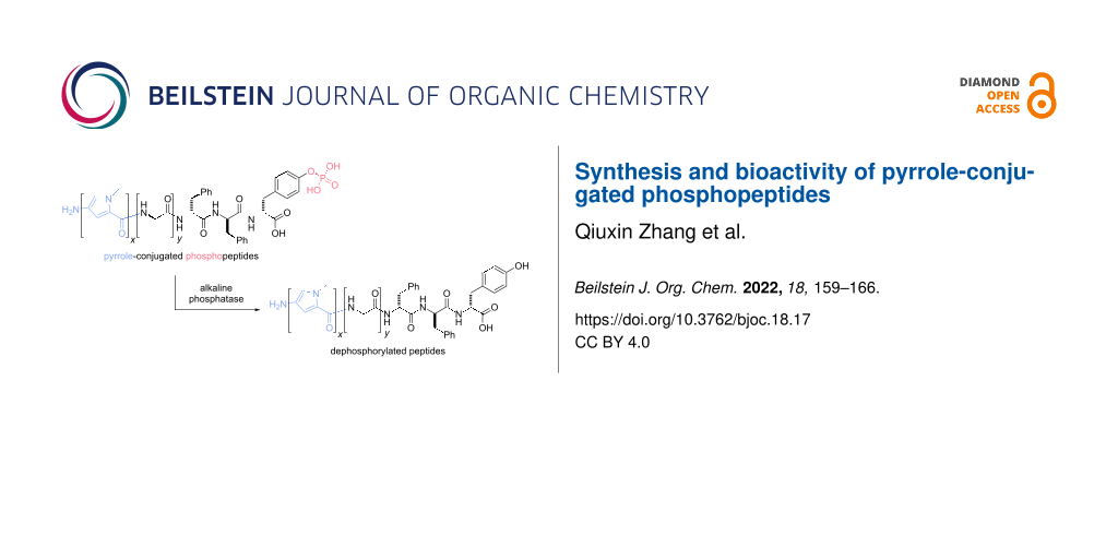
Abstract
Here we report the synthesis and effect on the cell viability of pyrrole-conjugated phosphopeptides. Encouraged by the selective inhibition of cancer cells by a naphthyl-capped phosphopeptide (Nap-ffpy, 1), we conjugated the heteroaromatic dipyrrole or tripyrrole motif at the N-terminal of short peptides containing phosphotyrosine or phosphoserine and examined the bioactivity of the resulting phosphopeptides (2–10). Although most of the phosphopeptides exhibit comparable activities with that of 1 against HeLa cells at 200 μM, they, differing from 1, are largely compatible with HeLa cells at 400 μM. Enzymatic dephosphorylation of 2–10, at 400 μM is unable to induce a dramatic morphological transition of the peptide assemblies observed in the case of 1. These results suggest that a heteroaromatic motif at the N-terminal of peptides likely disfavors the formation of extensive nanofibers or morphological changes during enzymatic self-assembly, thus provide useful insights for the development of phosphopeptides as substrates of phosphatases for controlling cell fate.

Graphical Abstract
Introduction
Biomacromolecular assemblies have received considerable attention recently in the field of biomaterials [1-7], among which peptides are of particular interest because of their unique merits, such as ease of design and tailoring (based on the known structures from proteins), good biocompatibility and degradability, and low immunogenicity. For example, recent works have demonstrated the potential of peptide assemblies for a wide range of applications, including drug delivery [8-11], collagen mimic [12], antibacterial [13,14], biomineralization [15,16], mimicry of amyloids [17], cell cultures [18], and tissue engineering [19]. Particularly, the use of enzyme-instructed self-assembly (EISA) [20,21] of peptide assemblies has expanded the applications of peptide assemblies, such as intracellular phase transition [22], molecular imaging [23-33], anisotropic hydrogels [34,35], targeting subcellular organelles [36-40], elimination of pluripotent stem cells (PSC) [41,42], and cancer therapy [43-54]. Being a multistep molecular process to generate non-diffusive peptide assemblies, EISA is a facile and useful approach to explore the emergent properties of peptide assemblies [55-59] in cellular environment. One of the most explored EISA processes is the use of alkaline phosphatase (ALP) to convert the micelles made of phosphopeptides to the nanofibers of peptides via enzymatic dephosphorylation [20,60]. While ALP-catalyzed EISA has received considerable exploration, the structures of the peptide substrates mainly have centered on naphthylacetyl-capped phosphopeptides (e.g., Nap-ffpy (1)) [36,61-66]. Considering naphthyl is an aromatic group, we decided to explore other aromatic N-terminal capping groups, such as heteroaromatic, N-terminal capping groups of phosphopeptides for EISA because they receive little exploration [67].
Our previous studies have shown nucleobases, as the heteroaromatic groups, are able to act as the N-capping group for EISA of phosphopeptides [67]. In this study, we chose to examine a different type of heteroaromatic group, pyrroles, because pyrrole is adaptable for solid-phase synthesis [68] so that it is feasible to conjugate multiple pyrroles to phosphopeptides. In addition, pyrrole has yet to be incorporated in peptides for EISA, though oligomeric pyrroles have been extensively explored for binding nucleic acids [68]. Based on a naphthyl-capped phosphopeptide (Nap-ffpy, 1), we conjugated heteroaromatic dipyrrole or tripyrrole motifs at the N-terminal of short peptides containing phosphotyrosine or phosphoserine and examined the bioactivity of the resulting phosphopeptides (2–10). Unexpectedly, enzymatic dephosphorylation of 2–10 rarely induces distinct morphological transitions of the peptide assemblies, such as the transition of nanoparticles to nanofibers, which is a transition observed in the case of 1 [43]. Incubating 2–10 with HeLa cells reveals that these phosphopeptides exhibit comparable inhibitory activity as that of 1 against the proliferation of HeLa cells at 200 μM. But 2–10 at 400 μM, differing drastically from 1, are largely compatible with HeLa cells, while 1, at 400 μM, significantly inhibits HeLa cells. These results suggest that morphological transition or extensive self-assembly triggered by enzymatic reactions likely is necessary for the inhibition of HeLa cells by EISA. This work, thus, provides useful insights for the development of phosphopeptide derivatives as enzyme substrates for controlling cell fate.
Results and Discussion
Molecular design
As illustrated in Scheme 1, the phosphopeptide (Nap-ffpy, 1) that inhibits HeLa cells consists of three segments, naphthylacetyl (Nap) at the N-terminal of the peptide, a self-assembling motif (ᴅ-phenylalanine-ᴅ-phenylalanine (ff)) [69] as the main backbone, and an enzymatic trigger ᴅ-phosphotyrosine (py). This design presents a substrate of alkaline phosphatase (ALP) for EISA. Based on this structure, a considerable number of studies have investigated the physical properties (enzyme-catalyzed hydrogelation or supramolecular structural transformation) [62,70] and biological properties (cell viability, biostability, and cell-selective growth inhibition) [43,62] of the naphthylacetyl N-terminal-capped phosphopeptides. These studies have produced a variety of phosphopeptides that selectively inhibit cancer cells [38,64,65,71,72]. Encouraged by the results from the naphthylacetyl capped phosphopeptides, we decided to use multiple N-methylpyrroles, as the heteroaromatic analog of naphthyl, to cap the N-terminal of phosphopeptides for EISA. According to this rationale, replacing the Nap capping group in 1 with a dipyrrole or a tripyrrole segment at the N-terminal generates 2a and 2b. Introducing two or three glycine residues between the pyrrole segment and the self-assembling segment produces 2c–h, which would help to understand the role of a spacer in the molecular structure. As the enantiomers of 2g and 2h, respectively, 3a and 3b offer an opportunity to assess the effects of stereochemistry of the pyrrole-capped phosphopeptides. Substituting the ᴅ-phosphotyrosine (py) in 2g and 2h, by ᴅ-phosphoserine (ps), affords 4a and 4b, respectively. Switching the ᴅ-phosphoserine in 4a and 4b by ʟ-phosphoserine (pS), creates 5a and 5b. ᴅ-Trialanine replaces triglycine in 2f–h to produce 6a–c, which should resist to proteases, such as polyglycine hydrolases [73], which are known to cleave at the glycine–glycine site. The addition of an arginine residue in the backbone of 2a leads to 7, which bears an additional positive charge compared to 2a. A ᴅ-tetraleucine (l4) replaces ᴅ-diphenylalanine (ff) in 2d and 2e to form 8a and 8b, respectively. Adding a Boc protecting group at the N-terminus of 2a produces 9. Attaching a guanidinoacetic acid motif to 2f and 2g results in 10a and 10b. 9 and 10 would help test the effects of additional N-terminal modifications. To examine the necessity of enzymatic dephosphorylation, we replaced ᴅ-phosphotyrosine (py) in 2g, 2h, 6a–c by ᴅ-tyrosine to yield 11a, 11b, 12a–c, respectively. Attaching N-methylpyrrole on the sidechain of NBD-ffky or NBD-ffkpy [66], another previously studied self-assembling peptide, produces 13 and 14. In addition, 15a–c consist of only pyrrole and glycine units, which should help delineate the roles of the self-assembling motif and the enzymatic triggers.
![[1860-5397-18-17-i1]](/bjoc/content/inline/1860-5397-18-17-i1.svg?scale=2.0&max-width=1024&background=FFFFFF)
Scheme 1: Molecular structures of the parent phosphopeptide 1 and its pyrrole-conjugated analogs 2–14.
Scheme 1: Molecular structures of the parent phosphopeptide 1 and its pyrrole-conjugated analogs 2–14.
Synthesis
The synthesis of the designed peptides combines solution synthesis of the enzyme trigger or the fluorophore with the solid-phase synthesis of the pyrrole-peptide conjugates. We used phosphorus pentoxide and phosphoric acid to react with ᴅ-tyrosine, ʟ-tyrosine, ʟ-serine, or ᴅ-serine to produce ᴅ-phosphotyrosine, ʟ-phosphotyrosine, ʟ-phosphoserine, or ᴅ-phosphoserine, respectively. After being protected by an Fmoc group, these phosphorylated amino acids are suitable for solid-phase synthesis. We also used solution-phase synthesis to synthesize NBD-β-alanine according to the reported procedures [66]. We used standard Fmoc solid-phase peptide synthesis (SPPS) to generate the peptide segments ((G/GG/GGG)ffpy for 2a–h, 9, 10a,b, GGGFFpY for 3a,b, GGGffps for 4a,b, GGGffpS for 5a,b, aaaffpy for 6a–c, rffpy for 7, GGllllpy for 8a,b, GGGffy for 11a,b, aaaffy for 12a–c, NBD-ffky for 13, NBD-ffkpy for 14, GG(G) for 15a–c). Then, we introduced the N-methylpyrrole (Py) units into the peptides obtained via solution-phase amide bond formation to produce 2a–h, 3a,b, 4a,b, 5a,b, 6a–c, 7, 8a,b, 11a,b, 12a–c, 13, 14, and 15a–c. We conjugated two Py units successively to the peptide ffpy and kept the Boc protecting group of the second Py unit to give a Boc-capped product, 9. We added guanidinoacetic acid to Py-GGGffpy and (Py)2-GGGffpy for making 10a and 10b, respectively. All the products were purified by HPLC.
Bioactivity
We examined the cytotoxicity of the synthesized compounds by incubating them with HeLa cells because HeLa cells overexpress ALP and are widely available. By incubating 200 μM of each compound with HeLa cells for 24 hours, we employed MTT assay to determine the cell viability of the HeLa cells. As shown in Figure 1, the MTT results indicate that most of the pyrrole-conjugated phosphopeptides, at 200 μM, are comparable with the activity of 1 against the HeLa cells. In terms of the pyrrole-conjugated ffpy series (2a–h), all of the compounds are rather compatible with the cells, with the cell viability ranging from 92.8% (2f) to 142.6% (2b). The incubation of the ʟ-enantiomers 3a and 3b with HeLa cells results in cell viabilities of 86.0% and 97.2%, respectively. While ʟ-phosphoserine peptides, 5a and 5b, hardly inhibit the HeLa cells, ᴅ-phosphoserine peptides, 4a and 4b, moderately inhibit the proliferation of the HeLa cells, among which 4a decreases cell viability to 69.2%. 6a–c, containing a ᴅ-trialanine motif and one to three pyrrole motifs at the N-terminal, hardly inhibit the HeLa cells. Compound 7, formed by the introduction of a ᴅ-arginine residue into 2a, exhibits similar cytotoxicity as that of 2a. 8a and 8b, formed by replacing the ff motif in 2d and 2e with l4, exhibit slight and little cytotoxicity, respectively. Capping the N-terminus with a Boc-protecting group (9) or a guanidinoacetic acid motif (10a and 10b) renders the molecules with higher cytotoxicity (resulting in cell viabilities of about 70%) than those of 2f and 2g. Compounds without phosphorylation (11a, 11b and 12a–c) show similar activity compared to their phosphorylated counterparts. These results indicate that these peptide assemblies are rather compatible with cells. Compounds 13 and 14, both possessing a pyrrole building block on the side chain, hardly inhibit the HeLa cells. While 15a and 15b exhibit slight cytotoxicity, 15c is less cytotoxic. This result agrees with the observation that dipyrrole-conjugated peptides, in general, exhibit higher inhibitory activity against HeLa cells than mono- or tripyrrole-conjugated peptides do. It is interesting that 4a exhibits higher inhibitory activity than that of 2g, despite that 4a bears ᴅ-phosphoserine and 2g bears ᴅ-phosphotyrosine. This result likely warrants further investigation on 4a. Another dipyrrole-conjugated peptide, 6b, was also selected for further study as a comparison to 4a since it shows good cell compatibility at 200 μM.
![[1860-5397-18-17-1]](/bjoc/content/figures/1860-5397-18-17-1.png?scale=2.0&max-width=1024&background=FFFFFF)
Figure 1: Cell viability of HeLa cells treated with 200 μM of each compound for 24 h.
Figure 1: Cell viability of HeLa cells treated with 200 μM of each compound for 24 h.
We also compared the cell viability of HeLa cells incubated with 1, 4a, and 6b at different concentrations for 3 days. As shown in Figure 2, the three compounds result in different cell viabilities versus concentration and incubation time. 1 hardly inhibits the proliferation of the cells at concentrations lower than 100 μM over the 3 days, while 200 μM of 1 begins to reduce cell viability on day 3. At the concentration of 400 μM, 1 drastically decreases the cell viability to 52.0% on day 1, while longer incubation time further inhibits cell proliferation, resulting in a cell viability of only 3.4% on day 3. This result agrees well with previous results [43,62]. Unlike 1, 4a slightly inhibits cell proliferation, with cell viability ranging from 89.6% to 72.3% with increasing concentrations from 20 to 400 μM. However, the inhibitory effect of 4a largely vanishes on day 2 and 3 of the incubation. This result implies that cells likely are able to degrade 4a overtime. Unlike 1 and 4a, 6b, exhibiting little cytotoxicity with increasing compound concentration or incubation time, is rather compatible to the cells. These results indicate that the replacement of Nap by pyrroles largely results in cell compatible phosphopeptide derivatives.
![[1860-5397-18-17-2]](/bjoc/content/figures/1860-5397-18-17-2.png?scale=2.0&max-width=1024&background=FFFFFF)
Figure 2: Cell viability of HeLa cells treated with 20 μM, 50 μM, 100 μM, 200 μM and 400 μM of 1, 4a and 6b for 1, 2, and 3 days.
Figure 2: Cell viability of HeLa cells treated with 20 μM, 50 μM, 100 μM, 200 μM and 400 μM of 1, 4a and 6b f...
Enzymatic conversion
To further investigate the reason for the different activities in cell assays among the three compounds, 1, 4a and 6b, we employed transmission electron microscopy (TEM) to study the morphologies of the assemblies of these compounds before and after enzymatic dephosphorylation. Previous literature has demonstrated that 1, as a precursor, is capable of forming hydrogels upon dephosphorylation catalyzed by ALP [43,62]. To be more specific, ALP enables a morphological transition from nanoparticles of 1 to networks of nanofibers of the corresponding dephosphorylated peptides, resulting in hydrogelation (Figure 3A) [62]. In comparison with 1, 4a, forming networks of nanoparticles at 400 μM and 200 μM, exhibits little change in the morphology of the assemblies after being incubated with 1 U/mL of ALP for 24 h (Figure 3B), although LC–MS confirms the enzymatic dephosphorylation of 4a (Supporting Information File 1, Figure S32). Similarly, 6b also forms dense nanoparticulate networks at 400 μM and 200 μM (Figure 3B). After the addition of 1 U/mL of ALP for 24 h, the nanoparticles remain, although the networks dissociate. Similarly, LC–MS confirms the enzymatic dephosphorylation of 6b (Supporting Information File 1, Figure S33) after the addition of ALP. In addition to the TEM images, we used circular dichroism (CD) to reveal the secondary structures of the assemblies before and after enzymatic action. The spectra show a significant change in the signal upon ALP treatment for 1, but merely slight changes for 4a and 6b, which are consistent with the TEM images (Supporting Information File 1, Figure S34). The differences in the morphological transition between 1 and 4a or 6b revealed by TEM and CD suggest that the shape-shifting (from nanoparticles to nanofibers) likely contributes to the cell death caused by 1 over days 2 and 3. These results together indicate the drastic morphological changes of phosphopeptides upon the addition of ALP may serve as a useful indicator to predict the cell compatibility or cytotoxicity of phosphopeptides.
![[1860-5397-18-17-3]](/bjoc/content/figures/1860-5397-18-17-3.png?scale=2.0&max-width=1024&background=FFFFFF)
Figure 3: A) TEM images of 1 before and after addition of ALP (0.5 U/mL) in PBS buffer (pH 7.4). Scale bar is 100 nm. Figure 3A was reprinted with permission from [62] https://pubs.acs.org/doi/10.1021/bm5010355, Copyright 2014 American Chemical Society. This content is not subjected to CC-BY 4.0. Permission requests related to the material excerpted should be directed to the ACS. B) TEM images of representative pyrrole-conjugated peptides 4a and 6b before and after the addition of ALP (1 U/mL) in PBS buffer (pH 7.4). Scale bar is 200 nm.
Figure 3: A) TEM images of 1 before and after addition of ALP (0.5 U/mL) in PBS buffer (pH 7.4). Scale bar is...
Conclusion
In summary, this work reports the design and synthesis of a series of pyrrole-conjugated phosphopeptides. These pyrrole-conjugated phosphopeptides are largely cell compatible and are unable to undergo drastic morphological transitions, such as from nanoparticles to nanofibers. These results, contrary to the result obtained from 1, suggest that the morphological transition of the peptide assemblies resulted from enzymatic reaction likely is responsible for disrupting cellular processes to inhibit cells. Other factors, such as endocytosis of the phosphopeptides [74] and the cellular localization of the peptide assemblies [75], certainly could contribute to the cell compatibilities of the pyrrole-conjugated phosphopeptides, which remain to be determined.
Supporting Information
| Supporting Information File 1: Experiment part. | ||
| Format: PDF | Size: 4.4 MB | Download |
References
-
Acar, H.; Srivastava, S.; Chung, E. J.; Schnorenberg, M. R.; Barrett, J. C.; LaBelle, J. L.; Tirrell, M. Adv. Drug Delivery Rev. 2017, 110–111, 65–79. doi:10.1016/j.addr.2016.08.006
Return to citation in text: [1] -
Kim, B. J.; Yang, D.; Xu, B. Trends Chem. 2020, 2, 71–83. doi:10.1016/j.trechm.2019.09.004
Return to citation in text: [1] -
Cheetham, A. G.; Chakroun, R. W.; Ma, W.; Cui, H. Chem. Soc. Rev. 2017, 46, 6638–6663. doi:10.1039/c7cs00521k
Return to citation in text: [1] -
Ulijn, R. V.; Jerala, R. Chem. Soc. Rev. 2018, 47, 3391–3394. doi:10.1039/c8cs90055h
Return to citation in text: [1] -
Gazit, E. Chem. Soc. Rev. 2007, 36, 1263–1269. doi:10.1039/b605536m
Return to citation in text: [1] -
Bietsch, J.; Olson, M.; Wang, G. Gels 2021, 7, 134. doi:10.3390/gels7030134
Return to citation in text: [1] -
Brito, A.; Abul-Haija, Y. M.; da Costa, D. S.; Novoa-Carballal, R.; Reis, R. L.; Ulijn, R. V.; Pires, R. A.; Pashkuleva, I. Chem. Sci. 2019, 10, 2385–2390. doi:10.1039/c8sc04361b
Return to citation in text: [1] -
Gao, Y.; Kuang, Y.; Guo, Z.-F.; Guo, Z.; Krauss, I. J.; Xu, B. J. Am. Chem. Soc. 2009, 131, 13576–13577. doi:10.1021/ja904411z
Return to citation in text: [1] -
Li, J.; Gao, Y.; Kuang, Y.; Shi, J.; Du, X.; Zhou, J.; Wang, H.; Yang, Z.; Xu, B. J. Am. Chem. Soc. 2013, 135, 9907–9914. doi:10.1021/ja404215g
Return to citation in text: [1] -
Cheetham, A. G.; Zhang, P.; Lin, Y.-a.; Lock, L. L.; Cui, H. J. Am. Chem. Soc. 2013, 135, 2907–2910. doi:10.1021/ja3115983
Return to citation in text: [1] -
Liang, C.; Yan, X.; Zhang, R.; Xu, T.; Zheng, D.; Tan, Z.; Chen, Y.; Gao, Z.; Wang, L.; Li, X.; Yang, Z. J. Controlled Release 2020, 317, 109–117. doi:10.1016/j.jconrel.2019.11.028
Return to citation in text: [1] -
Rele, S.; Song, Y.; Apkarian, R. P.; Qu, Z.; Conticello, V. P.; Chaikof, E. L. J. Am. Chem. Soc. 2007, 129, 14780–14787. doi:10.1021/ja0758990
Return to citation in text: [1] -
Salick, D. A.; Kretsinger, J. K.; Pochan, D. J.; Schneider, J. P. J. Am. Chem. Soc. 2007, 129, 14793–14799. doi:10.1021/ja076300z
Return to citation in text: [1] -
Chen, W.; Yang, S.; Li, S.; Lang, J. C.; Mao, C.; Kroll, P.; Tang, L.; Dong, H. ACS Appl. Mater. Interfaces 2019, 11, 28681–28689. doi:10.1021/acsami.9b09583
Return to citation in text: [1] -
Hartgerink, J. D.; Beniash, E.; Stupp, S. I. Science 2001, 294, 1684–1688. doi:10.1126/science.1063187
Return to citation in text: [1] -
Lee, S.-Y.; Gao, X.; Matsui, H. J. Am. Chem. Soc. 2007, 129, 2954–2958. doi:10.1021/ja0677057
Return to citation in text: [1] -
Burkoth, T. S.; Benzinger, T. L. S.; Urban, V.; Morgan, D. M.; Gregory, D. M.; Thiyagarajan, P.; Botto, R. E.; Meredith, S. C.; Lynn, D. G. J. Am. Chem. Soc. 2000, 122, 7883–7889. doi:10.1021/ja000645z
Return to citation in text: [1] -
Silva, G. A.; Czeisler, C.; Niece, K. L.; Beniash, E.; Harrington, D. A.; Kessler, J. A.; Stupp, S. I. Science 2004, 303, 1352–1355. doi:10.1126/science.1093783
Return to citation in text: [1] -
Tang, J. D.; Mura, C.; Lampe, K. J. J. Am. Chem. Soc. 2019, 141, 4886–4899. doi:10.1021/jacs.8b13363
Return to citation in text: [1] -
Yang, Z.; Gu, H.; Fu, D.; Gao, P.; Lam, J. K.; Xu, B. Adv. Mater. (Weinheim, Ger.) 2004, 16, 1440–1444. doi:10.1002/adma.200400340
Return to citation in text: [1] [2] -
Toledano, S.; Williams, R. J.; Jayawarna, V.; Ulijn, R. V. J. Am. Chem. Soc. 2006, 128, 1070–1071. doi:10.1021/ja056549l
Return to citation in text: [1] -
Yang, Z.; Liang, G.; Guo, Z.; Guo, Z.; Xu, B. Angew. Chem., Int. Ed. 2007, 46, 8216–8219. doi:10.1002/anie.200701697
Return to citation in text: [1] -
Dragulescu-Andrasi, A.; Kothapalli, S.-R.; Tikhomirov, G. A.; Rao, J.; Gambhir, S. S. J. Am. Chem. Soc. 2013, 135, 11015–11022. doi:10.1021/ja4010078
Return to citation in text: [1] -
Wang, H.; Chen, P.; Wu, H.; Zou, P.; Wu, J.; Liu, Y.; Liang, G. Anal. Chem. (Washington, DC, U. S.) 2019, 91, 14842–14845. doi:10.1021/acs.analchem.9b04788
Return to citation in text: [1] -
Chen, Z.; Chen, M.; Zhou, K.; Rao, J. Angew. Chem., Int. Ed. 2020, 59, 7864–7870. doi:10.1002/anie.201916352
Return to citation in text: [1] -
Nejadnik, H.; Ye, D.; Lenkov, O. D.; Donig, J. S.; Martin, J. E.; Castillo, R.; Derugin, N.; Sennino, B.; Rao, J.; Daldrup-Link, H. ACS Nano 2015, 9, 1150–1160. doi:10.1021/nn504494c
Return to citation in text: [1] -
Ye, D.; Shuhendler, A. J.; Cui, L.; Tong, L.; Tee, S. S.; Tikhomirov, G.; Felsher, D. W.; Rao, J. Nat. Chem. 2014, 6, 519–526. doi:10.1038/nchem.1920
Return to citation in text: [1] -
Hu, Y.; Zhang, J.; Miao, Y.; Wen, X.; Wang, J.; Sun, Y.; Chen, Y.; Lin, J.; Qiu, L.; Guo, K.; Chen, H.-Y.; Ye, D. Angew. Chem., Int. Ed. 2021, 60, 18082–18093. doi:10.1002/anie.202103307
Return to citation in text: [1] -
Yan, R.; Hu, Y.; Liu, F.; Wei, S.; Fang, D.; Shuhendler, A. J.; Liu, H.; Chen, H.-Y.; Ye, D. J. Am. Chem. Soc. 2019, 141, 10331–10341. doi:10.1021/jacs.9b03649
Return to citation in text: [1] -
Zhang, M.; Wang, C.; Yang, C.; Wu, H.; Xu, H.; Liang, G. Anal. Chem. (Washington, DC, U. S.) 2021, 93, 5665–5669. doi:10.1021/acs.analchem.1c00220
Return to citation in text: [1] -
Chen, P.; Wang, H.; Wu, H.; Zou, P.; Wang, C.; Liu, X.; Pan, Y.; Liu, Y.; Liang, G. Anal. Chem. (Washington, DC, U. S.) 2021, 93, 6329–6334. doi:10.1021/acs.analchem.1c00747
Return to citation in text: [1] -
Lin, Y.-X.; Qiao, S.-L.; Wang, Y.; Zhang, R.-X.; An, H.-W.; Ma, Y.; Rajapaksha, R. P. Y. J.; Qiao, Z.-Y.; Wang, L.; Wang, H. ACS Nano 2017, 11, 1826–1839. doi:10.1021/acsnano.6b07843
Return to citation in text: [1] -
Wang, C.; Du, W.; Zhang, T.; Liang, G. Anal. Chem. (Washington, DC, U. S.) 2020, 92, 15275–15279. doi:10.1021/acs.analchem.0c04227
Return to citation in text: [1] -
Li, I.-C.; Hartgerink, J. D. J. Am. Chem. Soc. 2017, 139, 8044–8050. doi:10.1021/jacs.7b04655
Return to citation in text: [1] -
Zhou, J.; Du, X.; Gao, Y.; Shi, J.; Xu, B. J. Am. Chem. Soc. 2014, 136, 2970–2973. doi:10.1021/ja4127399
Return to citation in text: [1] -
Feng, Z.; Wang, H.; Wang, S.; Zhang, Q.; Zhang, X.; Rodal, A. A.; Xu, B. J. Am. Chem. Soc. 2018, 140, 9566–9573. doi:10.1021/jacs.8b04641
Return to citation in text: [1] [2] -
Wang, H.; Feng, Z.; Tan, W.; Xu, B. Bioconjugate Chem. 2019, 30, 2528–2532. doi:10.1021/acs.bioconjchem.9b00524
Return to citation in text: [1] -
Wang, H.; Feng, Z.; Wang, Y.; Zhou, R.; Yang, Z.; Xu, B. J. Am. Chem. Soc. 2016, 138, 16046–16055. doi:10.1021/jacs.6b09783
Return to citation in text: [1] [2] -
He, H.; Wang, J.; Wang, H.; Zhou, N.; Yang, D.; Green, D. R.; Xu, B. J. Am. Chem. Soc. 2018, 140, 1215–1218. doi:10.1021/jacs.7b11582
Return to citation in text: [1] -
Zhou, L.; Lu, D.; Wang, Q.; Liu, S.; Lin, Q.; Sun, H. Biosens. Bioelectron. 2017, 91, 699–705. doi:10.1016/j.bios.2016.12.055
Return to citation in text: [1] -
Liu, S.; Zhang, Q.; Shy, A. N.; Yi, M.; He, H.; Lu, S.; Xu, B. J. Am. Chem. Soc. 2021, 143, 15852–15862. doi:10.1021/jacs.1c07923
Return to citation in text: [1] -
Kuang, Y.; Miki, K.; Parr, C. J. C.; Hayashi, K.; Takei, I.; Li, J.; Iwasaki, M.; Nakagawa, M.; Yoshida, Y.; Saito, H. Cell Chem. Biol. 2017, 24, 685–694.e4. doi:10.1016/j.chembiol.2017.04.010
Return to citation in text: [1] -
Kuang, Y.; Shi, J.; Li, J.; Yuan, D.; Alberti, K. A.; Xu, Q.; Xu, B. Angew. Chem., Int. Ed. 2014, 53, 8104–8107. doi:10.1002/anie.201402216
Return to citation in text: [1] [2] [3] [4] [5] -
Feng, Z.; Han, X.; Wang, H.; Tang, T.; Xu, B. Chem 2019, 5, 2442–2449. doi:10.1016/j.chempr.2019.06.020
Return to citation in text: [1] -
Zhou, J.; Du, X.; Yamagata, N.; Xu, B. J. Am. Chem. Soc. 2016, 138, 3813–3823. doi:10.1021/jacs.5b13541
Return to citation in text: [1] -
Tanaka, A.; Fukuoka, Y.; Morimoto, Y.; Honjo, T.; Koda, D.; Goto, M.; Maruyama, T. J. Am. Chem. Soc. 2015, 137, 770–775. doi:10.1021/ja510156v
Return to citation in text: [1] -
Li, J.; Shi, K.; Sabet, Z. F.; Fu, W.; Zhou, H.; Xu, S.; Liu, T.; You, M.; Cao, M.; Xu, M.; Cui, X.; Hu, B.; Liu, Y.; Chen, C. Sci. Adv. 2019, 5, eaax0937. doi:10.1126/sciadv.aax0937
Return to citation in text: [1] -
Yang, Z. M.; Xu, K. M.; Guo, Z. F.; Guo, Z. H.; Xu, B. Adv. Mater. (Weinheim, Ger.) 2007, 19, 3152–3156. doi:10.1002/adma.200701971
Return to citation in text: [1] -
Gao, J.; Zhan, J.; Yang, Z. Adv. Mater. (Weinheim, Ger.) 2020, 32, 1805798. doi:10.1002/adma.201805798
Return to citation in text: [1] -
Yao, Q.; Huang, Z.; Liu, D.; Chen, J.; Gao, Y. Adv. Mater. (Weinheim, Ger.) 2019, 31, 1804814. doi:10.1002/adma.201804814
Return to citation in text: [1] -
Li, L.-L.; Liu, W.-J.; Cai, Q.; Wang, H. J. Controlled Release 2017, 259, e82–e83. doi:10.1016/j.jconrel.2017.03.182
Return to citation in text: [1] -
Hu, X.; Yang, P.; He, J.; Liang, R.; Niu, D.; Wang, H.; Li, Y. J. Mater. Chem. B 2017, 5, 5931–5936. doi:10.1039/c7tb01268c
Return to citation in text: [1] -
Hu, L.; Li, Y.; Lin, X.; Huo, Y.; Zhang, H.; Wang, H. Angew. Chem., Int. Ed. 2021, 60, 21807–21816. doi:10.1002/anie.202103507
Return to citation in text: [1] -
Pires, R. A.; Abul-Haija, Y. M.; Costa, D. S.; Novoa-Carballal, R.; Reis, R. L.; Ulijn, R. V.; Pashkuleva, I. J. Am. Chem. Soc. 2015, 137, 576–579. doi:10.1021/ja5111893
Return to citation in text: [1] -
Wang, H.; Shi, J.; Feng, Z.; Zhou, R.; Wang, S.; Rodal, A. A.; Xu, B. Angew. Chem., Int. Ed. 2017, 56, 16297–16301. doi:10.1002/anie.201710269
Return to citation in text: [1] -
Yang, Z.; Liang, G.; Xu, B. Acc. Chem. Res. 2008, 41, 315–326. doi:10.1021/ar7001914
Return to citation in text: [1] -
Zhou, J.; Xu, B. Bioconjugate Chem. 2015, 26, 987–999. doi:10.1021/acs.bioconjchem.5b00196
Return to citation in text: [1] -
Yang, Z.; Ma, M.; Xu, B. Soft Matter 2009, 5, 2546–2548. doi:10.1039/b908206a
Return to citation in text: [1] -
Wang, H.; Feng, Z.; Del Signore, S. J.; Rodal, A. A.; Xu, B. J. Am. Chem. Soc. 2018, 140, 3505–3509. doi:10.1021/jacs.7b13307
Return to citation in text: [1] -
Feng, Z.; Wang, H.; Wang, F.; Oh, Y.; Berciu, C.; Cui, Q.; Egelman, E. H.; Xu, B. Cell Rep. Phys. Sci. 2020, 1, 100085. doi:10.1016/j.xcrp.2020.100085
Return to citation in text: [1] -
Wang, H.; Feng, Z.; Xu, B. Angew. Chem., Int. Ed. 2019, 58, 5567–5571. doi:10.1002/anie.201812998
Return to citation in text: [1] -
Shi, J.; Du, X.; Yuan, D.; Zhou, J.; Zhou, N.; Huang, Y.; Xu, B. Biomacromolecules 2014, 15, 3559–3568. doi:10.1021/bm5010355
Return to citation in text: [1] [2] [3] [4] [5] [6] [7] -
Yang, D.; He, H.; Kim, B. J.; Xu, B. Bioconjugate Chem. 2021, 32, 502–506. doi:10.1021/acs.bioconjchem.1c00032
Return to citation in text: [1] -
Tan, W.; Zhang, Q.; Wang, J.; Yi, M.; He, H.; Xu, B. Angew. Chem., Int. Ed. 2021, 60, 12796–12801. doi:10.1002/anie.202102601
Return to citation in text: [1] [2] -
He, H.; Liu, S.; Wu, D.; Xu, B. Angew. Chem., Int. Ed. 2020, 59, 16445–16450. doi:10.1002/anie.202006290
Return to citation in text: [1] [2] -
Wang, H.; Feng, Z.; Xu, B. J. Am. Chem. Soc. 2019, 141, 7271–7274. doi:10.1021/jacs.9b03346
Return to citation in text: [1] [2] [3] -
Li, X.; Kuang, Y.; Lin, H.-C.; Gao, Y.; Shi, J.; Xu, B. Angew. Chem., Int. Ed. 2011, 50, 9365–9369. doi:10.1002/anie.201103641
Return to citation in text: [1] [2] -
Dervan, P. B. Bioorg. Med. Chem. 2001, 9, 2215–2235. doi:10.1016/s0968-0896(01)00262-0
Return to citation in text: [1] [2] -
Reches, M.; Gazit, E. Science 2003, 300, 625–627. doi:10.1126/science.1082387
Return to citation in text: [1] -
Yang, Z.; Liang, G.; Ma, M.; Gao, Y.; Xu, B. Small 2007, 3, 558–562. doi:10.1002/smll.200700015
Return to citation in text: [1] -
Wang, J.; Tan, W.; Li, G.; Wu, D.; He, H.; Xu, J.; Yi, M.; Zhang, Y.; Aghvami, S. A.; Fraden, S.; Xu, B. Chem. – Eur. J. 2020, 26, 15116–15120. doi:10.1002/chem.202002974
Return to citation in text: [1] -
Huang, A.; Ou, C.; Cai, Y.; Wang, Z.; Li, H.; Yang, Z.; Chen, M. RSC Adv. 2016, 6, 32519–32522. doi:10.1039/c6ra01676f
Return to citation in text: [1] -
Naumann, T. A.; Naldrett, M. J.; Ward, T. J.; Price, N. P. J. Protein Sci. 2015, 24, 1147–1157. doi:10.1002/pro.2705
Return to citation in text: [1] -
He, H.; Guo, J.; Xu, J.; Wang, J.; Liu, S.; Xu, B. Nano Lett. 2021, 21, 4078–4085. doi:10.1021/acs.nanolett.1c01029
Return to citation in text: [1] -
Yang, D.; Kim, B. J.; He, H.; Xu, B. Pept. Sci. 2021, 113, e24173. doi:10.1002/pep2.24173
Return to citation in text: [1]
| 73. | Naumann, T. A.; Naldrett, M. J.; Ward, T. J.; Price, N. P. J. Protein Sci. 2015, 24, 1147–1157. doi:10.1002/pro.2705 |
| 66. | Wang, H.; Feng, Z.; Xu, B. J. Am. Chem. Soc. 2019, 141, 7271–7274. doi:10.1021/jacs.9b03346 |
| 66. | Wang, H.; Feng, Z.; Xu, B. J. Am. Chem. Soc. 2019, 141, 7271–7274. doi:10.1021/jacs.9b03346 |
| 1. | Acar, H.; Srivastava, S.; Chung, E. J.; Schnorenberg, M. R.; Barrett, J. C.; LaBelle, J. L.; Tirrell, M. Adv. Drug Delivery Rev. 2017, 110–111, 65–79. doi:10.1016/j.addr.2016.08.006 |
| 2. | Kim, B. J.; Yang, D.; Xu, B. Trends Chem. 2020, 2, 71–83. doi:10.1016/j.trechm.2019.09.004 |
| 3. | Cheetham, A. G.; Chakroun, R. W.; Ma, W.; Cui, H. Chem. Soc. Rev. 2017, 46, 6638–6663. doi:10.1039/c7cs00521k |
| 4. | Ulijn, R. V.; Jerala, R. Chem. Soc. Rev. 2018, 47, 3391–3394. doi:10.1039/c8cs90055h |
| 5. | Gazit, E. Chem. Soc. Rev. 2007, 36, 1263–1269. doi:10.1039/b605536m |
| 6. | Bietsch, J.; Olson, M.; Wang, G. Gels 2021, 7, 134. doi:10.3390/gels7030134 |
| 7. | Brito, A.; Abul-Haija, Y. M.; da Costa, D. S.; Novoa-Carballal, R.; Reis, R. L.; Ulijn, R. V.; Pires, R. A.; Pashkuleva, I. Chem. Sci. 2019, 10, 2385–2390. doi:10.1039/c8sc04361b |
| 15. | Hartgerink, J. D.; Beniash, E.; Stupp, S. I. Science 2001, 294, 1684–1688. doi:10.1126/science.1063187 |
| 16. | Lee, S.-Y.; Gao, X.; Matsui, H. J. Am. Chem. Soc. 2007, 129, 2954–2958. doi:10.1021/ja0677057 |
| 43. | Kuang, Y.; Shi, J.; Li, J.; Yuan, D.; Alberti, K. A.; Xu, Q.; Xu, B. Angew. Chem., Int. Ed. 2014, 53, 8104–8107. doi:10.1002/anie.201402216 |
| 44. | Feng, Z.; Han, X.; Wang, H.; Tang, T.; Xu, B. Chem 2019, 5, 2442–2449. doi:10.1016/j.chempr.2019.06.020 |
| 45. | Zhou, J.; Du, X.; Yamagata, N.; Xu, B. J. Am. Chem. Soc. 2016, 138, 3813–3823. doi:10.1021/jacs.5b13541 |
| 46. | Tanaka, A.; Fukuoka, Y.; Morimoto, Y.; Honjo, T.; Koda, D.; Goto, M.; Maruyama, T. J. Am. Chem. Soc. 2015, 137, 770–775. doi:10.1021/ja510156v |
| 47. | Li, J.; Shi, K.; Sabet, Z. F.; Fu, W.; Zhou, H.; Xu, S.; Liu, T.; You, M.; Cao, M.; Xu, M.; Cui, X.; Hu, B.; Liu, Y.; Chen, C. Sci. Adv. 2019, 5, eaax0937. doi:10.1126/sciadv.aax0937 |
| 48. | Yang, Z. M.; Xu, K. M.; Guo, Z. F.; Guo, Z. H.; Xu, B. Adv. Mater. (Weinheim, Ger.) 2007, 19, 3152–3156. doi:10.1002/adma.200701971 |
| 49. | Gao, J.; Zhan, J.; Yang, Z. Adv. Mater. (Weinheim, Ger.) 2020, 32, 1805798. doi:10.1002/adma.201805798 |
| 50. | Yao, Q.; Huang, Z.; Liu, D.; Chen, J.; Gao, Y. Adv. Mater. (Weinheim, Ger.) 2019, 31, 1804814. doi:10.1002/adma.201804814 |
| 51. | Li, L.-L.; Liu, W.-J.; Cai, Q.; Wang, H. J. Controlled Release 2017, 259, e82–e83. doi:10.1016/j.jconrel.2017.03.182 |
| 52. | Hu, X.; Yang, P.; He, J.; Liang, R.; Niu, D.; Wang, H.; Li, Y. J. Mater. Chem. B 2017, 5, 5931–5936. doi:10.1039/c7tb01268c |
| 53. | Hu, L.; Li, Y.; Lin, X.; Huo, Y.; Zhang, H.; Wang, H. Angew. Chem., Int. Ed. 2021, 60, 21807–21816. doi:10.1002/anie.202103507 |
| 54. | Pires, R. A.; Abul-Haija, Y. M.; Costa, D. S.; Novoa-Carballal, R.; Reis, R. L.; Ulijn, R. V.; Pashkuleva, I. J. Am. Chem. Soc. 2015, 137, 576–579. doi:10.1021/ja5111893 |
| 13. | Salick, D. A.; Kretsinger, J. K.; Pochan, D. J.; Schneider, J. P. J. Am. Chem. Soc. 2007, 129, 14793–14799. doi:10.1021/ja076300z |
| 14. | Chen, W.; Yang, S.; Li, S.; Lang, J. C.; Mao, C.; Kroll, P.; Tang, L.; Dong, H. ACS Appl. Mater. Interfaces 2019, 11, 28681–28689. doi:10.1021/acsami.9b09583 |
| 55. | Wang, H.; Shi, J.; Feng, Z.; Zhou, R.; Wang, S.; Rodal, A. A.; Xu, B. Angew. Chem., Int. Ed. 2017, 56, 16297–16301. doi:10.1002/anie.201710269 |
| 56. | Yang, Z.; Liang, G.; Xu, B. Acc. Chem. Res. 2008, 41, 315–326. doi:10.1021/ar7001914 |
| 57. | Zhou, J.; Xu, B. Bioconjugate Chem. 2015, 26, 987–999. doi:10.1021/acs.bioconjchem.5b00196 |
| 58. | Yang, Z.; Ma, M.; Xu, B. Soft Matter 2009, 5, 2546–2548. doi:10.1039/b908206a |
| 59. | Wang, H.; Feng, Z.; Del Signore, S. J.; Rodal, A. A.; Xu, B. J. Am. Chem. Soc. 2018, 140, 3505–3509. doi:10.1021/jacs.7b13307 |
| 12. | Rele, S.; Song, Y.; Apkarian, R. P.; Qu, Z.; Conticello, V. P.; Chaikof, E. L. J. Am. Chem. Soc. 2007, 129, 14780–14787. doi:10.1021/ja0758990 |
| 36. | Feng, Z.; Wang, H.; Wang, S.; Zhang, Q.; Zhang, X.; Rodal, A. A.; Xu, B. J. Am. Chem. Soc. 2018, 140, 9566–9573. doi:10.1021/jacs.8b04641 |
| 37. | Wang, H.; Feng, Z.; Tan, W.; Xu, B. Bioconjugate Chem. 2019, 30, 2528–2532. doi:10.1021/acs.bioconjchem.9b00524 |
| 38. | Wang, H.; Feng, Z.; Wang, Y.; Zhou, R.; Yang, Z.; Xu, B. J. Am. Chem. Soc. 2016, 138, 16046–16055. doi:10.1021/jacs.6b09783 |
| 39. | He, H.; Wang, J.; Wang, H.; Zhou, N.; Yang, D.; Green, D. R.; Xu, B. J. Am. Chem. Soc. 2018, 140, 1215–1218. doi:10.1021/jacs.7b11582 |
| 40. | Zhou, L.; Lu, D.; Wang, Q.; Liu, S.; Lin, Q.; Sun, H. Biosens. Bioelectron. 2017, 91, 699–705. doi:10.1016/j.bios.2016.12.055 |
| 74. | He, H.; Guo, J.; Xu, J.; Wang, J.; Liu, S.; Xu, B. Nano Lett. 2021, 21, 4078–4085. doi:10.1021/acs.nanolett.1c01029 |
| 8. | Gao, Y.; Kuang, Y.; Guo, Z.-F.; Guo, Z.; Krauss, I. J.; Xu, B. J. Am. Chem. Soc. 2009, 131, 13576–13577. doi:10.1021/ja904411z |
| 9. | Li, J.; Gao, Y.; Kuang, Y.; Shi, J.; Du, X.; Zhou, J.; Wang, H.; Yang, Z.; Xu, B. J. Am. Chem. Soc. 2013, 135, 9907–9914. doi:10.1021/ja404215g |
| 10. | Cheetham, A. G.; Zhang, P.; Lin, Y.-a.; Lock, L. L.; Cui, H. J. Am. Chem. Soc. 2013, 135, 2907–2910. doi:10.1021/ja3115983 |
| 11. | Liang, C.; Yan, X.; Zhang, R.; Xu, T.; Zheng, D.; Tan, Z.; Chen, Y.; Gao, Z.; Wang, L.; Li, X.; Yang, Z. J. Controlled Release 2020, 317, 109–117. doi:10.1016/j.jconrel.2019.11.028 |
| 41. | Liu, S.; Zhang, Q.; Shy, A. N.; Yi, M.; He, H.; Lu, S.; Xu, B. J. Am. Chem. Soc. 2021, 143, 15852–15862. doi:10.1021/jacs.1c07923 |
| 42. | Kuang, Y.; Miki, K.; Parr, C. J. C.; Hayashi, K.; Takei, I.; Li, J.; Iwasaki, M.; Nakagawa, M.; Yoshida, Y.; Saito, H. Cell Chem. Biol. 2017, 24, 685–694.e4. doi:10.1016/j.chembiol.2017.04.010 |
| 75. | Yang, D.; Kim, B. J.; He, H.; Xu, B. Pept. Sci. 2021, 113, e24173. doi:10.1002/pep2.24173 |
| 20. | Yang, Z.; Gu, H.; Fu, D.; Gao, P.; Lam, J. K.; Xu, B. Adv. Mater. (Weinheim, Ger.) 2004, 16, 1440–1444. doi:10.1002/adma.200400340 |
| 21. | Toledano, S.; Williams, R. J.; Jayawarna, V.; Ulijn, R. V. J. Am. Chem. Soc. 2006, 128, 1070–1071. doi:10.1021/ja056549l |
| 23. | Dragulescu-Andrasi, A.; Kothapalli, S.-R.; Tikhomirov, G. A.; Rao, J.; Gambhir, S. S. J. Am. Chem. Soc. 2013, 135, 11015–11022. doi:10.1021/ja4010078 |
| 24. | Wang, H.; Chen, P.; Wu, H.; Zou, P.; Wu, J.; Liu, Y.; Liang, G. Anal. Chem. (Washington, DC, U. S.) 2019, 91, 14842–14845. doi:10.1021/acs.analchem.9b04788 |
| 25. | Chen, Z.; Chen, M.; Zhou, K.; Rao, J. Angew. Chem., Int. Ed. 2020, 59, 7864–7870. doi:10.1002/anie.201916352 |
| 26. | Nejadnik, H.; Ye, D.; Lenkov, O. D.; Donig, J. S.; Martin, J. E.; Castillo, R.; Derugin, N.; Sennino, B.; Rao, J.; Daldrup-Link, H. ACS Nano 2015, 9, 1150–1160. doi:10.1021/nn504494c |
| 27. | Ye, D.; Shuhendler, A. J.; Cui, L.; Tong, L.; Tee, S. S.; Tikhomirov, G.; Felsher, D. W.; Rao, J. Nat. Chem. 2014, 6, 519–526. doi:10.1038/nchem.1920 |
| 28. | Hu, Y.; Zhang, J.; Miao, Y.; Wen, X.; Wang, J.; Sun, Y.; Chen, Y.; Lin, J.; Qiu, L.; Guo, K.; Chen, H.-Y.; Ye, D. Angew. Chem., Int. Ed. 2021, 60, 18082–18093. doi:10.1002/anie.202103307 |
| 29. | Yan, R.; Hu, Y.; Liu, F.; Wei, S.; Fang, D.; Shuhendler, A. J.; Liu, H.; Chen, H.-Y.; Ye, D. J. Am. Chem. Soc. 2019, 141, 10331–10341. doi:10.1021/jacs.9b03649 |
| 30. | Zhang, M.; Wang, C.; Yang, C.; Wu, H.; Xu, H.; Liang, G. Anal. Chem. (Washington, DC, U. S.) 2021, 93, 5665–5669. doi:10.1021/acs.analchem.1c00220 |
| 31. | Chen, P.; Wang, H.; Wu, H.; Zou, P.; Wang, C.; Liu, X.; Pan, Y.; Liu, Y.; Liang, G. Anal. Chem. (Washington, DC, U. S.) 2021, 93, 6329–6334. doi:10.1021/acs.analchem.1c00747 |
| 32. | Lin, Y.-X.; Qiao, S.-L.; Wang, Y.; Zhang, R.-X.; An, H.-W.; Ma, Y.; Rajapaksha, R. P. Y. J.; Qiao, Z.-Y.; Wang, L.; Wang, H. ACS Nano 2017, 11, 1826–1839. doi:10.1021/acsnano.6b07843 |
| 33. | Wang, C.; Du, W.; Zhang, T.; Liang, G. Anal. Chem. (Washington, DC, U. S.) 2020, 92, 15275–15279. doi:10.1021/acs.analchem.0c04227 |
| 62. | Shi, J.; Du, X.; Yuan, D.; Zhou, J.; Zhou, N.; Huang, Y.; Xu, B. Biomacromolecules 2014, 15, 3559–3568. doi:10.1021/bm5010355 |
| 19. | Tang, J. D.; Mura, C.; Lampe, K. J. J. Am. Chem. Soc. 2019, 141, 4886–4899. doi:10.1021/jacs.8b13363 |
| 34. | Li, I.-C.; Hartgerink, J. D. J. Am. Chem. Soc. 2017, 139, 8044–8050. doi:10.1021/jacs.7b04655 |
| 35. | Zhou, J.; Du, X.; Gao, Y.; Shi, J.; Xu, B. J. Am. Chem. Soc. 2014, 136, 2970–2973. doi:10.1021/ja4127399 |
| 62. | Shi, J.; Du, X.; Yuan, D.; Zhou, J.; Zhou, N.; Huang, Y.; Xu, B. Biomacromolecules 2014, 15, 3559–3568. doi:10.1021/bm5010355 |
| 18. | Silva, G. A.; Czeisler, C.; Niece, K. L.; Beniash, E.; Harrington, D. A.; Kessler, J. A.; Stupp, S. I. Science 2004, 303, 1352–1355. doi:10.1126/science.1093783 |
| 43. | Kuang, Y.; Shi, J.; Li, J.; Yuan, D.; Alberti, K. A.; Xu, Q.; Xu, B. Angew. Chem., Int. Ed. 2014, 53, 8104–8107. doi:10.1002/anie.201402216 |
| 62. | Shi, J.; Du, X.; Yuan, D.; Zhou, J.; Zhou, N.; Huang, Y.; Xu, B. Biomacromolecules 2014, 15, 3559–3568. doi:10.1021/bm5010355 |
| 17. | Burkoth, T. S.; Benzinger, T. L. S.; Urban, V.; Morgan, D. M.; Gregory, D. M.; Thiyagarajan, P.; Botto, R. E.; Meredith, S. C.; Lynn, D. G. J. Am. Chem. Soc. 2000, 122, 7883–7889. doi:10.1021/ja000645z |
| 22. | Yang, Z.; Liang, G.; Guo, Z.; Guo, Z.; Xu, B. Angew. Chem., Int. Ed. 2007, 46, 8216–8219. doi:10.1002/anie.200701697 |
| 43. | Kuang, Y.; Shi, J.; Li, J.; Yuan, D.; Alberti, K. A.; Xu, Q.; Xu, B. Angew. Chem., Int. Ed. 2014, 53, 8104–8107. doi:10.1002/anie.201402216 |
| 62. | Shi, J.; Du, X.; Yuan, D.; Zhou, J.; Zhou, N.; Huang, Y.; Xu, B. Biomacromolecules 2014, 15, 3559–3568. doi:10.1021/bm5010355 |
| 67. | Li, X.; Kuang, Y.; Lin, H.-C.; Gao, Y.; Shi, J.; Xu, B. Angew. Chem., Int. Ed. 2011, 50, 9365–9369. doi:10.1002/anie.201103641 |
| 20. | Yang, Z.; Gu, H.; Fu, D.; Gao, P.; Lam, J. K.; Xu, B. Adv. Mater. (Weinheim, Ger.) 2004, 16, 1440–1444. doi:10.1002/adma.200400340 |
| 60. | Feng, Z.; Wang, H.; Wang, F.; Oh, Y.; Berciu, C.; Cui, Q.; Egelman, E. H.; Xu, B. Cell Rep. Phys. Sci. 2020, 1, 100085. doi:10.1016/j.xcrp.2020.100085 |
| 36. | Feng, Z.; Wang, H.; Wang, S.; Zhang, Q.; Zhang, X.; Rodal, A. A.; Xu, B. J. Am. Chem. Soc. 2018, 140, 9566–9573. doi:10.1021/jacs.8b04641 |
| 61. | Wang, H.; Feng, Z.; Xu, B. Angew. Chem., Int. Ed. 2019, 58, 5567–5571. doi:10.1002/anie.201812998 |
| 62. | Shi, J.; Du, X.; Yuan, D.; Zhou, J.; Zhou, N.; Huang, Y.; Xu, B. Biomacromolecules 2014, 15, 3559–3568. doi:10.1021/bm5010355 |
| 63. | Yang, D.; He, H.; Kim, B. J.; Xu, B. Bioconjugate Chem. 2021, 32, 502–506. doi:10.1021/acs.bioconjchem.1c00032 |
| 64. | Tan, W.; Zhang, Q.; Wang, J.; Yi, M.; He, H.; Xu, B. Angew. Chem., Int. Ed. 2021, 60, 12796–12801. doi:10.1002/anie.202102601 |
| 65. | He, H.; Liu, S.; Wu, D.; Xu, B. Angew. Chem., Int. Ed. 2020, 59, 16445–16450. doi:10.1002/anie.202006290 |
| 66. | Wang, H.; Feng, Z.; Xu, B. J. Am. Chem. Soc. 2019, 141, 7271–7274. doi:10.1021/jacs.9b03346 |
| 43. | Kuang, Y.; Shi, J.; Li, J.; Yuan, D.; Alberti, K. A.; Xu, Q.; Xu, B. Angew. Chem., Int. Ed. 2014, 53, 8104–8107. doi:10.1002/anie.201402216 |
| 62. | Shi, J.; Du, X.; Yuan, D.; Zhou, J.; Zhou, N.; Huang, Y.; Xu, B. Biomacromolecules 2014, 15, 3559–3568. doi:10.1021/bm5010355 |
| 38. | Wang, H.; Feng, Z.; Wang, Y.; Zhou, R.; Yang, Z.; Xu, B. J. Am. Chem. Soc. 2016, 138, 16046–16055. doi:10.1021/jacs.6b09783 |
| 64. | Tan, W.; Zhang, Q.; Wang, J.; Yi, M.; He, H.; Xu, B. Angew. Chem., Int. Ed. 2021, 60, 12796–12801. doi:10.1002/anie.202102601 |
| 65. | He, H.; Liu, S.; Wu, D.; Xu, B. Angew. Chem., Int. Ed. 2020, 59, 16445–16450. doi:10.1002/anie.202006290 |
| 71. | Wang, J.; Tan, W.; Li, G.; Wu, D.; He, H.; Xu, J.; Yi, M.; Zhang, Y.; Aghvami, S. A.; Fraden, S.; Xu, B. Chem. – Eur. J. 2020, 26, 15116–15120. doi:10.1002/chem.202002974 |
| 72. | Huang, A.; Ou, C.; Cai, Y.; Wang, Z.; Li, H.; Yang, Z.; Chen, M. RSC Adv. 2016, 6, 32519–32522. doi:10.1039/c6ra01676f |
| 69. | Reches, M.; Gazit, E. Science 2003, 300, 625–627. doi:10.1126/science.1082387 |
| 62. | Shi, J.; Du, X.; Yuan, D.; Zhou, J.; Zhou, N.; Huang, Y.; Xu, B. Biomacromolecules 2014, 15, 3559–3568. doi:10.1021/bm5010355 |
| 70. | Yang, Z.; Liang, G.; Ma, M.; Gao, Y.; Xu, B. Small 2007, 3, 558–562. doi:10.1002/smll.200700015 |
| 68. | Dervan, P. B. Bioorg. Med. Chem. 2001, 9, 2215–2235. doi:10.1016/s0968-0896(01)00262-0 |
| 43. | Kuang, Y.; Shi, J.; Li, J.; Yuan, D.; Alberti, K. A.; Xu, Q.; Xu, B. Angew. Chem., Int. Ed. 2014, 53, 8104–8107. doi:10.1002/anie.201402216 |
| 67. | Li, X.; Kuang, Y.; Lin, H.-C.; Gao, Y.; Shi, J.; Xu, B. Angew. Chem., Int. Ed. 2011, 50, 9365–9369. doi:10.1002/anie.201103641 |
| 68. | Dervan, P. B. Bioorg. Med. Chem. 2001, 9, 2215–2235. doi:10.1016/s0968-0896(01)00262-0 |
© 2022 Zhang et al.; licensee Beilstein-Institut.
This is an open access article licensed under the terms of the Beilstein-Institut Open Access License Agreement (https://www.beilstein-journals.org/bjoc/terms), which is identical to the Creative Commons Attribution 4.0 International License (https://creativecommons.org/licenses/by/4.0). The reuse of material under this license requires that the author(s), source and license are credited. Third-party material in this article could be subject to other licenses (typically indicated in the credit line), and in this case, users are required to obtain permission from the license holder to reuse the material.