Univ. Brest, CNRS, CEMCA UMR 6521, 6 Avenue Victor Le Gorgeu, 29238 Brest, France
Corresponding author email
Associate Editor: K. N. Allen Beilstein J. Org. Chem.2023,19, 1299–1369.https://doi.org/10.3762/bjoc.19.96 Received 05 Apr 2023,
Accepted 17 Aug 2023,
Published 08 Sep 2023
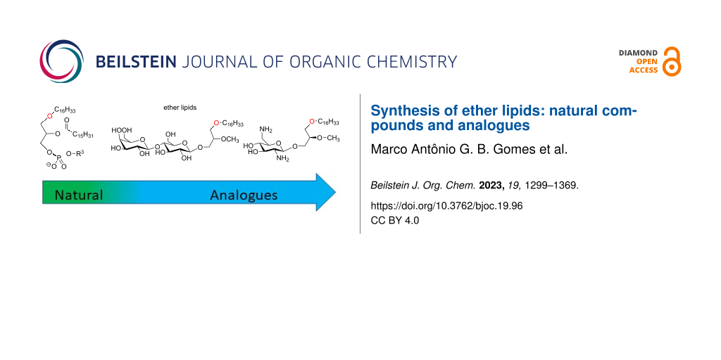
Ether lipids are compounds present in many living organisms including humans that feature an ether bond linkage at the sn-1 position of the glycerol. This class of lipids features singular structural roles and biological functions. Alkyl ether lipids and alkenyl ether lipids (also identified as plasmalogens) correspond to the two sub-classes of naturally occurring ether lipids. In 1979 the discovery of the structure of the platelet-activating factor (PAF) that belongs to the alkyl ether class of lipids increased the interest in these bioactive lipids and further promoted the synthesis of non-natural ether lipids that was initiated in the late 60’s with the development of edelfosine (an anticancer drug). More recently, ohmline, a glyco glycero ether lipid that modulates selectively SK3 ion channels and reduces in vivo the occurrence of bone metastases, and other glyco glycero ether also identified as GAEL (glycosylated antitumor ether lipids) that exhibit promising anticancer properties renew the interest in this class of compounds. Indeed, ether lipid represent a new and promising class of compounds featuring the capacity to modulate selectively the activity of some membrane proteins or, for other compounds, feature antiproliferative properties via an original mechanism of action. The increasing interest in studying ether lipids for fundamental and applied researches invited to review the methodologies developed to prepare ether lipids. In this review we focus on the synthetic method used for the preparation of alkyl ether lipids either naturally occurring ether lipids (e.g., PAF) or synthetic derivatives that were developed to study their biological properties. The synthesis of neutral or charged ether lipids are reported with the aim to assemble in this review the most frequently used methodologies to prepare this specific class of compounds.
Ether lipids (ELs) are natural compounds that feature a glycerol unit linked with an ether function to an alkyl (alkyl acyl ether lipid) or alkenyl (plasmalogen) lipid chain. For the alkenyl compounds, the vinyl ether function is characterized by a (Z)-configuration as shown in Figure 1. In addition, an acyl group is present on the secondary alcohol of the glycerol. This acyl group is constituted by a saturated or unsaturated lipid chain or, in the case of platelet-activating factor (PAF), by an acetyl group (R2 = CH3) [1]. The asymmetric carbon of the glycerol (sn-2 position) features a R configuration. The last substituent attached to the glycerol unit is a polar head group mostly constituted by a phosphatidylethanolamine group (PE) or a phosphocholine moiety (PC). ELs with either phosphatidylserine (PS) or phosphatidylinositol (PI) were also reported [2,3]. These two classes (alkyl and alkenyl) of tri-substituted glycerol ether lipids include a large number of compounds due to the possible structural variations at R1 (saturated and mono-unsaturated lipid chains) and R2 (a large variety of saturated, unsaturated and polyunsaturated lipid chains, Figure 1). These ether lipids constitute the majority of the ether lipids present in mammalians. Beside these two classes of compounds, there exists a multitude of other ether lipids that feature partly substituted glycerol. They correspond, for instance, to biosynthesized intermediates (e.g., 1-O-alkyl-glycerol-3-phosphate [4], lyso-PAF [5]) or neutral ether lipids (e.g., diacyl ether glycerol [6]). Ether glycerolipids are present in mammalian but also in anaerobic bacteria [7], archea (with an inverted stereochemistry at the sn-2 position of the glycerol) [8], protozoa [9], marine and land animals [10,11], but they are, according to the current scientific knowledge, absent in yeast [12] and plants [4].
Figure 1:
Chemical structure of some natural ether lipids (ELs).
Figure 1:
Chemical structure of some natural ether lipids (ELs).
It is estimated that ether lipids account for 10 to 20% of all the glycerophospholipids in humans. However, their tissue distribution is heterogeneous with abundant amount (up to 50% of the phosphoethanolamine based lipids) in central nervous system (mainly as PE-plasmalogen), in skeletal muscle, heart, kidney and lungs [13]. At a cellular scale, the biosynthesis of ELs is initiated in peroxisome and maturation is completed in the endoplasmic reticulum (ER). Then, ELs are spread off in the cell, to the plasma membranes but also in other biomembranes (membrane of mitochondria, ER, nucleus). The occurrence of ELs in tissues was first assessed by Snyder and Woods who reported that neutral ELs and phosphoglycero ELs were generally more abundant in cancer tissues than in normal human tissues [14]. It was also reported that the rate of neutral ether lipids in cancer cell line (quantified in vitro) was correlated to the tumorigenicity of the cancer cell lines in vivo [15]. However, the improvement of the analytical technics that now produces more accurate quantification of lipids with a distinction of neutral EL and phosphoglycero ELs produced contradictory data. Indeed, a recent study that focused on ether glycerophospholipids (alkyl and alkenyl ether lipids) indicates that in breast cancer cells (average of 9 breast cancer cell lines) the PC and PE ether lipids (alkyl and alkenyl together) were down regulated when compared to normal cells (MCF10A) with some variation in the different classes of lipid (PC, PE, PI, PS) [16]. However, alkylglyceronephosphate synthase (AGPS) is highly expressed in aggressive breast (231MFP), melanoma (C8161) and prostate cancer (PC3) cells compared with less aggressive cancer cells (MCF7, MUM2C, and LNCaP, respectively) suggesting that AGPS is an important player in the aggressiveness of cancers [17]. It was also suggested to use ELs as biomarkers for some human diseases like Parkinson disease [18], breast cancer [19] and in rectal adenocarcinoma [20] (in the last case by measuring lysophosphatidylcholine plasmalogen). Low levels of ether lipids are reported in inherited peroxisomal disorders (e.g., rhizomelic chondrodysplasia punctata or Zellweger syndrome) [21]. The severity of the phenotypes associated to peroxisomal disorders (e.g., brain abnormality, mental retardation, premature death) emphasizes the essential role of ether lipids in physiology of cells and tissues. This implication of ELs was also confirmed by producing glyceronephosphate O-acyltransferase (Gnpat) KO mouse which is a mouse model that stop the biosynthesis of ELs [22]. In that case, a reduction of the levels of various neurotransmitters were evidenced likely due to an alteration of the transport efficacy assumed by the synaptic vesicles. The phenotype of these KO mouse shows impaired social interactions and memory deficiency [23]. In another domain, it was reported that centenarians feature a specific profile of ELs in plasma with, in particular, higher level of O-alkyl form of phosphatidylcholine EL and a decreased level of phosphatidylethanolamine plasmalogen (alkenyl) [24]. It must be also noted that ELs are essential players of cell signaling [25]. Together, all these studies point out that ELs are essential for a multitude of biological functions [26] despite their mode of action is not yet fully understood.
From a molecular point of view, the ether function present in alkenyl ELs (plasmalogen) is highly sensitive to oxidation and it is reported that this function is even more reactive than unsaturated and polyunsaturated lipid chains [27]. Accordingly, plasmalogens could act as ROS scavenger and thus protect tissues (e.g., brain) from oxidative stress. The replacement of an ester bonding (present in diacyl glycero ether lipids) by an ether function has also some consequences on the biophysics of membranes. Two recent reviews by A. Koivuniemi [28] and by Jiménez-Rojo and Riezman [29] dedicated respectively to the effect of plasmalogen on biophysics of membranes and the molecular functions of ether lipids offer an interesting overview of the current knowledge of the effect of plasmalogen and ELs on membrane properties at a molecular scale. Shortly, according to molecular dynamics simulation, PE-plasmalogens form thicker, compressed and rigid bilayers when compared to PE-diacyl phosphoglycerolipids [30]. This is likely due to a reduction of the lateral area per molecule and an increase in lipid tail-ordering [31]. It must be however emphasis that the variability of the structure of the lipid chains can deeply influence the biophysical properties meaning that general conclusions could be modulated in function of the structure of these lipid chains. Another property of ELs is their capacity to favor inverted hexagonal phase and therefore to reduce the temperature of transition from Lα (lamellar-liquid crystalline phase) to HII phases (inverted hexagonal phase). This property, which was mainly assessed for PE-ELs, suggests that the presence of EL in biomembranes impacts their curvature. The effect of ELs on the biophysics of membranes suggests that ELs have an important role in some physiological phenomena like the sporulation processes of some prokaryotic cells [32], the epithelium to mesenchymal transition of breast epithelium cell lines [33], the fusion of membranes with extracellular or intracellular vesicles [34] including neurotransmission vesicles [35]. ELs could also be used as a probe to evaluate climate change [36].
All the articles and reviews reporting the description of natural ELs [37], their quantification [38], their biological functions and their role in physio-pathological situations [39,40] invited the researchers to propose new analogues of ether lipids and to study their effects on biological systems. Most of the time the synthetic analogues of ELs aimed to interact with biomembranes via supramolecular interactions with the lipids and proteins that are embedded in these membranes. We can hypothesis that their mechanism of action occurs via a direct interaction with membrane proteins or by a modification of the biophysical properties of the membranes. It must be noted that the design of new bioactive ELs is an alternative to others strategies (e.g., small molecules, antibodies) to modulate membrane proteins functions. Recent results from our group [41,42] and others renew the interest to develop new ether lipids with promising perspectives to address the modulation of membrane proteins that represent a pertinent strategy for some diseases like cancers. Beside the use of synthetic ether lipids for the prevention or the treatment of cancers via different mechanisms, it must be noted that some synthetic ether lipids (e.g., Ino-C2-PAF) have also the ability to regulate genes involved in innate and acquired immune response [43-45] and genes related to inflammation with possible application for the treatment of inflammatory skin diseases [46].
The goal of this review is to report the synthetic approaches of ELs (natural or artificial) that could be helpful to the scientific community for different reasons: 1) to have an overview on the methods already available, 2) to invite scientists that are working on synthesis methodology to apply their works to the design of ELs analogues, 3) the methods reported in this review can be useful tools for the scientific community working on the analysis of EL that require the preparation of standards for analytical purposes. The analysis and quantification of ether lipids is a very important field of research and development that was previously reviewed [47,48]. In this review, we focus on synthesis methodologies applied to prepare alkyl ether lipids. The synthesis methods specifically designed for the preparation of alkenyl ether lipids (plasmalogen) are not reviewed herein. For each compound, their synthesis is reported jointly, when appropriate, with some elements relative to their biological activity. The next sections also include some examples of non-phosphorus glycosylated antitumor ether lipids (GAEL) but more details on the biology of these ether lipids can be found elsewhere [49-51]. The synthesis of analogues of archaeal ether lipids is not included herein but the reader can refer to other articles and reviews dedicated to archaeal lipids [52,53]. It is worth to be noted that some analogues of ether lipids are free of any glycerol unit or ether functions. These molecules that belong to the alkylphosphocholine (APC) class of compounds (e.g., miltefosine) are not illustrated in this review because they were previously reviewed [54,55].
Review
1 Platelet Activating Factor (PAF) and PAF-analogues
The structure of PAF (1-O-alkyl-2-acetyl-sn-glyceryl-3-phosphorylcholine), which was reported in 1979 [56], features one ether function in sn-1 position, an acetyl group in sn-2 and a phosphocholine moiety as polar head group. In this section, we have included works that report the synthesis of PAF and, then, PAF-analogues. For the PAF-analogues, we have selected compounds that feature an acyl group in sn-2 position of the glycerol. Due to the number of PAF-analogues reported in the literature, we have done a selection, which is based on the methodology employed in order to have an overview of the most useful methods employed for the synthesis of PAF and PAF-analogues. The synthesis of alkyl EL involves the chemistry of glycerol or its direct precursors (e.g., glycidol, solketal, epichlorohydrin). The review of Lemaire et al., dedicated to the synthesis of glycerol ether, is complemental to this review article [57]. Of note, the review of Godfroid and Braquet attempted to decipher the binding site of PAF via a QSAR study [58].
1.1 Synthesis of PAF and some building blocks
The platelet activating factor (PAF (2.7), 1-O-octadecyl-2-O-acetyl-sn-glycero-3-phosphocholine; Figure 2) is a natural compound involved in many biological processes [59], including, for instance, its capacity to aggregate platelets, to induce hypotensive effects [60] or to mediate anaphylaxis and inflammation processes [61]. A first hemi-synthesis was reported by Demopoulos et al. [56] and by Blank et al. [62] and two formal syntheses were reported by Benveniste et al. [63,64]. The second formal synthesis starts from the glycerol ether lipid 2.1 that reacted with trityl chloride to yield 2.2. Then, the benzylation of the secondary alcohol produced 2.3. The primary alcohol was deprotected in acidic conditions to produce 2.4. The installation of the phosphocholine polar head group was achieved in two steps starting with the reaction of 2.4 with bromoethyl dichlorophosphate (2.5) to produce the phosphate derivative 2.6. The treatment of 2.6 with trimethylamine produced an ammonium salt. A treatment with silver carbonate was applied to remove any traces of bromide salts. Then, the secondary alcohol was deprotected by hydrogenolysis to produce 2.7 (lyso-PAF). Finally, the acetylation of the secondary alcohol produced the final compound 2.8 (PAF) (Figure 2). A critical step in this synthesis scheme is the use of bromoethyl dichlorophosphate, prepared from POCl3[65] and previously used for the synthesis of lecithin [66,67] and lecithin analogues [68], for the preparation of 2.6.
Figure 2:
Synthesis of lyso-PAF and PAF from 1-O-alkylglycerol [64].
Figure 2:
Synthesis of lyso-PAF and PAF from 1-O-alkylglycerol [64].
It must be noted that a long time before the discovery of the structure of PAF, Arnold, Weltzien and Westphal reported the synthesis of lyso-PAF starting from 1,3-benzylideneglycerol (3.1) [69] (Figure 3). 3.2 was prepared from 3.1 following the methodology reported by West et al. [70]. Then, 3.2 was deprotonated with sodium and the alcoholate reacted with 1-iodohexadecane to produce 3.3 in 50% yield. The phosphocholine moiety was incorporated by using 2-bromoethyl dichlorophosphate as a key reagent and following a previously reported sequence [67]. The debenzylation by using hydrogenolysis conditions produced 3.5 in 75% to 90% yield.
Figure 3:
Synthesis of lyso-PAF from 1,3-benzylideneglycerol 3.1[69].
Figure 3:
Synthesis of lyso-PAF from 1,3-benzylideneglycerol 3.1 [69].
In 1982, G. Hirth and R. Barner reported the synthesis of the two enantiomers of PAF [71]. The synthesis started from ᴅ-mannitol (4.1, Figure 4A). First, 4.1 was transformed to 1,2-isopropylidene-sn-glycerol (4.4) following a three-step sequence (45% yield over three steps) initially reported by H. Eilb [72]. Accordingly, ᴅ-mannitol was transformed in 1,2,5,6-diisopropylidene-ᴅ-mannitol (4.2) by reaction with acetone and ZnCl2. Of note, compound 4.2 was isolated with 5–10% of 1,2,3,4,5,6-triisopropylidene-ᴅ-mannitol. The oxidative cleavage of 4.2 with sodium periodate yields 4.3 that reacted immediately (one-pot procedure) with sodium borohydride to give 4.4. It must be noted that this sequence does not induce racemization and that 4.4 can be stored for months with 0.5% of solid KOH acting as a stabilizer [72]. Then, 4.4 was alkylated with stearyl tosylate to produce 4.5. The two alcohol functions of 4.5 were deprotected in acidic media to produce 3-O-octadecyl-sn-glycerol (4.6). The enantiomer of 4.6 was obtained from 4.4 by protecting the primary alcohol with a benzyl group to give 4.7. Then, the deprotection of the two alcohol functions with H2SO4 in water followed by the tosylation of the primary alcohol produced 4.8. The epoxidation of 4.8 occurred by reaction with t-BuOK in THF, thus producing 4.9 as a chiral electrophile. The regioselective opening of the epoxide is achieved by adding the octadecanol sodium salt. The intermediate was debenzylated by catalytic hydrogenolysis to produce 1-O-octadecyl-sn-glycerol (4.10). It must be noted that the authors, after the deprotection of the two alcohol functions of 4.7, attempted the direct alkylation of the primary alcohol with octadecyltosylate. However, a mixture of mono and dialkylation was formed and were separated by chromatography. Because 4.6 was obtained in better yields and in only 3 steps, the epimerization of 4.6 to 4.10 was also reported (Figure 4B). This epimerization is achieved in three-step sequence that starts with the double tosylation of 4.6 to produce 4.11. Then, the SN2 reaction with potassium acetate in DMSO produces the diester 4.12 with an inversion of the configuration of the chiral carbon atom. Then, 4.12 was hydrolyzed in the presence of KOH to produce 4.10. The installation of the phosphocholine group was achieved following two schemes: a) Starting from the diol 4.10 (Figure 4C), tritylation and benzylation produced 4.13. Then, the deprotection of the primary alcohol in acidic conditions allows introducing the phosphocholine polar head group by using POCl3 and the choline tosylate salt as reagents to yield 4.14. Finally, the debenzylation of the secondary alcohol and its acylation produce PAF 4.15. b) The second scheme is shorter (Figure 4D) and starts with the acylation of 4.16 to produce 4.17. Then, the debenzylation of the primary alcohol produced 4.18. Interestingly, the migration of the acetyl group from sn-2 to sn-3 position was not observed. Finally, the installation of the phosphocholine group was introduced following the same method to produce PAF 4.15. By using the same methodology, the epimer of PAF 4.15 was synthesized.
Figure 4:
A) Synthesis of the two enantiomers of octadecylglycerol (4.6 and 4.10) from ᴅ-mannitol (4.1); B) sequence for the epimerization of 4.6 to 4.10; C) installation of the phosphocholine moiety followed by acetylation; D) acetylation before the installation of the phosphocholine group [71].
Figure 4:
A) Synthesis of the two enantiomers of octadecylglycerol (4.6 and 4.10) from ᴅ-mannitol (4.1); B) s...
The group of Bittman reported in 1995 the synthesis of PAF with a four-step sequence starting from S-glycidol 5.1 in which the acylation of the sn-2 position was also achieved in the last step (Figure 5) [73]. DIBALH (diisobutylaluminium hydride) in toluene was added to hexadecanol in dichloromethane at 0 °C (Figure 5) to form in situ a lithium alcoholate. Then, S-glycidol was added at rt to produce in 50% yield the diol 5.2 after a regioselective opening of the epoxide. The lithium salts were removed by washing with potassium sodium tartrate (Seignette’s salt). Then, at low temperature an excess of 2-chloro-1,3,2-dioxaphospholane (5.3, 3.8 equiv) in the presence of diisopropylethylamine (DIPEA) reacted with the primary alcohol to produce, after an oxidation with Br2 and hydrolysis, the bromoethyl phosphate 5.4. Finally, the quaternarization with trimethylamine produced 5.5 and the acetylation produced 5.6 PAF.
Figure 5:
Four-step synthesis of PAF 5.6 from (S)-glycidol [73].
Figure 5:
Four-step synthesis of PAF 5.6 from (S)-glycidol [73].
The intermediate compounds like 6.2 (1-O-alkylglycerol) or the protected secondary alcohol 6.6, either as enantiopure or racemic forms, are key intermediates for several syntheses of alkyl ELs or analogues. It is worth noticing that rac-1-O-octadecylglycerol (6.2a) is some time identified in the literature as batyl alcohol and rac-1-O-hexadecylglycerol (6.2b) as chimyl alcohol. For the racemic form (Figure 6A), the usual synthesis starts from racemic solketal which is deprotonated with potassium [74], NaH [75], NaNH2[76] or KH and by using different solvents including benzene [74], toluene [76,77], THF [78], or DMF [75,79] and then alkylated with bromoalkyl [75,76] or mesylate lipid alcohol [74]. The same protocols (NaH, toluene or NaH, DMF) were applied to prepare enantiopure (R)-6.1 or (S)-6.1 from commercially available enantiopure solketal [75,76,78]. For the protected secondary alcohol of the glycerol derivative 6.6a (Figure 6B), a stereoselective synthesis, starting from either ᴅ- or ʟ-tartaric acids, produced first the intermediate 6.3 (ᴅ- or ʟ-threitol) that was then alkylated with mesityl lipid alcohol to produce 6.4[80,81]. The acetal protecting group was removed in acidic conditions and then the intermediate 6.5 was subjected to oxidative cleavage to yield an aldehyde that was reduced with NaBH4 to produce 6.6a,b (Figure 6B).
Figure 6:
Synthesis of 1-O-alkylglycerol A) from solketal, B) from ᴅ- or ʟ-tartaric acid and the intermediate 6.3[80].
Figure 6:
Synthesis of 1-O-alkylglycerol A) from solketal, B) from ᴅ- or ʟ-tartaric acid and the intermediate ...
If solketal is frequently used as starting material for the synthesis of EL, it must be noted that glycidol is another substrate of interest available as racemic or enantiopure form. In 1988, Bittman and Guivisdalsky reported a new approach for the synthesis of stereocontrolled 7.4 starting from allyl alcohol (Figure 7) [82]. The Sharpless asymmetric epoxidation of allyl alcohol followed by tosylation produced glycidyl tosylate 7.1a (Figure 7). The reaction of palmityl alcohol (C16H33-OH) in the presence of a catalytic amount of BF3 open regio- and stereoselectively the epoxide to produce 7.2a. Interestingly, the same reaction can be achieved on the substituted glycidol 7.1b,c with a yield of 70 to 80% and a regioselectivity that depends on the substituent (100% regioselectivity for R = Ts or mNO2-Ts and 90% of regioselectivity when R is tert-butyldiphenylsilane) [83,84]. Then, 7.2a was converted to the epoxide 7.5 by cyclisation in the presence of potassium carbonate in methanol, thus producing the interesting building block 7.5. A second option, optimized to avoid the formation of epoxide, used a hindered base and the reactive benzyltriflate as electrophile to achieve under mild conditions the benzylation of the alcohol function to yield 7.3. Then, the removing of the tosyl group required a two-step sequence. First, 7.3 reacted with cesium acetate and then the resulting ester was reduced with LiAlH4 to produce 7.4 with control of its stereochemistry.
Figure 7:
Synthesis of EL building blocks starting from substituted glycidol 7.1a–c[82].
Figure 7:
Synthesis of EL building blocks starting from substituted glycidol 7.1a–c [82].
Very recently, a new method was developed for the incorporation of the phosphocholine polar head group that makes use of the phosphoramidite 8.2 which is a weakly air sensitive reagent [85]. This method was applied for the synthesis of PAF as illustrated in Figure 8[86]. The alcohol 8.1 reacted with 8.2 in the presence of 1H-tetrazole to produce the trialkyl phosphite 8.3 that was oxidized with tert-butyl hydroperoxide to produce phosphate 8.4. Then, β-elimination of the cyanoethyl protecting group produced PAF with a global yield of 70%. The limit of this method arises from the instability of the precursor 8.1 for which the acyl group can shift easily from the sn-2 to the sn-3 position. Noteworthy, the preparation of 8.2 was achieved in 3 steps from phosphorus trichloride and by using choline tetraphenylborate (prepared from choline chloride) [87] instead of the choline p-toluenesulfonate salt to improve the solubility of the salt.
Figure 8:
Synthesis of PAF 8.5 by using phosphoramidite 8.2[86].
Figure 8:
Synthesis of PAF 8.5 by using phosphoramidite 8.2 [86].
1.2 Analogues of PAF with modification on sn-1 position
PAF is characterized by a C16 or C18 saturated lipid chain at the sn-1 position. A first analogue, reported by Hirth et al. in 1983, consisted in replacing this saturated lipid chains by the mono-unsaturated oleyl ((Z)-octadec-9-enyl) lipid chain (Figure 9) [88]. The synthesis starts from serine 9.1 and produce (R)-solketal (9.2) following a three-step protocol [89] that was recently revisited [90] (diazotation, esterification, acetalization). Then, the incorporation of the oleyl chain (C18H35) was achieved by the deprotonation of solketal in DMF followed by the addition of oleyl alcohol tosylate. 9.3 was isolated after the hydrolysis in acidic conditions of the acetal protecting group. The protection of the primary alcohol required a protecting group that can be deprotected without affecting the C=C double bond of the oleyl chain. Accordingly, the primary alcohol was protected with p-methoxydiphenylmethyl (MeO-trityl) in pyridine and then, esterified in sn-2 position with benzoyl chloride to produce 9.4. The deprotection of the primary alcohol under acidic conditions gave 9.5. The polar head group was installed by using the PCl3 method. Then, the benzoyl protecting group was removed (89%) and the hydroxy group at the sn-2 position was esterified with acetic anhydride (94%) to produce oleyl-PAF 9.7.
Figure 9:
Synthesis of oleyl-PAF 9.7 from ʟ-serine [88].
Figure 9:
Synthesis of oleyl-PAF 9.7 from ʟ-serine [88].
Different modifications involved the sn-1 position of the glycerol moiety. In 1984, Wissner et al., reported the incorporation of a phenyl group between the glycerol and the lipid chain. The lipid chain was bonded to the aromatic ring in either ortho-, meta-, and para-position [91]. The incorporation of a phenyl moiety starts with the reaction of the Grignard reagent formed from 4-bromoanisole (10.1, the other isomers 2- or 3-bromoanisole were also reported) with bromotetradecane in the presence of a copper salt (Figure 10). Then, the deprotection of the phenol function with BBr3 produced 10.2. The deprotonation of the phenol function with NaH in DMF and its reaction with solketal mesylate produced, after the deprotection of diol with HCl, the aryl ether glycerol 10.3. The protection of the sn-2 position with a benzyl group was achieved by a classical tritylation of the primary alcohol, benzylation of the secondary alcohol and removing the trityl protecting group. The low yield of this three-step sequence is due to the incorporation of the trityl group, which is reported with 42% yield. The next steps correspond to the adaptation of previously reported methods with the use of bromoethyl dichlorophosphate (10.5) as precursor of the phosphocholine polar head group. The last steps (debenzylation and acetylation) produced successively the lyso derivative 10.8 and the acetylated compound 10.9.
Figure 10:
Synthesis of racemic analogues of lyso-PAF 10.8 and PAF 10.9 featuring a phenyl group between the glycerol and the lipid chain (racemic synthesis) [91].
Figure 10:
Synthesis of racemic analogues of lyso-PAF 10.8 and PAF 10.9 featuring a phenyl group between the g...
The suppression of the ether function in position sn-1 of PAF or its replacement by a thioether function were also reported in 1984 with the aim to identify new compounds that could modulate platelet aggregation or featuring hypotensive effects. First, Wissner et al. reported the synthesis of racemic sn-1-deoxy-PAF 11.8 (Figure 11) [91]. First, n-octadecanoic acid chloride (11.1) reacted with tris[(trimethylsilyl)oxy]ethylene (11.2) [92] to produce, after acidic hydrolysis and subsequent decarboxylation, compound 11.3. Then, the phosphate moiety was introduced via the use of bromoethyl dichlorophosphate 11.4 to produce 11.5. The reduction of the ketone with NaBH4 produced 11.6, and then the incorporation of the trimethylammonium group produced deoxy-lyso-PAF 11.7. Finally, the acetylation of the secondary alcohol produced racemic deoxy-PAF 11.8. The biological evaluations of deoxy-PAF 11.8 have shown that it was less efficient than PAF to reduce blood pressure and to stimulate platelet aggregation [91].
Figure 11:
Synthesis of racemic deoxy-lyso-PAF 11.7 and deoxy-PAF 11.8[91].
Figure 11:
Synthesis of racemic deoxy-lyso-PAF 11.7 and deoxy-PAF 11.8 [91].
The synthesis of the racemic sn-1-thio-PAF 12.8 was reported by Maffrand et al. in 1984 (Figure 12) [93]. The sequence starts with the alkylation of the racemic thioglycerol with bromooctadecane in the presence of potassium hydroxide. Then, the protection of the primary alcohol was achieved either with trityl chloride or with dimethyl-tert-butylchlorosilane. The authors also attempted to use chlorotrimethylsilane but the protection was not regioselective (a mixture of primary and secondary protected alcohols was formed). The acylation of the secondary alcohol was then achieved with acetic anhydride in the presence of pyridine. Then, the deprotection of the trityl moiety of compound 12.4 by catalytic hydrogenation failed whereas heating it in 75% acetic acid solution produced the deprotected compound but migration of the acyl group from the sn-2 to the sn-3 position lead to an inseparable mixture of regioisomers. A selective desilylation of 12.5 was finally achieved with BF3·Et2O producing 12.6 without migration of the acyl group. Then, the phosphocholine polar head group was introduced with a sequential one-pot procedure using first 2-chloro-2-oxo-1,3,2-dioxaphospholane (12.7) to produce a cyclic phosphate as intermediate that subsequently reacted with trimethylamine to produce thio-PAF 12.8. The opening of dioxaphospholane with trimethylamine was initially reported by the group of P. Chabrier [94] and subsequently applied for the synthesis of diacyl-glycerophospholipids [95] and for the synthesis of ether lipids by J. Hajdu et al. [96].
Figure 12:
Synthesis of racemic thio-PAF 12.8[93].
Figure 12:
Synthesis of racemic thio-PAF 12.8 [93].
Wissner et al. modified the glycerol backbone by adding methylene units between either sn-1 and sn-2 positions or sn-2 and sn-3 positions [97]. The incorporation of one methylene unit is shown in Figure 13 as an illustration of all these possibilities. But-3-en-1-ol (13.1) was alkylated with bromohexadecane to produce the ether 13.2. The epoxidation of the carbon–carbon double bond with mCPBA produced the epoxide 13.3. Then, the addition of benzoic acid in the presence of acid catalysis produced an ester that was saponified to yield the diol 13.4. A three-step sequence is applied to produce compound 13.5 that features a secondary alcohol protected with a benzyl group. Then, the installation of the phosphocholine moiety (67%) followed by the deprotection of the secondary alcohol (100%) and its acetylation (53%) produced 13.6.
Figure 13:
Racemic synthesis of 13.6 to illustrate the modification of the glycerol backbone by adding a methylene unit [97].
Figure 13:
Racemic synthesis of 13.6 to illustrate the modification of the glycerol backbone by adding a methy...
Wissner et al. also reported the incorporation of a gem-dimethyl substituent on the glycerol backbone [97]. One illustration of this structural modification is shown in Figure 14. 2-Methylbut-3-en-2-ol (14.1) was used as substrate and the next steps were almost comparable to those reported in Figure 13. The key intermediate 14.4 was isolated after a three-step sequence and used to prepare 14.5.
Figure 14:
Racemic synthesis of 14.5 as an illustration of the introduction of methyl substituents on the glycerol backbone of PAF [97].
Figure 14:
Racemic synthesis of 14.5 as an illustration of the introduction of methyl substituents on the glyc...
1.3 Analogues of PAF with modification at the sn-2 position
The acylation of lyso-PAF with a series of functionalized carboxylic acid was reported in a series of articles from the group of Salomon [98,99]. This group aimed to identify natural compounds that could be formed by the oxidation of ether lipids featuring a polyunsaturated acyl chain in sn-2 position. This work includes, in addition to the oxidation of such type of polyunsaturated EL incorporated in liposomes [99], the formal synthesis of some oxidized derivatives. As an illustration, lyso-PAF 15.1 (extracted from egg albumin) was acylated using Steglich conditions with ω-unsaturated carboxylic acid to produce 15.2a[99] (Figure 15A and B) or with functionalized furane to produce 15.2b. The acylation was also achieved by reaction with cyclic acid anhydride to place in ω-position a carboxylic acid function as exemplified with 15.3a[98].
Figure 15:
Synthesis of functionalized sn-2-acyl chains of PC-EL; A) Steglich esterification or acylation reaction with cyclic acid anhydride; B) examples of reported compounds [98,99].
Figure 15:
Synthesis of functionalized sn-2-acyl chains of PC-EL; A) Steglich esterification or acylation reac...
The acetyl group present in the structure of PAF in position sn-2 of glycerol is readily hydrolyzed in vivo and in serum [100,101]. With the aim to produce more stable compounds, the modification of the sn-2 position of the glycerol was reported. A first option consisted in placing a carbamate function leading to the synthesis of methyl carbamoyl-PAF (1-O-hexadecyl-2-O-(N-methylcarbamoyl)-sn-glycero-3-phosphocholine, mc-PAF (16.3)). Its synthesis starts with the protonation of lyso-PAF to form 16.2 that subsequently reacted with methylisocyanate to produce mc-PAF 16.3 (Figure 16) [102]. The yield of this reaction was not reported. mc-PAF 16.3, which is indeed much more stable than PAF in serum, increases the formation of prostaglandin E2 from astrocyte cortical cell culture [103] and affect memory [104].
Figure 16:
Synthesis of racemic mc-PAF (16.3), a carbamate analogue of PAF [102].
Figure 16:
Synthesis of racemic mc-PAF (16.3), a carbamate analogue of PAF [102].
Ponpipom et al. have reported the synthesis of PAF-analogues featuring in sn-2 position either an azide, amine or acetamide group [79]. In each case, both enantiomers were reported. For the control of the chirality in position 2 of glycerol, (S)-solketal (17.1) was used as starting material to prepare first the hexadecylglycerol (R)-17.2 which was converted to its enantiomer following a five-step sequence (Figure 17A). First, tritylation and mesylation produced 17.3. Then, the nucleophilic substitution (SN2) reaction of benzoate with 17.3 produced the benzoate ester 17.4 with an inversion of configuration. Then, the two protecting groups (ester and trityl) were removed to produce (S)-17.6. The modification of the sn-2 position is illustrated in Figure 17B starting from the mesylate derivative (S)-17.3. Its reaction with sodium azide produced, following a SN2 reaction, the azido derivative 17.7. The deprotection of the alcohol function produced 17.8 that subsequently reacted with 2-chloro-2-oxo-1,3,2-dioxaphospholane (17.9) in the presence of trimethylamine to yield the phosphate 17.10 as an intermediate. Then, its reaction with trimethylamine produced the phosphocholine moiety and compound N3-PAF (17.11). Then, the amine (NH2-PAF) 17.12 was formed by catalytic hydrogenation and subsequently the (acetamido-PAF) 17.13 was formed by acetylation of the amine with acetic anhydride. It is worth noticing that acetamido-PAF 17.13 was previously reported following a different synthesis scheme starting from serine as chiral precursor [96,105]. Recently, is was reported that the acetamido-PAF 17.13 is an activator of the TRPV2 channel leading to constitutive Ca2+ entry thus influencing breast cancer cell migration [106].
Figure 17:
A) Synthesis of (R)-17.2 and (S)-17.6 starting from (S)-solketal (17.1); B) synthesis of N3-PAF (17.11), NH2-PAF (17.12) and acetamido-PAF (17.13) [79].
Figure 17:
A) Synthesis of (R)-17.2 and (S)-17.6 starting from (S)-solketal (17.1); B) synthesis of N3-PAF (17...
1.4 Analogues of PAF with modification at the sn-3 position
Finally, a series of PAF’s analogues were reported by changing the structure of the phosphocholine polar head group. Ohno et al. replaced the trimethylammonium moiety by either triethylammonium 18.2a, N-methylpiperidinium 18.2b, N-methylmorpholinium 18.2c, or N-methylpyrrolidinium 18.2d (Figure 18). 18.1 was used as substrate and was transformed in compounds 18.2a–d following a three-step sequence (introduction of the ammonium, debenzylation and acetylation) [81]. Two of these PAF-analogues (18.2b and 18.2d) were more efficient than PAF for platelet aggregation and for their ability to reduce hypertension.
Figure 18:
Modification of the phosphocholine polar head to produce PAF analogues [81].
Figure 18:
Modification of the phosphocholine polar head to produce PAF analogues [81].
Heymans et al. reported in 1985 a series of PAF-analogues featuring a polar head group in which the phosphate function was replaced by an ether function (Figure 19) [107]. The synthesis starts from the lipid alcohol 19.1 which was deprotonated and alkylated with a mesylamine to produce 19.2. Then, the formation of the ammonium by reaction with methyl iodide followed by the deprotection of the benzyl protecting group under acidic conditions and the acetylation produced the PAF-analogue 19.3. The analogue 19.5 was prepared from 19.2 by debenzylation using catalytic hydrogenation to produce 19.4 that was then acetylated to produce 19.5. 19.3 or 19.5 were not able to induce either platelet aggregation or bronco-constrictive activities.
Figure 19:
Racemic PAF analogues 19.3 and 19.5 characterized by the absence of the phosphate group [107].
Figure 19:
Racemic PAF analogues 19.3 and 19.5 characterized by the absence of the phosphate group [107].
A third modification of the phosphocholine polar head group consisted in replacing the ammonium moiety by myo-inositole-3,4,5-trisphosphate to prepare PIP3-PAF (Figure 20) [108]. The synthesis reported by Wang et al. in 2001 started from the enantiopure diol 20.1, protected with a para-methoxybenzyl (PMB) group at the sn-3 position. 20.1 was selectively alkylated on the primary alcohol to produce 20.2 via the use of dibutyltin oxide as selective reagent for the alkylation of diols [109]. For this reaction, CsF was added to increase the reactivity of the alkyl bromide, likely by a combined effect that includes the interaction of the cesium cation with the halogen atom and the activation of the Sn–O bond of the stannylene acetal via a pentacoordinated intermediate with the fluoride anion [110]. The acetylation of the secondary alcohol and the deprotection of the primary alcohol with 2,3-dichloro-5,6-dicyano-1,4-benzoquinone (DDQ) produced 20.4. Then, the incorporation of the phosphoinositol moiety was achieved by using phosphoramidite chemistry. First, the alcohol 20.4 reacted with O-benzyl-N,N,N’,N’-tetraisopropylphosphorodiamidite to produce the phosphoramidite 20.5 that subsequently reacted with dibenzyl-tris(dibenzylphosphate) myo-inositol (20.6). The oxidation of the phosphite intermediate with m-CPBA followed by the catalytic hydrogenolysis of the benzyl protecting groups produced PIP3-PAF (20.7).
PAF and PAF-analogues that feature an acyl or more generally an ester group in sn-2 position are unstable in physiological environment. The introduction of a second ether function in sn-2 position was applied to increase the stability of such type of compounds in biological media. The initial investigations evaluated the capacity of PAF analogues, featuring higher physiological stability, to aggregate platelets [111]. All these characteristics led to the development of edelfosine and other EL analogues. It is worth noting, that the review articles of Houlihan et al. [112], Brachwitz and Vollgraf [113] and Principe and Braquet [114] reporting the biological effects of phospholipid antitumor agents but without discussing the synthesis approaches, are complementary to the synthesis aspects presented in the upcoming section.
2.1 Synthesis of edelfosine
Edelfosine (1-O-octadecyl-2-O-methylglycero-3-phosphocholine) ET-18-OCH3 or ET-16-OCH3 is an alkyl ether lipid with a methoxy group in sn-2 position and a sn-3 phosphocholine moiety. The length of the saturated lipid chain is composed by either 16 or 18 carbon atoms in the backbone and the stereochemistry of the carbon atom in sn-2 position can be racemic or stereocontrolled. Edelfosine was first synthesized by G. Kny in 1969 [115] but to the best of our knowledge, R. Berchtold reported in 1982 the first synthesis in large quantities with a control of the chirality at the sn-2 position (Figure 21) [116]. The synthesis starts from (S)-1,2-isopropylideneglycerol (21.1). The deprotonation of the primary alcohol with sodium amide followed by the benzylation of the sodium alcoholate produced 21.2 in 95% yield. The deprotection of the acetal was achieved with acetic acid in a mixture of isopropanol and water to give 21.3. Then, a critical step is the alkylation of the primary alcohol in the presence of the unprotected secondary alcohol. This step was achieved by the deprotonation of 21.3 followed by the reaction with 1-bromooctadecane. Compound 21.4 was purified by chromatography on silica gel followed by a recrystallization in acetone. Deprotonation of the secondary alcohol present in 21.4 followed by the addition of iodomethane as electrophile produced 21.5 in 89% yield. The debenzylation of the sn-3 alcohol was achieved by catalytic hydrogenolysis to give 21.6. Then, the phosphocholine moiety was introduced by using 2-bromoethyl phosphorodichloridate as key reagent to give 21.7 that subsequently reacted with trimethylamine to produce 21.8 (edelfosine; Et-18-OCH3).
Figure 21:
Large-scale synthesis of C18-edelfosine (21.8) [116].
Figure 21:
Large-scale synthesis of C18-edelfosine (21.8) [116].
In 1987 [117] and 1988 [118], J. Hadju and S. K. Bhatia reported a stereocontrolled synthesis of edelfosine starting from the commercial isopropylidene-ʟ-glyceric acid methyl ester (22.1, Figure 22). The synthesis starts with a trans-acetalization step to remove the acetal protecting group thus producing 22.2. Then, the primary alcohol was protected by reaction with tritylpyridinium tetrafluoroborate salt to produce 22.3. In the next step, the secondary alcohol was methylated with iodomethane in the presence of silver salts (AgBF4) and silver base (Ag2CO3) to give 22.4. Alcohol 22.5 was isolated after the reduction of the ester group of 22.4. Then, the C16 alkyl chain was introduced to form 22.6 by the reaction of the alcoholate formed by deprotonation of 22.5 and hexadecyl mesylate. Then, the trityl group was removed under acidic conditions in a mixture of methanol and chloroform to give 22.7. The last two steps consist in introducing the phosphocholine moiety. A first step consists in the reaction of 2-chloro-2-oxo-1,2,3-dioxaphospholane (22.8) with 22.7 to yield 22.9. Then, the heterocycle was opened by reaction with trimethylamine to produce 22.10. However, the last step features the lower yield (54%) of this 8-step synthesis.
Figure 22:
Synthesis of C16-edelfosine (22.10) starting from isopropylidene-ʟ-glyceric acid methyl ester (22.1) as a chiral substrate [118].
Figure 22:
Synthesis of C16-edelfosine (22.10) starting from isopropylidene-ʟ-glyceric acid methyl ester (22.1...
In 1994, Bittman et al. reported an alternative strategy to introduce the phosphocholine moiety by the preparation of a cyclic phosphite as a key intermediate [119]. This one-pot three-step sequence starts with the reaction of 23.1 with chlorophosphite 23.2 in the presence of diisopropylethylamine (DIPEA, Figure 23). Then, the intermediate 23.3 reacts with Br2 to produce the intermediate 23.4. Finally, the addition of trimethylamine in an aqueous-organic medium produces edelfosine (23.5).
Figure 23:
Phosphocholine moiety installation by the use of chlorophosphite 23.2 as key reagent [119].
Figure 23:
Phosphocholine moiety installation by the use of chlorophosphite 23.2 as key reagent [119].
For most of the syntheses of edelfosine reported above, the phosphocholine polar head group was introduced during the last steps. Accordingly, the glycerol moiety functionalized with a lipid chain (C16 or C18) and a methoxy group, respectively, in sn-1 and sn-2 position of glycerol (alkyl-methoxy-glycerol – AMG) constitutes an important building unit. The synthesis of this intermediate was prepared as a racemic (Figure 24) or stereocontrolled form. One strategy to prepare rac-AMG starts from 24.1 (24.1 can be prepared from glycerol) [120,121]. Methylation of 24.1 is readily achieved by the deprotonation of the secondary alcohol with NaH followed by the methylation with iodomethane. Then, the cleavage of the acetal occurs by reaction with BH3·THF to give 24.3. Then, the primary alcohol was alkylated with the lipid chain (e.g., C16H33) to produce 24.4. Finally, the benzyl protecting group was removed by catalytic hydrogenolysis to produce 24.5.
Figure 24:
Synthesis of rac-1-alkyl-2-O-methylglycerol (AMG) [120].
Figure 24:
Synthesis of rac-1-alkyl-2-O-methylglycerol (AMG) [120].
In addition to the stereocontrolled synthesis of AMG reported in Figure 21[116] and Figure 22[118], another possibility uses dimethyl-ᴅ-tartrate (25.1) as chiral precursor (Figure 25) [81]. This 8-step synthesis starts with the protection of the diol to form the benzylidene tartrate 25.2. Then, a reductive cleavage of the acetal and the reduction of the two ester functions produced 2-O-benzyl-ᴅ-threitol (25.3) in nearly quantitative yield. The acetalization of the gem-diol produce 25.4 that was deprotonated with KH and alkylated with hexadecyl mesylate to produce 25.5. The deprotection of the secondary alcohol under catalytic hydrogenolysis conditions produced 25.6. Then, the deprotonation of 25.6 followed by the alkylation of the alcoholate with iodomethane produced 25.7. The oxidative cleavage of the gem-diol with lead acetate produced an aldehyde that was reduced with sodium borohydride to give the alcohol 25.9.
Figure 25:
Synthesis of stereocontrolled 1-alkyl-2-O-methyl glycerol 25.9 (AMG) from dimethyl ᴅ-tartrate [81].
Figure 25:
Synthesis of stereocontrolled 1-alkyl-2-O-methyl glycerol 25.9 (AMG) from dimethyl ᴅ-tartrate [81].
In view of the remarkable effects of edelfosine on cancer cells [122], its action as a proapoptotic agent [123,124] and its effect on lipid raft [125,126] or its action against leishmania [127,128], the synthesis of analogues of edelfosine are numerous and aimed to improve its efficacy and/or the selectivity that could also reduce side effects. In the next section, we report a selection of the molecular modifications that are classified depending on the position of the glycerol moiety (sn-1 lipid chain; sn-2; sn-3 polar head group) where the molecular structure is altered. However, the incorporation of a saccharide unit or an inositol moiety is included in subsequent sections.
Modulation sn-1: In 1986, Morris-Natschke et al. [129,130] reported a racemic synthesis of thioether analogues of edelfosine using thioglycerol as precursor. The reaction started with the S-alkylation of thioglycerol by bromo- or iodoalkyl chains as previously reported [131]. Then, the primary alcohol was protected with a trityl group to form 26.1 (Figure 26A). The secondary alcohol was first deprotonated with sodium hydride and alkylated with iodomethane to give 26.2. 26.3 was isolated after the deprotection of the primary alcohol with BF3. Then, the installation of the phosphocholine polar head used POCl3 and choline tosylate to produce in low yield the phosphocholine thioether lipid 26.4. In this study, the authors reported that compound 26.4 has similar cell toxicity than edelfosine-C18 on HL-60 leukemic cells [129,130]. The authors also reported that the sulfone 26.5 (Figure 26B) had similar cytotoxicity than edelfosine-C18. In 1987, the group of E. J. Modest reported that the thioether 26.4 featured comparable cytotoxicity than C18-edelfosine on two leukemic cell lines (HL60 and K562) [132]. Finally, it must be noted that choline tetraphenylborate salt was advantageously used for the preparation of glycerophospholipids by Harbison and Griffin [133]. This salt of choline is poorly hygroscopic and is more soluble in organic media like pyridine.
Figure 26:
A) Racemic synthesis of thioether 26.4[129,130], B) structure of sulfone analogue 26.5[129].
Figure 26:
A) Racemic synthesis of thioether 26.4 [129,130], B) structure of sulfone analogue 26.5 [129].
A stereocontrolled synthesis of 26.4 was reported by Hajdu and Bhatia in 1988. The sequence starts from 27.1 that was prepared from ʟ-glyceric acid (Figure 27) [118]. Then, the free alcohol was converted as an efficient leaving group by reaction with 4-nitrobenzenesulfonyl chloride in the presence of dimethylaminopyridine (DMAP). Then, 27.2 reacted with potassium thioacetate to produce the thioester 27.3. Its reduction with lithium aluminium hydride produced the free thiol 27.4 that was used as nucleophile on octadecyl iodide to install the C18 lipid chain. The deprotection of the primary alcohol produced 27.6 that, in a two-step sequence, was used to install the phosphocholine moiety to produce 27.8.
Figure 27:
Stereocontrolled synthesis of C18-edelfosine thioether analogue 27.8[118].
Figure 27:
Stereocontrolled synthesis of C18-edelfosine thioether analogue 27.8 [118].
The double structural modification of edelfosine that consists to link the lipid chain via a thioether function and to replace the phosphate moiety by a thiophosphate was reported by Markowska et al. in 1993 [134]. As detailed in Figure 28, the synthesis starts with a Mitsunobu esterification of 28.1 with thioacetic acid to produce the thioester 28.2. Then, the reduction with lithium aluminium hydride produced the thiol 28.3. Finally, the phosphocholine moiety was introduced by using 28.4 followed by the opening of the intermediate heterocycle with trimethylamine to produce 28.5.
Figure 28:
Synthesis of thioether 28.4 that include a thiophosphate function [134].
Figure 28:
Synthesis of thioether 28.4 that include a thiophosphate function [134].
It must be noted that C. Piantadosi, S. Morris-Natschke et al. reported in 1990 [135] a series of alkyl thioethers with additional modifications in position sn-3. First, the thioether 29.1 (Figure 29A) was mesylated to 29.2. Then, a bromine atom was introduced via a nucleophilic substitution with LiBr in acetone to form 29.3. Finally, the primary bromide was used to introduce different ammonium salts as illustrated with compound 29.4. A variation of this sequence consisted in placing an ether function in position sn-3 (Figure 29B). Accordingly, a Williamson ether synthesis was applied to 29.1 with bromoethanol protected with a THP group to produce 29.5. Then, the deprotection of the THP group, the installation of a mesyl group, the exchange with LiBr and the final reaction with trimethylamine produced 29.6.
Figure 29:
Synthesis of ammonium thioether 29.4 and 29.6[135].
Figure 29:
Synthesis of ammonium thioether 29.4 and 29.6 [135].
The replacement of the oxygen atom located in sn-1 position by a methylamino group corresponds to a compound known as BN52211 (30.6, Figure 30). This compound was used in many studies for its antitumor cytotoxicity [136] or its immunologic properties [137]. To the best of our knowledge, the synthesis of this compound is only reported in a patent published in 1991 by P. Braquet et al. [138]. The reaction starts with the deprotonation of 30.1 to produce the alcoholate that was alkylated with iodomethane. Then, the reductive opening reaction of the 1,3-dioxane heterocycle in the presence of BH3 as reducing agent produced 30.2. The mesylation of 30.2 followed by the nucleophilic addition of N-methyloctacedylamine produced 30.3. The debenzylation of 30.3 to produce 30.4 followed by the installation of the phosphocholine moiety by using the chlorophosphate 30.5 yield 30.6 (BN52211).
Figure 30:
Synthesis of the N-methylamino analogue of edelfosine 30.6 (BN52211) [138].
Figure 30:
Synthesis of the N-methylamino analogue of edelfosine 30.6 (BN52211) [138].
The replacement of the oxygen atom at the sn-1 position of the glycerol by a methylene unit (sn-1-desoxy glycerol derivatives) was reported by Bonjouklian et al. in 1986 [139]. These authors reported the synthesis of edelfosine analogues with saturated, unsaturated or polyunsaturated lipid chains. For the analogues with a saturated lipid chain (Figure 31A), the reaction started from the diol 31.1. A four-step sequence, which is not detailed in their publication, produced the intermediate 31.2. Then, the phosphocholine moiety was introduced by using POCl3 and choline tosylate to produce 31.3. For the unsaturated and polyunsaturated analogues, the reaction started with the metalation of tert-butyl methyl ether according to the conditions reported by Corey and Eckrich [140], followed by the nucleophilic addition on unsaturated or polyunsaturated aldehyde to produce, as an example, 31.5. Then, the alkylation of the secondary alcohol with iodoethane and the transformation of the t-Bu ether in acetyl ester following the method of Ganem and Small [141], produced, after saponification, the key intermediate 31.6.
Figure 31:
Synthesis of 1-desoxy analogues of edelfosine; A) with a saturated alkyl chain; B) synthesis of the precursor 31.6 with a mono-unsaturated lipid chain [139].
Figure 31:
Synthesis of 1-desoxy analogues of edelfosine; A) with a saturated alkyl chain; B) synthesis of the...
In 1993, Pinchuk reported a stereocontrolled synthesis of both enantiomers of analogues of edelfosine featuring a C18:1 mono-unsaturated lipid chain [142]. This synthesis starts from ᴅ-mannitol that was protected with 4-methoxybenzaldehyde to produce 32.1 (Figure 32) [143]. Then, the methylation of the two alcohol functions produced 32.2. A regioselective reductive cleavage of the acetal was achieved by using either sodium cyanoborohydride and trifluoroacetic acid to yield the thermodynamic product 32.4 or sodium cyanoborohydride and trimethylchlorosilane (a bulkier reagent) that favor the formation of the kinetic product 32.3 (Figure 32). Then, a quite similar sequence (the order was different) can be applied to produce one of the enantiomers (R)-32.8 or (S)-32.8. As exemplified for (S)-32.8 in Figure 32, the oxidative cleavage of 32.4 with sodium periodate and the reduction of the aldehyde intermediate with sodium borohydride yields the alcohol 32.5 that was deprotonated with NaH and subsequently alkylated with oleyl alcohol mesylate. The deprotection of the primary alcohol with cerium salts, produced the key intermediate 32.6. Finally, the phosphocholine group was installed via the use of the 2-chloro-2-oxo-1,2,3-dioxaphospholane (32.7) to produce (S)-32.8.
Figure 32:
Stereocontrolled synthesis of edelfosine analogue (S)-32.8 featuring a C18:1 lipid chain [142].
Figure 32:
Stereocontrolled synthesis of edelfosine analogue (S)-32.8 featuring a C18:1 lipid chain [142].
The last two examples illustrated the incorporation of a mono-unsaturated lipid chain in the structure of edelfosine analogues. Another type of modification involved the lipid chains. In 1989 the group of Counsell reported analogues of edelfosine with an iodophenyl group positioned in ω-position of the lipid chain. These structures were designed for bio-imaging purposes [144]. In 1995, Mauleón et al. [145], extended this study by reporting the incorporation of a phenyl group in ω-position or by adding an alkyl group, a ketone or an alcohol function as illustrated in Figure 33. The idea of these modifications was to gain in selectivity because previous works demonstrated that for PAF a straight saturated alkyl chains was required for its biological effects [58]. The synthesis started by the deprotonation of the alcohol 33.1 that reacted with the bromoalkyl 33.2 that features a phenyl group in ω-position to produce 33.3. Then, the debenzylation and the installation of the phosphocholine polar head group using 2-bromoethyl dichlorophosphate (33.4) produced 33.5. Two analogues, among many others, were prepared following the same method featuring a branched lipid chain (33.6) or a lipid chain with a ketone function (33.7) as shown in Figure 33B.
Figure 33:
Synthesis of edelfosine analogues with modulation of the lipid chain; A) illustration with the synthesis of 33.4; B) structures of 33.5 with a methyl group and 33.6 with a ketone [145].
Figure 33:
Synthesis of edelfosine analogues with modulation of the lipid chain; A) illustration with the synt...
The replacement of the sn-1 ether linkage by a carbamate moiety was proposed by the company Takeda Chemicals Ind. [146,147]. The reaction starts with the conversion of the fatty acid (palmitic acid (34.1) in the example shown in Figure 34) into a reactive lipid isocyanate via the Curtius rearrangement with diphenylphosphoryl azide (DPPA). Then, the isocyanate generated in situ reacted with 2-methoxypropane-1,2-diol to produce the carbamate 34.2. The use of a glycerol building unit already methylated in sn-2 position as precursor is interesting since it renders the synthesis more convergent. This approach is adapted when the stereocontrol of the sn-2 position is not required. Then, the phosphocholine polar head group was introduced in a two-step sequence by using 2-bromoethyl phosphorodichloridate (34.3) to give 34.5 in two steps. Following this synthesis scheme Tsushima et al. [147], also introduced different ammonium salts (e.g., pyridinium, thiazolium). This series of molecules were evaluated as antifungal compounds. The compounds featuring a C14 lipid chain and a phosphocholine polar head group were the most efficient compounds.
Figure 34:
Synthesis of phospholipid featuring a carbamate function to link the lipid chain to the glycerol unit [146].
Figure 34:
Synthesis of phospholipid featuring a carbamate function to link the lipid chain to the glycerol un...
The replacement of the alkyl chain present in sn-1 position by a sesquiterpene linked to the glycerol moiety via a carbonate tether and the incorporation of one unsaturated lipid chain via an ether function at the sn-2 position was described by Gil-Mesón et. al, in 2016 [148]. The goal of this study was to design sesquiterpene bioconjugates having a structure inspired from ether lipids like edelfosine. The strategy started from the protected glycerol 35.1 that was deprotonated with sodium amide and alkylated with bromooctadecane according to a Williamson reaction to produce 35.2 (Figure 35). After deprotection of the primary alcohol function, 35.3 was monoprotected with tert-butyldimetylsilyl (TBS) to produce 35.4 that was treated with diphosgene in the presence of N,N-dimethylaniline providing the intermediate 35.5 featuring a chloroformate moiety at the sn-1 position. Then, the sesquiterpene moiety 35.6 was incorporated by the reaction of its alcohol function with the chloroformate 35.5 in the presence of DMAP and DIPEA. To finalize the synthesis, the TBS protecting group was removed and, then, the phosphocholine polar head group was installed by using the POCl3 method. The phosphate intermediate 35.9 received the choline group in a reaction with choline tetraphenylborate and 2,4,6-triisopropylbenzenesulfonyl chloride (TPS) acting as a coupling agent providing the sesquiterpene conjugate derivative 35.10.
Figure 35:
Synthesis of sesquiterpene conjugates of phospho glycero ether lipids [148].
Figure 35:
Synthesis of sesquiterpene conjugates of phospho glycero ether lipids [148].
Modification at the sn-2 position: Different modifications at the position sn-2 of the glycerol were reported. Bittman et al. reported in 1987 the synthesis of one analogue of edelfosine featuring one additional methyl group on the glycerol moiety as illustrated by the compounds 36.7 and 36.10 (Figure 36) [149]. The aim of the synthesis of these compounds was to evaluate the consequences of steric hindrance on the biological activity. They reported that 36.10 was neither an inhibitor of acetyl-CoA-dependent acyltransferase nor a toxic compound against human leukemic cells (HL-60). However, the methoxy analogue 36.7 exhibited high cell toxicity against HL-60 thus emphasis the importance of the methoxy group on cancer cell cytotoxicity. The synthesis of 36.7 and 36.10 starts with the alkylation of 2-methylprop-2-enol (36.1) to produce 36.2. The bis-hydroxylation of the C–C double bond was achieved with hydrogen peroxide in the presence of formic acid to produce 36.3. Then, tritylation of the primary alcohol produced 36.4 which is an intermediate for the synthesis of both 36.7 and 36.10. For the synthesis of 36.7, the tertiary alcohol was methylated to produce 36.5. Then, the detritylation and the installation of the phosphocholine polar head group produced 36.7. For the synthesis of 36.10, the benzyl ether 36.8 was prepared and then, the trityl protecting group was removed. Then, the installation of the phosphocholine moiety and the debenzylation produced 36.10 featuring an unsubstituted alcohol.
Figure 36:
Racemic synthesis of methyl-substituted glycerol analogues 36.7 and 36.10: A) synthesis of diether 36.7; B) synthesis of lyso-analogue 36.10[149].
Figure 36:
Racemic synthesis of methyl-substituted glycerol analogues 36.7 and 36.10: A) synthesis of diether ...
Ilmofosine is an analogue of edelfosine featuring two structural modifications. First, the C16 lipid chain is attached via a thioether linkage instead of an ether linkage for edelfosine. The second is related to the presence of a methoxymethyl fragment instead of a methoxy group present in edelfosine. Ilmofosine was tested in a phase I clinical trial and proved to be acceptable at a dose of 450 mg/m2 but no effect on the solid tumor was noticed [150] and a phase II clinical trial in non-small cell bronchogenic carcinoma had no effect on tumor regressions [151]. This compound is also efficient to prevent in vitro degranulation of mast cells [152]. Of note, the enrichment of plasma membrane with polyunsaturated lipids (e.g., DHA) increased the sensibility of leukemic cells to ilmofosine [153]. The first synthesis of ilmofosine [154] was improved by Bittman et al. [155] by using 2-hydroxymethylacrylate 37.2 which was prepared on large scale from ethyl acrylate 37.1 (Figure 37) [156]. The bromination of 37.2 and the incorporation of the lipid chain via a nucleophilic substitution with hexadecylthiol produce the lipid derivative 37.4. The reduction of the ester function to a primary alcohol produces 37.5. The alcohol function was methylated with iodomethane after a deprotonation with sodium hydride. Then, the hydroboration and oxidative treatment with sodium perborate produced a primary alcohol. The installation of the phosphocholine group was achieved by using the POCl3 method to produce ilmofosine (37.6) in 37% overall yield.
Figure 37:
Racemic synthesis of ilmofosine (37.6) [155,156].
Figure 37:
Racemic synthesis of ilmofosine (37.6) [155,156].
The group of Bittman also reported a stereocontrolled synthesis of an oxygenated analogue of ilmofosine [157]. As shown in Figure 38A, the reaction started with the formation of a 1,3-cyclic stannoxane which is open and mono-alkylated to produce the ether lipid 38.2 on a large scale (12 g). Then, the methylation of the primary alcohol was achieved in the presence of tetrabutylammonium hydrogenosulfate as phase transfer catalyst to give 38.3. The hydroboration of 38.3 with diisopinocampheylborane ((+)-Ipc2BH) produced 38.4 with high ee (84% ee). Then, the phosphocholine moiety was installed in 56% yield by using the method using 2-chloro-1,3,2-dioxaphospholane 2-oxide (38.5) to produce 38.6. 38.6 and ilmofosine were evaluated as antiproliferative agent on epithelial cancer cell lines (MCF-7, A549, A427). Almost identical in vitro cytotoxicity was observed for ilmofosine and (R)- or (S)-38.6 emphasis that both the chirality and the nature of the heteroatom (oxygen versus sulfur) have no influence on the cytotoxicity. The 2-fluoro analogue of 38.8 was reported by Burchardt et al. (Figure 38B) [158]. The key step is a bromofluorination of 38.2. Then, the methylation of the alcohol function produced 38.8 which was subsequently functionalized with a phosphocholine polar head group.
Figure 38:
A) Stereoselective synthesis of 38.5 via a stereoselective hydroboration reaction; B) synthesis of fluorinated intermediate 38.8[157,158].
Figure 38:
A) Stereoselective synthesis of 38.5 via a stereoselective hydroboration reaction; B) synthesis of ...
The modification of the glycerol moiety by the incorporation of a tetrahydrofuran heterocycle was reported in 1987 by Houlihan et al. (Figure 39) [159]. 2-Furoic acid (39.1) was reduced into 2-tetrahydrofuroic acid and esterified to produce 39.2. Then, the reduction of the ester function with DIBALH and the hydroxymethylation produced the diol 39.3. The mono-alkylation of the diol produced in modest yield the ether lipid 39.4. The installation of the phosphocholine polar head group, via the method of Chabrier et al. [94], produced SRI62-834 (39.6). It is reported that this compound featured similar cytotoxicity than edelfosine on cancer cell lines [136] and was also an inhibitor of PAF. Of note, the two enantiomers of SRI62-834 show the same cytotoxicity as the racemic form on HT29 and HL60 cancer cells [136].
Figure 39:
Racemic synthesis of SRI62-834 (39.6) featuring a spiro-tetrahydrofurane heterocycle in position 2 of the glycerol backbone [159].
Figure 39:
Racemic synthesis of SRI62-834 (39.6) featuring a spiro-tetrahydrofurane heterocycle in position 2 ...
The installation of an imidazole or triazole heterocycle in sn-2 position in place of the secondary alcohol was reported in 1997 by H. K. Nair et al. (Figure 40) [160]. Starting from rac-1-O-n-octadecyl-2-p-toluenesulfonyl-3-O-tritylglycerol (40.1), the imidazole moiety was incorporated via a nucleophilic substitution (Figure 40). Then, the deprotection of the primary alcohol to 40.3 and the installation of the phosphocholine moiety produced the imidazole derivative 40.5. The same protocol was used to prepare an analogue with a triazole moiety in position sn-2. These two analogues were tested as cytotoxic compounds against cancer cells. The authors reported that 40.5 and its triazole analogue featured similar cytotoxicity against MDA-MB-231, HL60 or HT29 than edelfosine (IC50 = 2–6 µM).
Figure 40:
Racemic synthesis of edelfosine analogue 40.5 featuring an imidazole moiety in sn-2 position [160].
Figure 40:
Racemic synthesis of edelfosine analogue 40.5 featuring an imidazole moiety in sn-2 position [160].
The replacement of the methoxy group in sn-2 position of edelfosine by a fluorine atom was reported by Brachwitz et al. in 1982 (Figure 41A) [161] Starting from glycerol ether lipid 41.1, the secondary alcohol was activated with a tosyl moiety as leaving group. Then, the fluorine atom was introduced by nucleophilic substitution using tetrabutylammonium fluoride to give 41.3. After removing the trityl group to yield 41.4[162], the phosphocholine moiety was introduced in two steps using 2-bromoethyl dichlorophosphate as reagent [161]. 41.6 was tested as antiproliferative compounds against Ehrlich ascites carcinoma cells and with an IC50 of 7 µM was a little less efficient than edelfosine (IC50 = 4.5 µM). The same synthetic route was used by Brachwitz et al. to place a 2,2,2-trifluoroethyl group in the sn-2 position (Figure 41B) [163]. The incorporation of the trifluoroethyl group was achieved by reaction of the ether lipid 41.2 with 2,2,2-trifluoroethanol in the presence of sodium hydroxide and with tetrabutylammonium salt as phase transfer catalysis (PTC). 41.8 exhibited almost the same cytotoxicity as 41.6 on Ehrlich ascites carcinoma cells with IC50 = 9.5 µM [164].
Figure 41:
Racemic synthesis of fluorine-functionalized EL: A) Synthesis of 41.6 and B) synthesis of 41.8[161-163].
Figure 41:
Racemic synthesis of fluorine-functionalized EL: A) Synthesis of 41.6 and B) synthesis of 41.8 [161-163].
In 1989, Nomura et al. reported the synthesis of ether lipids with a double modification on the glycerol moiety [165]. The authors introduced different modifications in position 2 of the glycerol including a keto ester (42.6, Figure 42A), a carboxylic acid function (42.7, Figure 42B) or a carbamate or thiocarbamate moiety (42.8 and 42.9). In the meantime, position 3 of the glycerol is constituted by a zwitterionic group with a decyl chain acting as a linker between the phosphate and the ammonium salt. The synthesis of one analogue is illustrated in Figure 42A. Starting from the alcohol 42.1, the first step consists in the installation of the polar head group that uses phosphorus oxychloride and the ammonium salt 42.2 that was previously prepared from decane-1,10-diol. Then, 42.3 was debenzylated by catalytic hydrogenolysis to give 42.4. Finally, the secondary alcohol opens the lactone of the ketene 42.5 to offer the analogue 42.6 with a β-keto ester moiety at position 2 of the glycerol.
Figure 42:
A) Synthesis of the β-keto-ester 42.6 that also features a decyl linker between the phosphate and the ammonium salt; B) structures of three further analogues 42.7, 42.8, and 42.9 prepared from the same substrate 42.4[165].
Figure 42:
A) Synthesis of the β-keto-ester 42.6 that also features a decyl linker between the phosphate and t...
Modification at the sn-3 position: The modifications in position sn-3 of the glycerol correspond to alternatives to the phosphocholine moiety or to introduce some structural alterations corresponding for instance to transform the zwitterion polar head group into a cationic moiety. Of note, the replacement of the zwitterion polar head group by a non-ionic polar group (e.g., saccharide) is mainly reported in the next section.
With the aim to prepare ether lipids resistant to phospholipase C, the replacement of the phosphate group by a phosphonate was reported by R. Bittman et al. [166,167]. The enantioselective sequence starts with the addition of hexadecanol on glycidol tosylate 43.1 assisted by boron trifluoride (Figure 43A). The resulting secondary alcohol 43.2 placed in basic media produced the epoxide 43.3. Then, the key step of the sequence involved the nucleophilic addition of the lithium salt of dimethyl methylphosphonate 43.4 on the epoxide 43.3 in the presence of boron trifluoride to give 43.5. Diazomethane was employed to achieve the methylation of the secondary alcohol to form 43.6. Then, the phosphonic acid function was prepared by reaction with bromotrimethylsilane according to the McKenna method [168]. Finally, 43.7 resulted from the coupling of the phosphonic acid function with choline tosylate that was promoted with trichloroacetonitrile according to the method of Rosenthal [169,170]. The synthesis of the thio-analogue 43.11 (Figure 43B) used the thioether 43.8 as starting material that was previously prepared from glycidol and hexadecylthiol [169]. The epoxide 43.9 was prepared by a Mitsunobu reaction. Then, the incorporation of the dimethyl phosphonate and the installation of the choline moiety used the same methodology than for 43.6 and 43.7. The cytotoxicity of 43.7 and 43.11 was comparable to those of edelfosine on WEHI-3B cells. In vivo, these two compounds prolonged the survival of CD1 mice implemented with L1210 tumors.
Figure 43:
Synthesis of phosphonate-based ether lipids; A) edelfosine phosphonate analogue 43.7 and B) thioether analogue 43.11[166,167].
Figure 43:
Synthesis of phosphonate-based ether lipids; A) edelfosine phosphonate analogue 43.7 and B) thioeth...
Another strategy to introduce a hydrolytically stable phosphonate function was reported by Bittman and Arthur in 2004 by using the Wadsworth–Horner–Emmons (WHE) reaction (Figure 44) [171]. Starting from the enantiomerically controlled diether 44.1, a two-step one-pot synthesis combining Swern oxidation and WHE reaction produced the vinylphosphonate 44.2 (R or S). The transformation of the phosphonate to phosphonic acid with bromotrimethylsilane and then the coupling of the phosphonic acid with the choline moiety produced the enantiopure derivatives 44.3 or 44.4. Interestingly, a singularity of these two enantiomers is that they exhibit different biological effects on PKB (protein kinase B) while similar efficacy was reported on MAPK (mitogen-activated protein kinase) and JNK (c-Jun-NH2-terminal kinase).
Figure 44:
Enantioselective synthesis of phosphonates 44.3 and 44.4[171].
Figure 44:
Enantioselective synthesis of phosphonates 44.3 and 44.4 [171].
Still with the aim to limit the action of phospholipase, Regan and Markoulides reported in 2015 the replacement of the phosphate group by a phosphinate moiety meaning that the phosphorus atom is bonded to two stable P–C bonds thus rendering the molecule resistant to both phospholipase C and D [172]. The sequence starts from hexadecanol, which was allylated to 45.2 (Figure 45). Then, the ozonolysis of 45.2 in reductive medium produced aldehyde 45.3. The nucleophilic addition of vinylmagnesium bromide produced 45.4 that was methylated on the secondary alcohol to give 45.5. The radical addition of sodium hypophosphite, used in excess, produced in good yield the phosphinic acid 45.6. The double silylation of 45.6 produced a phosphorus(III) intermediate that reacted with acrylonitrile (Michael addition) and produced, after hydrolysis of the remaining silylphosphinate, 45.7. The methylation of 45.7 with trimethyl orthoformate produced the methyl phosphinate 45.8. Then, the reduction of the nitrile into primary amine 45.9 was achieved by hydrogenation in the presence of Raney-nickel catalyst. Finally, the per-methylation of the primary amine and the hydrolysis of methylphosphinate produced the zwitterion 45.10. Of note, the demethylation of the phosphinate was also achieved in the presence of iodotrimethylsilane but in that case the demethylation of the methoxy group was also observed producing a lyso-phosphinate derivative. The biological evaluation of 45.10 was not reported.
Figure 45:
Racemic synthesis of phosphinate-based ether lipid 45.10[172].
Figure 45:
Racemic synthesis of phosphinate-based ether lipid 45.10 [172].
As reported above, in vivo and/or clinical trials revealed that edelfosine and different analogues exhibited elevated toxicity. One strategy, reported by Stekar et al. [173], investigated a modification that consisted in the replacement of the phosphocholine polar head group of edelfosine by a phospho-arsonium polar head group. The synthesis starts from glycerol that was protected as 1,3-benzylideneglycerol (46.1, Figure 46). Then, the methylation of the alcohol function was achieved with dimethyl sulfate to give 46.2, which was subsequently deprotected to give 2-O-methylglycerol (46.3). This compound was alkylated with an octadecyl mesylate via a nucleophilic substitution to produce 46.4. Then, the zwitterion polar head group was introduced by reaction of 46.4 successively with POCl3 in the presence of pyridine, with arsonium choline bromide and hydrolysis to produce 46.5. The toxicity of 46.5 was studied and compared to edelfosine. In vitro toxicity revealed almost identical toxicity but acute in vivo toxicity assays indicated that 46.5 was less toxic than edelfosine (the LD50 is 1.7 mmol/kg for 46.5 and 0.8 mmol/kg for edelfosine). The low toxicity of arsonium-based cationic lipids was also confirmed later in other studies dedicated to the development of gene synthetic carriers [174,175].
Figure 46:
Racemic synthesis of edelfosine arsonium analogue 46.5[173].
Figure 46:
Racemic synthesis of edelfosine arsonium analogue 46.5 [173].
The replacement of the trimethylammonium moiety of edelfosine by a dimethylammonium was reported by Hajdu and Bhatia and follows the same methodology used for the synthesis of edelfosine [118]. The difference occurs at the very last step by the reaction of the cyclic phosphate 47.1 with dimethylamine (Figure 47). This reaction suggests that the cyclic phosphate can be open by a series of amines or nucleophiles that could produce a series of edelfosine analogues.
Figure 47:
Synthesis of edelfosine dimethylammonium analogue 47.2[118].
Figure 47:
Synthesis of edelfosine dimethylammonium analogue 47.2 [118].
In line of the modification of the amine moiety of the polar head group of edelfosine, Lemmen and Stumpf [176] reported in 1990 a strategy to introduce a N-methylphosphoethanolamine moiety (Figure 48). The reaction of the diether 48.1 with 2-chloro-3-methyl-1,3,2-λ3-oxazaphospholane (48.2) in the presence of triethylamine followed by the opening of the heterocyclic intermediate with water in the presence of tetrazole produced the intermediate 48.3 featuring a dialkyl phosphite function. Finally, 48.3 was oxidized in situ with tert-butyl hydroperoxide to give 48.4.
Figure 48:
Synthesis of rac-C18-edelfosine methylammonium analogue 48.4[176].
Figure 48:
Synthesis of rac-C18-edelfosine methylammonium analogue 48.4 [176].
The replacement of the trimethylammonium moiety, which is present in the phosphocholine polar head group of edefosine, with a modified ammonium salt was reported, for instance, by Fournier et al. in 1994 [177]. Starting from the enantiopure phosphate 49.1 (Figure 49A), the ammonium salts 49.2 and 49.3 were obtained by reaction with either N-methylpyrrolidine or N-methylmorpholine used in excess (the yields of this reaction were not specified). In the meantime, these authors reported a ramification of the ethylene linker between the ammonium and the phosphate function with a methyl group to prepare analogues with a methylcholine polar head group. For that purpose, the synthesis used the phosphate 49.4 as starting material which was previously reported by Hong et al. [178]. The coupling between 49.4 and the methylcholine 49.5 or 49.6 proceeded in the presence of mesitylsulfonyl chloride. Interestingly, 49.2 and 49.3 presented similar or higher cytotoxicity than edelfosine ((R)-, (S)- or rac-edelfosine) on the three leukemic cell lines tested (CEM, HUT78, and Namalwa) [177]. Regarding the methylcholine derivatives, the presence of the methyl in alpha position to the nitrogen atom (49.8) was more cytotoxic than in β-position (49.7).
Figure 49:
A) Synthesis of edelfosine N-methylpyrrolidinium analogue 49.2 or N-methylmorpholinium analogue 49.3 cations; B) illustration of the synthesis of ether lipids with methylphosphocholine polar head group [177].
Figure 49:
A) Synthesis of edelfosine N-methylpyrrolidinium analogue 49.2 or N-methylmorpholinium analogue 49.3...
The replacement of the phosphocholine (PC) polar group of edelfosine with phosphoethanolamine (PE) or phosphatidylserine (PS) was reported. For the PE derivative [179] the synthesis starts from 2-O-methylglycerol (50.1, Figure 50A) that was alkylated with 1-bromotetradecane to produce 50.2. Then, the incorporation of the polar head group required two steps. First, 2-phthalimidoethyl phosphorodichloridate (50.3) was reacted with 50.2 and then water was added to hydrolyze the remaining P–Cl bond. Finally, the primary amine was deprotected with hydrazine to give 50.4. Of note, in this publication, Nomura et al. have also reported analogues with a variation of the lipid chain length and have also replaced the trimethylammonium group with a pyridinium moiety as illustrated with 50.5. They have also reported sn-2 desoxy analogues as illustrated with 50.6. All these compounds were tested for their antimicrobial activities. For the PS derivative, Brachwitz et al. [180] reported an enzymatic method to prepare phosphatidylserine analogue 50.9. 50.9 was tested as inhibitor of Ehrlich ascites tumor cell growth (IC50 = 30 µM).
Figure 50:
A) Synthesis of edelfosine’s analogue 50.4 with a PE polar group; B) illustration of a pyridinium derivative 50.5 and a desoxy analogue 50.6; C) enzymatic synthesis of edelfosine phosphatidylserine analogue 50.9[179,180].
Figure 50:
A) Synthesis of edelfosine’s analogue 50.4 with a PE polar group; B) illustration of a pyridinium d...
In 1983, Takeda Chemicals reported the synthesis and the biological effect (inhibitor of PAF) of 51.4 (CV-3988) that features a methoxy group in position 2 of glycerol, an octadecyl lipid chain linked to glycerol via a carbamate function and a polar group constituted by a phosphate and a thiazolium moiety (Figure 51A) [146]. The synthesis of 51.4 started with the incorporation of the lipid chain via the formation of a carbamate function (51.2). Then, the installation of the polar group used 2-bromoethyl dichlorophosphate (51.3) to introduce the phosphate moiety and then the reaction with thiazole produced the thiazolium compound 51.4 (CV 3988). The lipid diether with a thiazolium moiety was also reported in 1992 by Wissner et al. (Figure 51B) [181]. These analogues are also featuring an aromatic ring between the thiazolium and the phosphate moiety. Starting from the diether 51.5, the reaction with the dichlorophosphate 51.6[182] produced intermediate 51.7. Then, the reaction with thiazole produced the thiazolium 51.8.
Figure 51:
A) Synthesis of 51.4 featuring a thiazolium cationic moiety; B) synthesis of thiazolium-based EL 51.8[146,181].
Figure 51:
A) Synthesis of 51.4 featuring a thiazolium cationic moiety; B) synthesis of thiazolium-based EL 51...
After having illustrated the modification of the cationic moiety present in edelfosine, another series of compounds consisted in removing the phosphate group, thus leading to cationic ether lipids or protonable ether lipids. In 1990, E. J. Modest et al. reported the synthesis of a series of cationic ether lipids starting from the diether 52.1 (Figure 52) [183]. The transformation of 52.1 into its bromide analogue 52.2 was achieved in a two-step sequence using mesylation and reaction with lithium bromide [135]. Finally, the introduction of the ammonium salt was achieved by reaction with either trimethylamine in a sealed tube or with triethylamine to give respectively 52.3 and 52.4. With the aim to keep present the oxygen atom in position 3 of the glycerol, the introduction of a 2-bromoethyl moiety was achieved to prepare 52.5 in a five-step sequence. Then, the same reaction protocol producing the ammonium salts was applied to give 52.6[135]. Of note, a series of compounds having some structural analogy with 52.6 (the difference comes from the presence of an ester function in position 2 of glycerol; i,e., acetyl) were reported by Wissner et al. [182].
Figure 52:
Synthesis of cationic ether lipids 52.3, 52.4 and 52.6[135,183].
Figure 52:
Synthesis of cationic ether lipids 52.3, 52.4 and 52.6 [135,183].
Another series of compounds devoid of the phosphate group was reported by Takeda Chemicals Industries in 1989 [184]. The synthesis of one typical example, featuring a carbamate linkage between the lipid chain and glycerol, a methoxy group in position 2 of glycerol and an ammonium salt linked via an acetylated carbamate function in position 3 of glycerol, is presented in Figure 53. The synthesis used 53.1 as starting material that was prepared according to a previous reported method [147]. 53.1 reacted with phenyl chloroformate to give the phenyl carbonate 53.2. N,N-Dimethylethylenediamine was added to 53.2 to produce the carbamate 53.3. Then, a regioselective acylation of the carbamate function present in position 3 of glycerol was achieved with acetic anhydride in the presence of pyridine. Finally, the methylation of the tertiary amine with iodomethane produced the ammonium salt 53.5 (the yield was not indicated for this step).
Figure 53:
Synthesis of cationic carbamate ether lipid 53.5[184].
Figure 53:
Synthesis of cationic carbamate ether lipid 53.5 [184].
The replacement of the carbamate function that links the cationic moiety to the glycerol backbone in position 3 of compound 53.5 with a sulfonamide group was reported by Kamata et al. (Figure 54) [185]. This sequence started from the diether 54.1 that was transformed in the primary amine 54.2 by a sequence using a Mitsunobu reaction (to introduce the phthalimide moiety) followed by reaction with hydrazine. Then, the amine 54.2 reacted with 3-chloropropanesulfonyl chloride to produce 54.3. The exchange of the chloride with iodide was achieved with sodium iodide in methyl ethyl ketone used as solvent. Finally, the reaction of 54.4 with trimethylamine produced 54.5. It must be noted that the authors used 54.4 to introduce a thiazolium salt instead of the trimethylammonium moiety. In this paper, the authors also reported a large series of analogues of 54.5 in which the ether function present in position 1 of the glycerol was replaced by a carbamate function and with different cationic groups (e.g., thiazolium, pyridinium, quinolinium) located close to the sulfonamide tether as illustrated with 54.6.
Figure 54:
Synthesis of cationic sulfonamide 54.5[185].
Figure 54:
Synthesis of cationic sulfonamide 54.5 [185].
The compound ONO-6240 (55.1) and SRI-63-119 (55.2, Figure 55) developed respectively by ONO Pharmaceuticals and Sandoz are other examples of ether lipids without phosphate groups. To the best of our knowledge, their synthesis is not reported in the literature. These compounds were reported to be PAF antagonists [186,187].
Figure 55:
Chemical structure of ONO-6240 (55.1) and SRI-63-119 (55.2).
Figure 55:
Chemical structure of ONO-6240 (55.1) and SRI-63-119 (55.2).
The precedent figures illustrated the suppression of the phosphate moiety present in edelfosine thus producing cationic ether lipids. Another structural modification consisted in removing the cationic moiety and to substitute the phosphate function in order to isolate non-ionic ether lipids as reported by McGuigan et al. in 1995 [188]. These non-ionic ether lipids were prepared by using 56.1 as starting material. The reaction of 56.1 with the appropriate chlorophosphate in pyridine produced a series of ether lipids. Some of these compounds (56.2–56.9) are presented in Figure 56. This series of compounds were tested in vitro as inhibitors of the incorporation of tritiated thymidine in DNA of CNCM-I222 cells. For this in vitro assay, compound 56.9 exhibited the highest inhibition at 10 and 100 µM.
Figure 56:
Synthesis of non-ionic ether lipids 56.2–56.9[188].
Figure 56:
Synthesis of non-ionic ether lipids 56.2–56.9 [188].
Another series of compounds came from the replacement of the phosphocholine moiety of edelfosine by fragments previously identified for their biological effects. The first goal of these hybrid structures was to combine the biological effect of ether lipids with those of another drug and more specifically antiviral drugs. In 1997, G. D. Kini et al. reported the conjugation of ether lipids with foscarnet [189]. For this purpose, the primary alcohol present in 57.1 was first protected with a trityl group to give 57.2 (Figure 57). Then, the secondary alcohol was methylated with iodomethane and the primary alcohol was deprotected with trifluoroacetic acid to give 57.3. The coupling of 57.3 with methyl phosphonoformate 57.4 in the presence of DCC yields 57.5. Finally, the saponification of the ester function produced the ether lipid conjugate 57.6 as its disodium salt.
Figure 57:
Synthesis of ether lipid conjugated to foscarnet 57.6[189].
Figure 57:
Synthesis of ether lipid conjugated to foscarnet 57.6 [189].
W. E. Berdel et al. reported in 1986 the conjugation of ether lipids with nucleotides derived from arabinofuranosylcytosine [178]. As detailed in Figure 58A, benzylglycerol 58.1 was alkylated with bromooctadecane in the presence of sodium amide, methylated at the position 2 of the glycerol and finally the primary alcohol was deprotected by catalytic hydrogenolysis to give 58.2. Then, the reaction of 58.2 with phenyl phosphonodichloridate followed by hydrolysis and hydrogenolysis to remove the phenyl group in the presence of PtO2 as catalyst produced 58.3. The coupling reaction of 58.3 with 1-β-ᴅ-arabinofuranosylcytosine-5'-monophosphoromorpholidate 58.4 gives 58.5 with an unspecified yield. The incorporation of AZT (3’-azido-3’-deoxythymidine) directly bonded to the phosphate group present in edelfosine was reported by T. Mavromoustakos et al. in 2001 (Figure 58B) [190]. First, the authors used a method initially reported by Hajdu and Bhatia [118] (Figure 22) to prepare 58.6 in 49% yield from ʟ-glyceric methyl ester acetonide. Then, the trityl group of 58.6 was removed in acidic media to give 58.7. The primary alcohol of 58.7 reacted with 2-chlorophenyl bis(1H-1,2,4-triazol-1-yl)phosphinate (58.8) in the presence of pyridine to give, after hydrolysis in presence of triethylamine, the phosphate salt 58.9. Then, the coupling of 58.9 with AZT in the presence of 1-(mesitylene-2-sulfonyl)-3-nitro-1H-1,2,4-triazole (MSNT) used as coupling agent produced 58.11. In the final step, the O-chlorophenyl group was removed by hydrolysis in the presence of pyridine to give 58.12.
Figure 58:
A) Synthesis of ether lipid conjugated to arabinofuranosylcytosine; B) synthesis of AZT conjugated to ether lipid [178,190].
Figure 58:
A) Synthesis of ether lipid conjugated to arabinofuranosylcytosine; B) synthesis of AZT conjugated ...
In 2019, Y. Xia et al. reported the conjugation of edelfosine to quercetin that belongs to flavonoids [191]. Quercetin is known for its antioxidant properties. The synthesis starts from the ether lipid 59.1 that was alkylated with an excess of 1,3-dibromopropane to produce 59.2 (Figure 59). Then, the incorporation of the benzyl-protected quercetin (59.3) via the formation of an ether linkage yields 59.4. After debenzylation, the final ether lipid conjugate of quercetin 59.5 was isolated. Of note, compound 59.5, at the difference of quercetin, was soluble in a mixture of methanol and dichloromethane. 59.5 was tested in vitro on a panel of cancer cells. The IC50 concentrations were lower for 59.5 than for edelfosine indicating an improved antiproliferative activity. Although edelfosine exhibited a membrane damage as revealed by lactate dehydrogenase released, this situation was not observed for 59.5. The authors reported that 59.5 induced apoptosis.
Figure 59:
Synthesis of quercetin conjugate to edelfosine [191].
Figure 59:
Synthesis of quercetin conjugate to edelfosine [191].
The interest in glyco ether lipids increased over the last 20 years. This family of compounds that features a glycerol ether lipid backbone and at least one saccharide moiety was also identified by some authors as glycosylated antitumor ether lipids (GAELs). To the best of our knowledge this acronym was introduced by R. Bittman et al. in 1996 [192]. Nevertheless, if most of the biological studies of this type of compounds are currently focused on cancer treatment applications, it would not be surprising that other applications could emerge. In this section we have included the synthesis of ether lipids featuring at least one saccharide unit. Accordingly, the two coming sub-sections correspond to the incorporation of a saccharide moiety either in positions 2 and 3 of glycerol. Of note, the phosphocholine moiety present in edelfosine can still be present in the structure of the saccharide analogues (e.g., Glc-PAF) or absent (e.g., ohmline). The analogues possessing either an inositol or an aminoglycoside moiety are presented in the subsequent sections.
3.1 Saccharide unit in position 2 of glycerol
As reported above, edelfosine and some analogues were identified for their anticancer properties or for their utility for other pathologies. Unfortunately, the toxicity of these compounds has limited applications. With the aim to propose new analogues with reduced toxicity or side effects, the groups of J. Mulzer and W. Reutter, have reported in 1995 the incorporation of a saccharide unit in the structure of amphiphilic compounds that still featuring a phosphocholine moiety. First, they have reported a 1-acylglycero-sn-3-phosphocholine derivative with a glucose moiety in sn-2 position (Glc-PC). They have shown that this compound was a non-toxic (at 10 µM) inhibitor of cell proliferation and that the antiproliferative properties were not mediated by protein kinase C [193]. Then, they reported one analogue of Glc-PC but featuring one ether function in sn-1 position instead of an acyl linkage (Figure 60) [194]. This synthesis starts from (S)-solketal (60.1), which was alkylated with allyl bromide to produce 60.2. Then, the deprotection of the acetal and the protection of the primary alcohol as a benzoic ester produced 60.3. The glycosylation reaction involving the secondary alcohol and tetrabenzyl-β-ᴅ-glucopyranosyl fluoride (60.4) produced 60.5. The saponification reaction liberates the primary alcohol that was alkylated with different alkyl bromides including 1-bromohexadecane as illustrated in Figure 60 to afford 60.6. The alcohol function protected with an allyl group was deprotected in the presence of palladium used as catalyst to yield 60.7. Then, the installation of the phosphocholine polar head group and the debenzylation of the protected alcohol functions of glucose produced the glucose analogue of PAF 60.8 (Glc-PAF) (the acetyl group of PAF was replaced by the glucopyranosyl moiety). Of note, a review dedicated to the biological effects of Glc-PC and 60.8 (Glc-PAF) and other analogues was published by Danker et al. in 2010 [195].
In 2020, the groups of G. Arthur and F. Schweizer from the University of Manitoba reported a series of ether lipids functionalized with an ʟ-rhamnose moiety [196]. As detailed in Figure 61, 61.1 was tosylated on the primary alcohol to give 61.2. Then, the azide function was introduced with sodium azide in DMF to produce 61.3. The glycosylation step used the method of B. Fraser-Reid [197] with the glycoside donor 61.4 and the acceptor 61.3 in the presence of N-iodosuccinimide (NIS) as coupling reagent and with silver triflate as catalyst. The resulting compound 61.5 was treated with sodium methoxide in methanol to remove the acetyl groups to give 61.6. The application of Staudinger’s reaction conditions with trimethylphosphine as reagent produced the amino derivative 61.7. Then, coupling of the amino group of 61.7 with carboxylic acid 61.8 with TBTU as coupling reagent produced amide 61.9. In the meantime, the authors prepared 61.10 following a similar methodology to incorporate an ʟ-glucopyranoside moiety. 61.7 and 61.9 were tested as cytotoxic compounds on epithelial cancer cell lines. 61.7 proved to be the most efficient. Investigations related to the mechanism of action concluded that cell death was not due to a membrane lysis-based mechanism. In vivo studies on chicken embryo model of high-grade ovarian cancer proved the efficacy of compound 61.7 (ʟ-Rham) [198].
Figure 61:
A) Synthesis of amino ether lipid 61.7 functionalized with a rhamnose unit and its amide analogue 61.9; B) structure of ʟ-glucose-based derivative 61.10, an analogue of 61.7[196].
Figure 61:
A) Synthesis of amino ether lipid 61.7 functionalized with a rhamnose unit and its amide analogue 6...
The synthesis of glycerol-based lipids functionalized with a saccharide moiety have interested many chemists from the beginning of the 70’s [199]. In the 80’s, the synthesis of glycerol monoether [200] or diether with two lipid chains [201,202] and with different saccharide moieties (e.g., glucopyranosyl [203], 2-desoxy-2-fluoromannopyranosyl [204]) was reported. One of the first studies that reports the synthesis of glycerol diether including a methoxy group in position 3 of the glycerol was published by N. Weber and H. Benning in 1986 [205]. The initial idea was to replace the ionic phosphocholine group present in the structure of edelfosine with non-ionic polar fragments. As shown in Figure 62A, the glycosylation reaction used the Knoenigs–Knorr method using acetobromo-α-ᴅ-glucose 62.2 and the ether lipid 62.1. The acetylated β-ᴅ-glucopyranosyl ether lipid 62.3 was isolated and then, the acetyl groups were removed to give 62.4. Of note, the same methodology was applied to prepare the disaccharide derivative 62.5 (Figure 62B) that features a maltose unit as neutral polar group. In this study, the authors have applied the synthesis from racemic or stereocontrolled substrate 62.2. In a subsequent study, these authors reported that the analogue of 62.4 possessing an octadecyl lipid chain labeled with 14C was rapidly metabolized in vitro (Ehrlich ascites tumor cells). They concluded that the glycosidic bond was cleaved by the action of a β-glycosidase [206]. Of note, R. W. Franck and C. H. Marzabadi reported the analogue of 62.4 with a 2-desoxyglucose moiety [207,208].
Figure 62:
A) Synthesis of glucose ether lipid 62.4; B) structure of ether lipid 62.5 possessing a maltose unit [205].
Figure 62:
A) Synthesis of glucose ether lipid 62.4; B) structure of ether lipid 62.5 possessing a maltose uni...
In 1991, the group of M. Liefländer reported the synthesis of neutral analogues of edelfosine with the replacement of the phosphocholine polar group with either glucuronic acid methyl ester 63.8, cellobiose 63.9 or maltose 63.10 (Figure 63A and B) [209]. The synthesis (Figure 63A), illustrated with the preparation of glucuronic acid methyl ester 63.8, started with the synthesis of the diether 63.5 which is a key intermediate. Its synthesis used 3-O-benzyl-sn-glycerol as starting material followed an unusual method because others, requiring protecting groups, did not produced satisfactory yields according to the authors. Here, the reaction of the diol 63.1 with dibutyltin oxide produced the tin acetal 63.2. This compound reacted selectively in sn-1 position with 1-bromohexadecane to produce, after workup, the diether 63.3. The next two steps were achieved following a previously reported protocol [71] to give 63.5. Then, the last two steps were carried out using their own protocol published earlier to give 63.8[200]. The cellobiose analogue 63.9 and the maltose analogue 63.10 were prepared following the same methodology.
Figure 63:
A) Synthesis of glucuronic methyl ester 63.8; B) structure of cellobiose 63.9 and maltose 63.10 analogues prepared following the same protocol [209].
Figure 63:
A) Synthesis of glucuronic methyl ester 63.8; B) structure of cellobiose 63.9 and maltose 63.10 ana...
In 1994, the group of Bittman reported briefly the synthesis of 64.7 (no experimental parts) and in detail its biological effect that was compared to edelfosine [210]. The chemistry part of this work was more detailed in a second paper published in the same year [211]. They reported that edelfosine was an antineoplastic agent by limiting the number of colonies forming of murine WEHI-3B cells at 1 µM whereas 64.7 exerted any action up to 20 µM. They also reported that both compounds have a lysis effect on erythrocytes but with different kinetics and dose effect. The synthesis of 64.7 was inspired from a previous work reporting the synthesis of cerebrosides [212]. The anomeric acetyl group of peracetylated maltose 64.1 was quantitatively deprotected with hydrazine in acetic acid (Figure 64). Then, the deprotonation of the alcohol with butyllithium and the reaction of the alcoholate with tetramethyldiamidophosphorochloridate (64.3) produced the phosphorodiamidate 64.4[212]. Then, the glycosylation of alcohol 64.5 with 64.4 catalyzed with trimethylsilyltriflate produced 64.6[211]. 64.6 was deacetylated in methanol in the presence of potassium hydroxide to yield the maltose derivative 64.7. The same methodology was applied to prepare 64.8 that features a lactose moiety in place of the maltose unit.
Figure 64:
A) Synthesis of maltosyl glycerolipid 64.7; B) structure of lactose analogue 64.8 prepared following the same protocol [210,211].
Figure 64:
A) Synthesis of maltosyl glycerolipid 64.7; B) structure of lactose analogue 64.8 prepared followin...
The group of Bittman reported in 1996 a series of glycosylated antitumor ether lipids (GAELs) featuring either a glucose or a mannose moiety [192]. First, they reported an asymmetric synthesis of the aglycone part starting with the asymmetric bis-hydroxylation of allyl 4-methoxyphenyl ether 65.1 to produce 65.2 (Figure 65A). The opening of the 1,2-O-stannylidene intermediate, previously reported by Liefländer [209], is regioselective but it is not fully controlled leading to the formation of 65.3 (90%) and 65.4 (8%), required a separation by chromatography. Then, the methylation of the sn-2 alcohol function produced 65.5 and the deprotection of the primary alcohol with cerium(IV) ammonium nitrate (CAN) produced the key intermediate 65.6 in 95% yield. The saccharide units (glucose or mannose) were activated in anomeric position with a trichloroacetimidate group. The glycosylation reaction was achieved with trimethylsilyl trifluoromethanesulfonate (TMSOTf) used as catalyst and in the presence of molecular sieves (3 Å) (Figure 65B). The assistance of the adjacent acetyl group explained the formation of the β-anomer for the glucose derivative and α-anomer for mannose. For the glucose derivative (65.8), the authors removed the acetyl group in position 2 via the preparation of a xanthate and its reduction with tributyltin in the presence of AIBN, followed by debenzylation produced 65.9 (Figure 65C). The methylation of the intermediate secondary alcohol produced 65.10. By using the same protocol, the mannose derivatives 65.11, 65.12 and 65.13 were characterized. From this study, the authors reported that 65.9 was more efficient than edelfosine to inhibit the growth of MCF-7 human breast cancer cells. All the other compounds were less efficient.
Figure 65:
A) Asymmetric synthesis of the aglycone moiety starting from allyl 4-methoxyphenyl ether; B) glycosylation reaction to produce 65.8; C) structure of glycol ether lipids [192].
Figure 65:
A) Asymmetric synthesis of the aglycone moiety starting from allyl 4-methoxyphenyl ether; B) glycos...
In 2011, the group of C. Vandier have reported for the first time that edelfosine inhibited the activity of SK3/KCa2.3 channels at 1 µM which is a non-toxic concentration (toxicity was observed at a concentration higher than 3 µM) [213]. Nevertheless, with the aim to evaluate less toxic compounds, we reported the synthesis of analogues of edelfosine in which the phosphocholine polar head group was replaced by a mono or disaccharide unit [41]. As an example, the synthesis of 66.4 (also identified as ohmline) starts (Figure 66) from the racemic (or chiral) alcohol 1-O-hexadecyl-2-O-methylglycerol (66.1). The glycosylation proceeds with hepta-O-acetyllactose-1-O-trichloroacetimidate (66.2) in the presence of boron trifluoride etherate to produce 66.3 that features a β-glycosidic linkage. Then, the deprotection of the alcohol function with sodium methanolate in methanol produced ohmline in 88% yield. A similar protocol was applied to the mono and disaccharides thus producing compounds 66.5 (glucose), 66.6 (galactose) and 66.7 (melibiose). These compounds were able to inhibit the activity of SK3 ion channels [214] and to reduce SK3-dependent cancer cell motility. The most efficient one is 66.4 (ohmline) that reduced SK3 current by 73% at 10 µM and SK3-dependent cancer cell motility by 50% (24 h at 300 nM). Noteworthy, ohmline which is non-toxic at 10 µM is also one of the most selective agents for the inhibition of SK3 [50] with only a limited effect on the other isoforms SK1 (20% of inhibition at 10 µM) and no effect on SK2 and IKCa (SK4). It must be noted, that the chirality in position sn-2 of the glycerol has no effect on the modulation of SK3 in vitro [41]. Finally, ohmline was also tested in vivo and proved its efficacy to prevent the occurrence of bone metastasis in a breast cancer model (murine experiments) [42]. Of note, the incorporation of a pyrene fluorescent group in omega position of the lipid chain of ohmline was prepared following the same methodologies as employed to prepare ohmline [215].
Figure 66:
A) Synthesis of ohmline possessing a lactose moiety. B) Structure of other glyco glycero lipids prepared following the same method [41].
Figure 66:
A) Synthesis of ohmline possessing a lactose moiety. B) Structure of other glyco glycero lipids pre...
With the aim to improve the water solubility of ohmline and also to evaluate the effect of a negative charge localized in the polar head group, we designed analogues possessing a phosphate moiety placed between glycerol and the saccharide or disaccharide unit [216]. The synthesis, illustrated with lactose in Figure 67A, consisted in first introducing the phosphate moiety at the anomeric position of lactose heptaacetate (67.1) by using phosphorus trichloride followed by a hydrolysis step to produce the salt 67.2. The incorporation of the lipid moiety (1-O-hexadecyl-2-O-methyl-rac-glycerol 67.3) was achieved via a mixed anhydride with pivaloyl chloride which is, for such type of lipid moiety, the best coupling agent [217]. Then, the di-substituted phosphite was oxidized with I2 in the presence of pyridine and water to produce 67.4. Finally, deacetylation produced the lactose phosphate glycerol ether lipid 67.5. A similar procedure was applied to prepare the maltose and melibiose derivatives 67.6 and 67.7 (Figure 67B). The lactose derivative 67.5 was indeed more water soluble than ohmline. This series of compounds exhibited almost the same activity than ohmline to reduce the activity of SK3 at 10 µM and to reduce SK3-dependent cell motility at 300 nM. This study also demonstrates that both the structure of the disaccharide unit and the stereochemistry of the glycoside linkage have, in this series of compounds, a limited incidence on the biological effects. More recently, we have reported via 2H NMR using deuterated ohmline that it has a tropism for membranes. This behavior is easily explained by its amphiphilic structure. Furthermore, the incorporation of ohmline in model membranes impact the order parameters especially for the rigid membrane featuring high ratios of cholesterol [218]. In addition, A. M. Bouchet, via molecular dynamics simulations reported that the presence of ohmline in model biomembranes induces a modification of the position of cholesterol [218]. Altogether, these results suggest that ohmline could act on SK3 channels function not via a direct interaction between SK3 proteins but via a local modulation of the biophysical properties of the plasma membrane. This selectivity of action (e.g., ohmline has no effect on SK2 channels) could be explained by the different interaction of membrane proteins with its lipid environment. Interestingly, we have also reported in the context of colon cancer cells that ohmline modifies the anti-EGFR mAbs action mediated by the control of calcium signaling [219].
Figure 67:
A) Synthesis of lactose-glycerol ether lipid 67.5; B) analogues possessing a maltose (67.6) or melibiose moiety (67.7) [216].
Figure 67:
A) Synthesis of lactose-glycerol ether lipid 67.5; B) analogues possessing a maltose (67.6) or meli...
The incorporation of the (1→6)-digalactosyl moiety in place of the lactosyl group present in ohmline was inspired from a study reporting that some plants, under phosphate deprivation, induced the biosynthesis of (1→6)-digalactosyldiacylglycerol (DGDG) in place of phosphatidylcholinediacylglycerol [220]. We proposed two distinct syntheses for the preparation of digalactosyl ether lipids [221]. The common point of these two strategies was based on the glycosylation reaction of the galactosyl intermediate 68.1 (Figure 68). According to the first approach (Figure 68A), we used the classical protecting groups (benzyl, acetyl, trityl) to prepare in three steps the galactosyl derivative 68.2. The second galactosyl unit 68.3 was prepared in two steps (56% yield) from the expensive commercial compound methyl α-ᴅ-galactopyranoside. Then, the anomeric position of 68.3 was transformed in α-iodo intermediate 68.4 that reacted with 68.2 to produce 68.5. The acetyl and benzyl groups were removed to produce the (1→6)-digalactosylglycerol ether lipid 68.6. Finally, 68.6 was isolated in 8 steps with a global yield of 5%. The second approach for the synthesis of 68.6 (Figure 68B), consisted in using trimethylsilyl as protecting group. Accordingly, 68.1 was per-silylated and then the silylated primary alcohol was deprotected with acetic acid in a mixture of acetone and methanol to produce 68.7. The second galactosyl moiety 68.8 was prepared in one step (silylation) from galactose. The anomeric position was iodinated with trimethylsilyl iodide to produce 68.9 which was directly engaged in the coupling reaction with alcohol 68.7 to produce 68.10. The deprotection of the seven alcohol functions was achieved by using an acidic resin to produce 68.6 in 77% yield. According to this second synthesis scheme, 68.6 was prepared in 5 steps in 25% global yield. The evaluation of 68.6 for the modulation of SK3 ion channels indicated that it was much less efficient than ohmline [221].
Figure 68:
Synthesis of digalactosyl EL 68.6, A) by using trityl, benzyl and acetyl protecting groups, B) by using trimethylsilyl as protecting group [221].
Figure 68:
Synthesis of digalactosyl EL 68.6, A) by using trityl, benzyl and acetyl protecting groups, B) by u...
The methodology using trimethylsilyl as protecting group was also employed to prepare another series of disaccharide-based ether lipids. This study aimed to evaluate the effect of (1→6) versus (1→4) disaccharide linkage, to study the stereochemistry of the glycosidic linkage for ohmline (α versus β linkage) and to study the effect of the order of the saccharide units (Glc-Gal versus Gal-Glc) [222]. As an example (Figure 69), α-ohmline was prepared by the per-silylation of lactose to produce 69.1. Then, the mono-iodination at the anomeric position with trimethylsilyl iodide produced an electrophile that reacted with the lipid alcohol 69.2. Then, the deprotection of the seven alcohol functions produced 69.3 (α-ohmline). The other molecules prepared in this study are presented in Figure 69B. The biological evaluation indicated that α-ohmline was completely inefficient to inhibit SK3. Among the other compounds, we found that 69.6 was as efficient as ohmline while 69.3 was inefficient. More recently, we replaced the oxygen atom localized at the sn-1 position of the glycerol unit of ohmline by a sulfur atom at the same position or close to this position. We found that some of these sulfur analogues of ohmline were activators of SK3 channels. This gain of function was applied to vasorelaxation assays [223].
Figure 69:
A) Synthesis of α-ohmline; B) structure of disaccharide ether lipids prepared by using similar methodologies [222].
Figure 69:
A) Synthesis of α-ohmline; B) structure of disaccharide ether lipids prepared by using similar meth...
In 2015, the group of N. G. Morozova reported in a communication the synthesis of analogues of ohmline in which the methoxy group in position 2 of glycerol was replaced by an ethoxy group and other derivatives with a carbamate linker between the glycerol and the saccharide unit [224]. The synthesis started from the diether 70.1 that was glycosylated with a bromide glycosyl donor like hepta-acetylated lactose 70.2 in the presence of mercury salts (Figure 70). Then, deacetylation produced 70.3. A second approach consisted in adding a linker between the saccharide unit (e.g., lactose) and the glycerol moiety. To this end, 70.1 reacted with carbonyldiimidazole (CDI) to produce 70.4. Then, the addition of amino-alcohol (e.g., aminoethanol) produced 70.5. Finally, the glycosylation and the deprotection of the alcohol functions produced 70.6. In this communication, the yield and spectroscopic characterizations of compound 70.4 are not reported.
Figure 70:
Synthesis of lactose ether lipid 70.3 and its analogue 70.6 featuring a carbamate function as linker [224].
Figure 70:
Synthesis of lactose ether lipid 70.3 and its analogue 70.6 featuring a carbamate function as linke...
The incorporation of a rhamnose moiety in sn-3 position of glycerol was reported by F. Schweizer and G. Arthur in 2020 [196]. The glycosylation of the diether 71.1 with phenyl 2,3,4-triacetyl-1-thio-α-ʟ-rhamnopyranoside (71.2) in the presence of silver triflate and N-iodosuccinimide produced the rhamnopyranoside derivative 71.3. Deprotection of the alcohol functions produced 71.4 (Figure 71). This neutral ether lipid 71.4 proved to be less toxic against cancer cell lines than other derivatives that included in their structure a protonable amino group.
Figure 71:
Synthesis of rhamnopyranoside diether 71.4[196].
Figure 71:
Synthesis of rhamnopyranoside diether 71.4 [196].
With the aim to produce glyco ether lipids featuring higher lipophilicity and possibly higher stability in biological media, R. Bittman et al. reported in 1990 the synthesis of the α-thioglucopyranoside 72.5 (Figure 72) [225]. The glycosylation occurred between the tosylated diether 72.1 and 2,3,4,6-tetra-O-acetyl-1-mercapto-β-ᴅ-glucopyranose (72.2) in the presence of DBU (1,8-diazabicyclo(5.4.0)undec-7-ene) to produce a mixture of the α- and β-thioglycoside 72.3 and 72.4 that were separated by flash chromatography. The deprotection of the alcohol groups of 72.3 was achieved with barium oxide in methanol to give α-thioglycoside 72.5. The evaluation of 72.5 and 72.6 indicated that 72.5, used at 40 µM, limited strongly the incorporation of [3H]thymidine into DNA of WEHI 3B and C653 cell lines while 72.6 was almost inefficient. However, 72.5 was less efficient than edelfosine.
Figure 72:
Synthesis of 1-O-hexadecyl-2-O-methyl-3-S-(α-ᴅ-1'-thioglucopyranosyl)-sn-glycerol (72.5) [225].
Figure 72:
Synthesis of 1-O-hexadecyl-2-O-methyl-3-S-(α-ᴅ-1'-thioglucopyranosyl)-sn-glycerol (72.5) [225].
Still with the aim to produce metabolically stable glyco ether lipids, the groups of R. W. Franck, R. Bittman and G. Arthur reported in 1999 the synthesis of the C-glycoside 73.10 that features a 2-desoxyglucopyranoside moiety [226]. The synthesis of the ether lipid fragment (Figure 73A) starts from butanetriol 73.1 that was selectively protected by reaction with benzaldehyde in the presence of orthoformiate to produce the acetal 73.2. The alkylation of the remaining alcohol function with 1-bromo-hexadecane produced the protected ether lipid 73.3. The opening of the 1,3-dioxane acetal following the Hanessian–Hullar reaction produced the bromo derivative 73.4 that was used as electrophile in the second part of the synthesis (Figure 73B). The starting saccharide material was the benzylated glucal 73.5 that was hydrohalogenated and subsequently transformed in the thioacetate 73.6. In situ generation of a thiol intermediate from 73.6 and its reaction with 73.4 produced the 2-desoxythioglucopyranoside 73.7. The deprotection of the secondary alcohol by trans-esterification with sodium methanolate and the subsequent methylation of the alcoholate allowed the replacement of the benzoyl group by a methoxy group. Then, reaction with magnesium monoperoxyphthalate (MMPP) produced sulfone 73.8. The application of the Ramberg–Bäcklund reaction that involved dibromotetrafluoroethane and KOH/Al2O3 lead to the elimination of SO2 and the formation of alkene 73.9 as a mixture of diastereoisomers. Finally, ionic hydrogenation reduced the alkene and a subsequent catalytic hydrogenolysis lead to the debenzylated C-glycoside 73.10.
Figure 73:
A) Preparation of lipid intermediate 73.4; B) synthesis of 2-desoxy-C-glycoside 73.10[226].
Figure 73:
A) Preparation of lipid intermediate 73.4; B) synthesis of 2-desoxy-C-glycoside 73.10 [226].
N. G. Morozova et al., after reporting the incorporation of a cationic group (imidazolium) between position 3 of glycerol and a saccharide moiety [227], reported in 2019 the functionalization of the position 6 of a saccharide moiety (galactose, mannose) with a cationic group (pyridinium or N-methylimidazolium) [228]. The synthesis of the pyridinium derivative is outlined in Figure 74. The synthesis started with the mesylation in position 6 of the galactosyl derivative 74.1. Then, the pyridinium salt was formed by refluxing 74.2 with pyridine. In the last step the acetyl protecting groups were removed to produce 74.3.
Figure 74:
Synthesis of galactose-pyridinium salt 74.3[228].
Figure 74:
Synthesis of galactose-pyridinium salt 74.3 [228].
4 Inositol and analogous derivatives of glycerol ether lipids
Phosphoinositides constitute a class of lipids that are involved in a multitude of biochemical processes [229]. These biological roles of phosphoinositides incited chemists and biologists to incorporate phosphatidylinositol or inositol moieties in the structure of glycerol ether lipids.
4.1 Incorporation of an inositol moiety or analogues in position 2 of glycerol
K. Danker et al. reported in 2006 the synthesis of one analogue of Glc-PAF in which the glucosyl unit was replaced by a myo-inositol moiety (Figure 75) [230]. The synthesis starts from the ether lipid 75.1 which was converted into the tetrahydropyrane protected primary alcohol 75.2. Then, the allylation of the sn-2 position produced 75.3, which, after a sequence including ozonolyze and reduction, produced 75.4. This sequence allows the incorporation of an ethylene glycol spacer at the sn-2 position. Then, the tosylation of the primary alcohol gave 75.5 and the incorporation of racemic 2,3,4,5,6-penta-O-benzylinositol (75.6) produced 75.7. The cleavage of the THP protecting group and the installation of the phosphocholine polar head group produced 75.9. The debenzylation by catalytic hydrogenolysis produced Ino-C2-PAF (75.10). This compound featured a low toxicity (non-toxic up to 5 µM) on keratinocyte cell line (HaCaT cells) but prevent its proliferation.
Figure 75:
Synthesis of myo-inositol derivative Ino-C2-PAF (75.10) [230].
Figure 75:
Synthesis of myo-inositol derivative Ino-C2-PAF (75.10) [230].
4.2 Incorporation of an inositol moiety or analogues in position 3 of glycerol
The groups of K. S. Ishaq and E. J. Modest reported in 1989 the synthesis of analogues of edelfosine in which the choline unit was replaced by a myo-inositol moiety (compound 76.10, Figure 76) [231]. First, the synthesis required to prepare the benzylated phospho-myo-inositol 76.7 as reported in Figure 76A. This sequence follows the method initially reported by R. Gigg [232]. myo-Inositol was first protected in positions 1 and 2 by reaction with acetone dimethylacetal in acidic conditions to produce 76.2. Then, the benzylation of the remaining alcohol functions and the deprotection of the acetal in acidic conditions produced the tetrabenzylinositol 76.3. Then, an allylation reaction proceeds in position 1 selectively to produce 76.4. The benzylation of the position 2 and the deallylation via an isomerization of the allyl group to a prop-1-enyl moiety with potassium tert-butylate followed by hydrolysis in acidic conditions produced pentabenzylinositol 76.5. The incorporation of the phosphate moiety was achieved with a two-step sequence using first a chlorophosphate and second the deprotection of the trichloroethyl groups with zinc in methanol to produce 76.6 and subsequently its pyridinium salt 76.7. The synthesis of the ether lipid is reported in Figure 76B. The diol 76.8, prepared following the method of J. R. Surles et al. [233], was mono-alkylated with an octadecyl lipid chain to produce 76.9. Then, the coupling of 76.9 with the protected inositol 76.7 in the presence of 2,4,6-triisopropylbenzenesulfonyl chloride (TPS) followed by debenzylation by catalytic hydrogenolysis produced 76.10. The authors reported that 76.10 inhibited protein kinase C.
Figure 76:
A) Synthesis of myo-inositol phosphate building block 76.7; B) synthesis of myo-inositolphosphate diether lipid 76.10[231].
Figure 76:
A) Synthesis of myo-inositol phosphate building block 76.7; B) synthesis of myo-inositolphosphate d...
In 1998, the groups of G. Powis and A. P. Kozikowski reported the synthesis of phosphatidyl-3-desoxyinositol ether lipid 77.4 and its phosphonate analogue 77.9 (Figure 77). The use of the 3-desoxyinositole moiety aimed to develop antiproliferative compounds without affecting the signaling roles of phosphatidyl inositol glycerol lipids [234]. For the preparation of 77.4 (Figure 77A), the reaction started from 2,4,5,6-tetra-O-benzyl-3-desoxy-myo-inositol (77.1) that reacted with N,N-diisopropyl-O-benzylphosphorodiamidite to produce a phosphoramidite as intermediate. This intermediate reacted with diether lipid 77.2 and the oxidation of the phosphite produced phosphate 77.3. The final step consisted in removing the five benzyl groups by hydrogenolysis catalyzed with palladium hydroxide on charcoal to give 77.4. For the phosphonate derivative 77.9 (Figure 77B) the synthesis used also 77.1 as substrate. The coupling of 77.1 with the ammonium salt of monobenzylphosphite in the presence of pivaloyl chloride (Pv) produced H-phosphonate 77.5. Then, its deprotonation with sodium hydride and methylation of the intermediate produced methylphosphonate 77.6. The deprotonation of 77.6 with butyllithium and the reaction of the intermediate with triflate 77.7 produced diether 77.8. Finally, the benzyl groups were removed by catalytic hydrogenolysis. It must be noted that the same groups reported in 2000 the synthesis of 77.10 which is an analogue of 77.4 with a 3,4-desoxy-myo-inositol moiety as shown in Figure 77C[235]. These groups reported that 77.10 was 18-fold more efficient than 77.4 in the inhibition of PI3-K.
Figure 77:
A) Synthesis of phosphatidyl-3-desoxy-inositol 77.4; B) synthesis of phosphono-3-desoxyinositol 77.9; C) structure of the analogue 3,4-desoxyinositol 77.10[234,235].
Figure 77:
A) Synthesis of phosphatidyl-3-desoxy-inositol 77.4; B) synthesis of phosphono-3-desoxyinositol 77.9...
The incorporation of a phosphorylated inositol moiety in the structure of ether lipids was reported several times. In 1997, the group of J. R. Falck reported in a communication the synthesis of the diether glycero lipid 78.1 with a phosphatidylinositol diphosphate (PIP2) as polar-head group (Figure 78A) [236]. In 2001, the group of Chen reported in detail the synthesis of phosphatidyl-myo-inositol-3,4,5-trisphosphate derivative 78.9 (Figure 78B) [108]. The synthesis started from 78.2 (glycerol protected with a para-methoxybenzyl group in position sn-3). The incorporation of the hexadecyl lipid chain was achieved by using tin acetal [209] to produce 78.3. The methylation of the secondary alcohol produced 78.4 and the deprotection of the sn-3 alcohol with 2,3-dichloro-5,6-dicyano-para-benzoquinone (DDQ) produced alcohol 78.5. The reaction of 78.5 with N,N-diisopropyl-O-benzylphosphorodiamidite produced phosphoramidite 78.6. Its reaction with 2,6-O,O-dibenzyl-myo-inositol-3,4,5-tris(dibenzylphosphate) 78.7 followed by the oxidation of the phosphite group to a phosphate group produced 78.8. Finally, the catalytic hydrogenolysis of 78.8 produced the final product 78.9. The authors reported that the diether 78.9 had a lower affinity for serum proteins compared to its diacyl analogue. 78.9 was also identified for its capacity to stimulate Ca2+ influx in T cells.
Figure 78:
A) Structure of diether phosphatidyl-myo-inositol-3,4-diphosphate 78.1; B) synthesis of phosphatidyl-myo-inositol-3,4,5-triphosphate 78.9[108,236].
Figure 78:
A) Structure of diether phosphatidyl-myo-inositol-3,4-diphosphate 78.1; B) synthesis of phosphatidy...
In 2000, the group of A. P. Kozikowski reported further modifications on the position 3 of myo-inositol by placing a hydroxymethyl group in place of the hydroxy function as illustrated with the structure of 79.4 (Figure 79A) [237]. In the meantime, this group also reported the replacement of phosphate linkage between the diether glycerol unit and the modified myo-inositol moiety as illustrated with compound 79.7 (Figure 79B). The synthesis of both compounds 79.4 and 79.7 required first the preparation of the analogue of myo-inositol 79.1 as shown in Figure 79. The synthesis of 79.4 used the phosphorodiamidite strategy to link 79.1 to the diether glycerol 79.2. The final oxidation of the phosphite produced phosphate 79.3. Finally, the debenzylation is achieved by catalytic hydrogenolysis to produce 79.4. For the preparation of carbonate 79.7, the carbonate linker is introduced via the reaction with 1,1’-carbonyldiimidazole to produce in a two-step sequence carbonate 79.6. The final debenzylation produced the analogue 79.7. With the aim to improve the metabolic stability of diether-PI analogues, A. P. Kozikowski reported other analogues featuring an alkylation in position 2 of myo-inositol as illustrated with 79.8 (Figure 79C) or with an isobutyl group as shown with 79.9[238]. With the aim to prepare analogues that could mimic 3-phosphorylated phosphatidylinositol, A. P. Kozikowski’s group reported compounds with a hydroxymethylphosphonic acid moiety in position 3 as illustrated with compound 79.10[239]. The complete biological studies of these diether analogues of diacylglycerolphosphatidylinositol (DAG-PI) were reported in a first study indicating that the desoxy analogues in position 3 and alkylated in position 2 and with a phosphate linker (e.g., 79.8) were the most efficient compounds to inhibit the serine/threonine kinase Akt [240]. In another study, it was reported that some diether analogues of DAG-PI were able, simultaneously, to inhibit Akt and to activate AMP-activated protein kinase (AMPK) [241].
Figure 79:
A) Synthesis of diether-phosphatidyl derivative 79.4 featuring a hydroxymethyl group in place of a hydroxy group present in myo-inositol; B) synthesis of analogue 79.7 possessing a carbonate moiety in place of the phosphate linkage; C) structure of diether DAG-PI analogues prepared following a similar synthetic scheme [237-239].
Figure 79:
A) Synthesis of diether-phosphatidyl derivative 79.4 featuring a hydroxymethyl group in place of a ...
The anticancer properties of glyco-glycero ether lipids invited chemists to modulate the structure of the saccharide moiety leading to the incorporation of aminosaccharides either in position 2 or 3 of glycerol.
5.1 Glycero ether lipids with aminoglycosides in position 2 of glycerol
The group of K. Danker reported in 2005 the synthesis of glucosamine glycerophospho ether lipids [78]. The synthesis used (R)-solketal (80.1) as starting material (Figure 80). The incorporation of the lipid chain attached to the glycerol via an ether function produced 80.2. Then, the acetal was cleaved in acetic acid in the presence of water and the deprotected primary alcohol was protected with tert-butyldimethylsilyl chloride (TBDMSCl) to produce 80.3. The incorporation of the protected glucosamine 80.4 (the amine is protected with dimethylmaleimide (DMM) and the three alcohol functions were acetylated) activated at the anomeric position as tricholoroacetimidate was achieved by using trimethylsilyl trifluoromethanesulfonate as catalyst (TMSOTf; 1 mol %) to produce the intermediate 80.5. Then, the TBDMS protecting group was removed to produce 80.7. The authors mentioned some difficulties to remove the TBDMS protecting group by using classical conditions (TBAF or HF-pyridine) and reported that FeCl3·H2O (2 equivalents) in dichloromethane was efficient to produce 80.6. The incorporation of the phosphocholine moiety was achieved by using POCl3 as phosphorus source and by a successive addition of alcohol 80.7, choline tosylate (solubilized in a large amount of pyridine) and finally by the hydrolysis of the last P–Cl bond to produce 80.8. The control of the temperature is essential to avoid the disubstitution of POCl3 as it was previously reported for the synthesis of a phosphocholine polar head group [88] or for the design of trisubstituted phosphates [242] or phosphoramidates [243]. Finally, the deprotection of the amine was achieved by the treatment with sodium hydroxide followed by the addition of hydrochloric acid at a controlled pH (pH 5). The final compound Glc-amine-PAF (80.9) was isolated in 27% overall yield (135 mg).
5.2 Glycero ether lipids with aminoglycosides in position 3 of glycerol
The incorporation of an aminosaccharide moiety in position 3 of glycerol was more extensively studied than its incorporation in position 2 (last section).
In 1991, S. Adam and F. Kaufmann reported the synthesis of compound 81.4 (Figure 81) [244] with the aim to propose new antiproliferative ether lipids. 81.4 was prepared in two steps from the ether lipid building block 81.1. First, the glycosilation reaction of 81.1 with the protected glucosamine 81.2 which was activated at the anomeric position was achieved in the presence of silver triflate (Figure 81A). The resulting glyco ether lipid 81.3 was deprotected with sodium methanolate to produce 81.4. In addition, the authors substituted the aminoglycoside moiety in position 3 by the incorportation of propionic acid and a dipeptide fragment to produce respectivelly 81.5 and 81.6 (Figure 81B). It must be noted that the glycosylation reaction involving 81.1 and 81.2 was also reported by R. Bittman et al. with zinc chloride used as catalyst [245]. The β-stereoisomer was formed, however, the addition of trityl chloride and a longer reaction time (36 h) produced the α-stereoisomer. This result indicates that the β-isomer is formed under kinetic control and the α-isomer corresponds to the thermodynamic product.
Figure 81:
Synthesis of glucosamine ether lipid 81.4 and its analogues functionalized in position 3 of the aminoglycoside moiety [244].
Figure 81:
Synthesis of glucosamine ether lipid 81.4 and its analogues functionalized in position 3 of the ami...
In 1996 the group of Arthur and Bittman reported the synthesis of the fully deprotected aminoglycosyl ether lipid 82.5 (Figure 82) [246]. The glycosylation reaction involving 82.1 and 82.2 was achieved in the presence of zinc chloride and trityl chloride used in stoichiometric quantities. Then, the deprotection of acetyl ester functions was achieved with KOH in methanol at 20 °C to produce 82.4. Finally, the deprotection of the primary amine was achieved with KOH in refluxed methanol solution to produce 82.5. In 2011, the same group reported more hydrophobic analogues of 82.5 featuring an N-benzylated amine prepared by reductive amination of 82.5 in methanol [247]. In another study, the same group reported that 82.5 induced the formation of cytoplasmic acidic vacuoles [248]. They have also reported that 82.5 induced cell death by a mechanism independent of autophagy and caspase activation. 82.5 is likely to induce cell death via a permeabilization of lysosomal membrane leading to release hydrolases into the cytosol [249].
Figure 82:
Synthesis of fully deprotected aminoglucoside ether lipid 82.5[246].
With the aim to reduce the degradation of aminoglycosides (e.g., 82.5) with glucosidase, the synthesis of S- and C-glycosides was reported by the group of Bittman and Arthur (Figure 83) [250]. The synthesis started with the synthesis of the iodo ether lipid 83.4 from the protected ether lipid 83.1 that was prepared from 1,2,4-butanetriol [226]. The deprotection of 83.1 under acidic conditions followed by the silylation of the primary alcohol with TBMSCl produced 83.2. Then, methylation of the secondary alcohol and deprotection of the primary alcohol produced 83.3. The application of the Appel reaction in the presence of triphenylphosphine and iodine produced 83.4. The selective deacetylation of the thioacetyl group of 83.5 in the presence of hydrazine, produced in situ a thiol that was used as nucleophilic species to react with iodide 83.4 to produce the thioglycoside 83.6. The key step for the preparation of the C-glycoside involved a Ramberg–Bäcklund rearrangement of a sulfone that requires KOH. These reaction conditions were not compatible with the presence of the acetyl groups on the glucosamine moiety. Accordingly, the protecting groups were modified. First, the acetyl ester protecting groups were removed by reaction of guanidine in ethanol. The 4,6-diol was protected by trans-acetalization using benzylidene acetal to produce 83.7. The remaining alcohol function was protected with TBDMS and the thioglycoside was oxidized to a sulfone with magnesium monoperoxyphthalate (MMPP) to produce 83.8. The application of the Ramberg–Bäcklund rearrangement was achieved in the presence of dibromotetrafluoroethane as halogenating agent. The alkene 83.9 (Z-stereoisomer only) was hydrogenated and then the protecting groups were removed in two steps to produce 83.12.
Figure 83:
Synthesis of C-aminoglycoside 83.12 using Ramberg–Bäcklund rearrangement as a key step [250].
Figure 83:
Synthesis of C-aminoglycoside 83.12 using Ramberg–Bäcklund rearrangement as a key step [250].
In 2013, the groups of Arthur and Schweizer studied the role of the anomeric linkage, the cationic charge and the glycerol moiety on the antitumor activity of glyco glycero ether lipids [251]. In this study they have included the aminoglycoside 84.1 (β-anomer) and its thio-analogue 84.2, the α-aminoglycoside 84.3 and its thio-analogue 84.4, the α-glycoside with an azido group 84.5 or a guanidinium moiety 84.6 in position 3 of the saccharide unit and an analogue without the glycerol moiety 84.7 (Figure 84A). The thio-analogue 84.2 was prepared from the diether 84.8 (Figure 84B). The Appel reaction produced the iodide 84.9. Then, the thiol 84.10 was prepared in two steps using potassium thioacetate as source of sulfur. The glycosylation reaction of 84.10 with glucosamine 84.11 protected on the amine with a phthalimide group and with four acetyl groups on the alcohol functions produced the protected aminothioglycoside 84.12. Then, the deprotection of both the alcohol and primary amine was achieved with methylamine in ethanol to produce 84.2 (Figure 84B). The α-glycosides 84.4, 84.5, and 84.6 were prepared from glucosamine hydrochloride 84.13 (Figure 84C). The introduction of the azido group was achieved by an adaptation of the method initially reported by Vasella et al. [252], to produce, after protecting the alcohol function with acetyl groups, the azido derivative 84.14. The activation of the anomeric position with thiophenol produced 84.15 that was engaged in the glycosylation reaction with alcohol 84.8 and catalyzed with silver triflate. The two diastereoisomers 84.16 and 84.17 were separated and 84.17 was used as key intermediate for the preparation of compounds 84.3, 84.5, and 84.6. The α-thioglycoside was prepared from azido derivative 84.14 (Figure 84D). First, iodination of the anomeric position in the presence of aluminum and iodine [253] produced glycosyl iodide 84.18. Then, the glycosylation reaction involving the thiol 84.10 and 84.18 in the presence of silver triflate produced exclusively the α-azidothioglycoside 84.19. This compound was the precursor of compound 84.4. Of note, the glycosylation reaction involving thiol 84.10 and the azidoglucoside activated in the anomeric position with a trichloroacetimidate was also reported by Schweizer and Arthur [254]. In that case a mixture of α- and β-stereoisomers was isolated and separated to produce both anomers of amino-thioglycoside 84.2 and 84.4.
Figure 84:
A) List of the most important glyco lipids and amino glyco lipids included in the study of Arthur and Schweizer; B) synthesis of the β-amino-thioglycoside 84.2; C) synthesis of the key intermediates 84.16 and 84.17 for the preparation of the α-stereoisomers 84.3, 84.5, 84.6; D) synthesis of the precursor 84.19 for the synthesis of the α-aminothioglycoside 84.4[251].
Figure 84:
A) List of the most important glyco lipids and amino glyco lipids included in the study of Arthur a...
In 2014 the group of Schweizer and Arthur reported the synthesis of mannosamine ether lipid 85.6 (Figure 85) [254]. The synthesis starts with the replacement of the amine group by an azido group using azide triflate as reagent. Then, the acetylation of the alcohol function produced 85.2. The activation of the anomeric position with thiophenol in the presence of Lewis acid produced 85.3 as a pure anomer (α). The glycosylation reaction involving the ether alcohol 85.4 and 85.3 in the presence of silver triflate produced 85.5. The deprotection of the alcohol function and the reduction of the azide group into an amine produced the mannosamine ether lipid 85.6. It was observed that 85.6 had a weak effect on the viability of different epithelial cancer cell lines.
Figure 85:
Synthesis of mannosamine ether lipid 85.6[254].
Figure 85:
Synthesis of mannosamine ether lipid 85.6 [254].
With the aim to avoid the degradation of glycoside ether lipids that can occur in vivo with glucosidases, the groups of Arthur and Schweizer reported the synthesis of glucosamine ether lipid derivatives featuring the non-natural ʟ-glucosamine (Figure 86A) [255]. The synthesis started from ʟ-mannose (86.1) that was transformed into 86.2 after 7 steps (17% yield). Then, 86.2 reacted with sodium azide to produce 86.3. The glycosylation reaction of 86.3 with 86.4 in the presence of silver triflate produced the intermediate 86.5. Then, the deprotection of the benzoyl group with methanolate produced 86.6. An acidic treatment removed the acetal to produce 86.7. Finally, the reduction of the azide moiety by hydrogenolysis catalyzed with palladium on charcoal produced 86.8. The authors also reported the synthesis of analogues of 86.8 having on the ʟ-glucosamine moiety a second amino group (Figure 86B). The intermediate 86.9 was prepared from ʟ-mannose in 7 steps (18% yield). 86.9 was mesylated in positions 2 and 6 to produce 86.10. The incorporation of two azide groups was obtained by reaction of 86.10 with sodium azide to produce 86.11. Then, the glycosylation reaction in the presence of 86.4 and silver triflate produced a mixture of anomers. The anomer α-86.12 was separated and engaged in a deprotection of the benzoyl group to produce 86.13. Then, the Staudinger reaction with trimethylphosphine produced the diamino derivative 86.14. Finally, the debenzylation of the protected alcohol was achieved by hydrogenolysis catalyzed by palladium on charcoal to produce 86.15. The authors reported that 86.14 was one of the most efficient compounds to kill cancer stem cells in this series of compounds. The mechanism of action does not involve apoptosis pathways nor a membrolytic effect. Some results showing cytoplasmic vacuolization suggest cell death by methuosis.
Figure 86:
A) Synthesis of glucosamine ether lipids with a non-natural ʟ-glucosamine moiety; B) synthesis of ether lipids with a 2,6-diamino-ʟ-glucosyl moiety [255].
Figure 86:
A) Synthesis of glucosamine ether lipids with a non-natural ʟ-glucosamine moiety; B) synthesis of e...
In a series of diamino ether lipids, the derivatives 87.1–87.4 (Figure 87A) were identified by the group of Arthur and Schweizer as the most efficient anticancer agents against several epithelial cancer cells [256]. The synthesis of 87.1 is reported in Figure 87B. Glucosamine was transformed in compound 87.5 in 3 steps following a reported methodology [251]. Then, 87.5 was deacetylated to produce 87.6. The primary alcohol was selectively tosylated to produce 87.7 and then, the azido group was introduced to give the diazide derivative 87.8. The two remaining unprotected primary alcohol were then acetylated to give 87.9. The glycosylation reaction was achieved by reaction of 87.9 with 87.10 in the presence of silver triflate and N-iodosuccinimide (NIS). A mixture of anomers was formed and the α-anomer 87.11 was isolated in 33% yield. The deacetylation produced 87.13. Then, the Staudinger reaction was applied in the presence of trimethylphosphine to produce 87.1. For the synthesis of the β-anomer 87.2, the strategy was different (Figure 87C). Starting from glucosamine hydrochloride 87.14, the amine was protected as naphthalimide (NPhth) and then, the alcohol functions were acetylated to produce the fully protected intermediate 87.15. The introduction of the phenylthiol moiety at the anomeric position followed by removing the acetyl protecting groups produced 87.16. A three-step sequence (tosylation, azidation, acetylation) allowed to introduce an azido group in place of the primary alcohol and to protect the two remaining alcohol functions as acetyl esters 87.17. The glycosylation reaction with 87.10 produced the β-anomer 87.18 in 47% yield. Then, the deprotection of the amine and alcohols was achieved with ethylenediamine in butanol and the two azido groups were converted in an amine under Staudinger conditions to produce 87.2. 87.3 was synthesized following the same methods as used for the synthesis of 87.2. The synthesis of 87.4 is detailed in Figure 88A. The synthesis started by a Mitsunobu reaction using diisopropylazodicarboxylate (DIAD), triphenylphosphine and trimethylsilylazide as nucleophile. As previously reported by Bittman et al., this Mitsunobu reaction applied to 1,2-diols is regioselective in favor of the formation of 2-azido-1-ol [257]. The application of these conditions to compound 88.1 produced the 2-azido derivative 88.2 in 50% yield. Then, the glycosylation reaction with 88.3 in the presence of silver triflate and N-iodosuccinimide produced the glyco ether lipid 88.4. The reduction of the azido group to an amino group under Staudinger conditions produced 88.5. Then, the alcohol and amino groups were deprotected with ethylenediamine in butanol to produce 87.4.
Figure 87:
A) Structure of the most efficient anticancer agents 87.1–87.4 featuring a diamino glyco ether lipid structure; B) synthesis of the α-diamino glyco ether lipid 87.1; C) synthesis of the β-diamino glyco ether lipid 87.2[256].
Figure 87:
A) Structure of the most efficient anticancer agents 87.1–87.4 featuring a diamino glyco ether lipi...
A double glycosylation of the glycerol ether lipid 88.6 with the donor 88.7 was also reported (Figure 88B). The resulting product 88.8 was deprotected with ethylenediamine to produce 88.9. Then, the two azido groups were reduced with trimethylphosphine to produce the tetraamino derivative 88.10. This compound 88.10, likely due to its higher hydrophilic properties that could reduce cell absorption, proved to be less toxic for cancer cells when compared to compounds 87.2 and 87.3[256].
A similar double glycosylation but with mono-amino glycoside was also reported by Arthur and Schweizer [258]. The singularity of the biological action of this class of compounds (amino glycoside ether lipids) that kill cancer cells by methuosis (apoptosis independent pathway) was recently reviewed [259].
The incorporation of a third amino function within the structure of mono-amino-glycoside ether lipids was reported by the groups of Arthur and Schweizer (Figure 89) [260]. 89.1 that was previously reported [258] was used as starting material. The two acetylated alcohol functions were deprotected by trans-esterification using methanolate as nucleophile. Then, the reduction of the azido group by palladium-catalyzed hydrogenation produced 89.2 (Figure 89). The reductive amination of 89.2 with 12-azidododecanal produced 89.3. The deprotection of the primary amine protected as naphthalimide (NPhth) was achieved with ethylenediamine in butanol. Then, the azido group was reduced to a primary amine by palladium hydrogenation to give the final compound 89.4. 89.4 was identified as the most efficient compound in a series of 8 compounds that were evaluated for their cytotoxicity on 6 human epithelial cancer cell lines. Noteworthy, 89.4 was more efficient than doxorubicin and cisplatin against BT474 cancer stem cells and was also very efficient against triple negative breast cancer (TNBC) cell lines. In vitro experiments revealed that 89.4 did not activate caspase nor had a membranolytic mode of action.
Figure 89:
Synthesis of triamino ether lipid 89.4[260].
Figure 89:
Synthesis of triamino ether lipid 89.4 [260].
The groups of G. Arthur and F. Schweizer also reported the synthesis of amino-glycosyl ether lipids conjugated with chlorambucil which is an alkylating agent [261]. The conjugation was achieved in positions 2 or 6 of the 2,6-diaminoglucopyranoside moiety via an amide linkage. An illustration of this conjugation in position 6 is depicted in Figure 90. Starting from 90.1, the primary amine was deprotected with ethylenediamine in butanol to produce 90.2. Then, the primary amine was protected as a tert-butoxycarbamate (BOC) to produce 90.3. The hydrogenation of the azide function produced the amine 90.4 that was engaged in an amidation reaction with chlorambucil (90.5) in the presence of DIPEA, TBTU in dimethylformamide to produce 90.6. In the last step, the BOC protecting group was removed with trifluoroacetic acid (TFA) to produce 90.7. The viability of six epithelial cancer cells (e.g., MDA-MB-231, PC3) was evaluated in the presence of 90.7. The authors reported that 90.7 was more cytotoxic than the same compound which lack the chlorambucil moiety.
Figure 90:
Synthesis of chlorambucil conjugate 90.7[261].
Figure 90:
Synthesis of chlorambucil conjugate 90.7 [261].
This review reports the synthesis of a multitude of ether lipids featuring different lipid chains, polar head groups and possessing a glycerol unit or molecular fragment mimicking it. Each synthesis is singular but some synthesis steps and synthesis strategies are common and some are more frequently employed. In this section we propose to summarize first the main strategies to prepare the glycero lipid building blocks and then the methods used to introduce the polar head groups (e.g., phosphocholine or a saccharide unit).
As depicted in Figure 91A, one of the most frequently used sequence starts from solketal (racemic or chiral). The lipid chain (here a palmitic lipid chain) was first introduced and the two alcohol functions are deprotected under acidic conditions. Then, the selective protection of the primary alcohol can be achieved with trityl chloride in pyridine [139] or in dichloromethane in the presence of DMAP [189]. The methylation of the secondary alcohol followed by the deprotection of the trityl group produced the key intermediate 91.3. The limitation of this approach can come from the stability of the trityl group when the intermediate compound must be purified by chromatography on silica gel. An alternative strategy (Figure 91B) uses tert-butyldimethylsilyl (TBDMS) as protecting group. The methylation of the secondary alcohol is also readily alkylated by using iodomethane in the presence of Ag2O [215]. Another strategy uses dibutyltin oxide to produce the tin acetal 91.7 as intermediate. Interestingly, this acetal can be regioselectively alkylated with a lipid chain to produce, at a multi-gram scale, the lipid glycerol derivative 91.8[209]. This efficient method was globally less employed. This is likely explained by the use of tin species that are known to be difficult to remove completely from the final compounds.
Figure 91:
Three main methods for the preparation of glycerol ether lipid 91.3; A) from solketal and via a tritylation step; B) by using TBDMS as protecting group and a methylation step using silver oxide; C) by using dibutyltin oxide.
Figure 91:
Three main methods for the preparation of glycerol ether lipid 91.3; A) from solketal and via a tri...
For the installation of a phosphocholine polar head group four classical methods are presented in Figure 92. The first method uses POCl3 as electrophile followed by the reaction with choline tosylate in the presence of pyridine (Figure 92A). This reaction was applied to the preparation of PAF [71]. The difficulty with this method comes from the sensitivity of POCl3 with water and from the low solubility of choline tosylate. An alternative method that involved well soluble reagents uses 2-chloro-2-oxo-1,2,3-dioxaphospholane (92.3, Figure 92B). The intermediate 92.4 is readily opened thanks to the nucleophilicity of trimethylamine. This method was applied for the preparation of edelfosine [118]. An alternative of this method uses 2-chloro-1,3,2-dioxaphospholane (92.6). The bromination of the intermediate 92.7 produces the dibromo derivative 92.8 that was converted into racemic edelfosine after a reaction with trimethylamine (Figure 92C) [119]. Finally, the use of 2-bromoethyl phosphorodichloridate produces the monobromo derivative 92.10 that was subsequently transformed in edelfosine (Figure 92D) [116]. It must be noted that all these methods implied water sensitive reagents and the purification of the final compounds is not so easy.
Figure 92:
Four different methods for the installation of the phosphocholine polar head group; A) method using POCl3 and choline tosylate; B) method using 2-chloro-2-oxo-1,2,3-dioxaphospholane; C) method using a cyclic chlorophosphite; D) method using 2-bromoethyl phosphorodichloridate.
Figure 92:
Four different methods for the installation of the phosphocholine polar head group; A) method using...
Most recent works have revealed the interest to replace the phosphocholine polar head group by a saccharide moiety or an aminosaccharide group. Two examples of the incorporation of a saccharide polar head group are illustrated in Figure 93. For the incorporation of a lactose unit (a monosaccharide group can be introduced according to the same methodology), the O-glycosylation reaction involves the alcohol 93.1 and the protected lactose 93.2 activated at the anomeric position with a trichloroacetimidate (Schmidt’s method) [262]. This protocol was applied to prepare ohmline [41]. For the incorporation of the aminoglycoside moiety, a classical method for the O-glycosylation consists to engage the protected chloroglucosamine 93.6 in the presence of Lewis acid. This sequence was used to prepare compound 93.8[244].
Figure 93:
Illustration of two methods for the installation of saccharides or aminosaccharides; A) O-glycosylation reaction used to prepare ohmline; B) O-glycosylation involving protected chloroglucosamine.
Figure 93:
Illustration of two methods for the installation of saccharides or aminosaccharides; A) O-glycosyla...
Ether lipids constitute a class of natural compounds that are present in many human organs and their distribution/expression is altered in some pathological diseases (e.g., cancer). The exact roles of this class of lipids is not yet fully understood and the diversity of their structure due to the possible variation of the lipid chains present in sn-1 and sn-2 might influence their biological roles. The progress in analytical methods to determine more accurately the tissues distribution of each type of ether lipids is needed to correlate their structure on the physiological or pathological effect of this class of compounds. Beside the need to better understand the biological role of ether lipids, the development of synthetic analogues of ether lipids was developed thanks to organic chemists and biologists. This type of research, developed jointly by academics and private companies focused on the introduction of a second ether function in position sn-2 of glycerol in order to produce more biologically stable compound analogues of PAF. The identification of the anticancer effect of edelfosine invited to propose new structures and to develop new synthetic methodologies to produce ether lipid analogues. It must be noted that the initial works of H. Eibl, O. Westphal, and I. Benveniste followed by the important contributions of the groups of R. Bittman, G. Arthur and F. Schweizer were essential to this field of research. The works aiming to decipher the mechanism of action of edelfosine illustrated by the key contributions of the group of F. Mollinedo. This group demonstrated that edelfosine is a pro-apoptotic agent and they also demonstrated the role of the lipid raft in edelfosine action [263] that opened the way to lipid raft-targeted therapy [264]. More recent developments of ether lipids were based on one side by the synthesis of non-toxic analogues of edelfosine by the replacement of the phosphocholine moiety by a saccharide or disaccharide unit (ohmline). The group of C. Vandier brought a decisive contribution by demonstrating the effect of edelfosine and ohmline as modulators of the SK3 ion channel and having an influence on SK3-dependent cancer cell migration encountered in some cancer cells. The capacity of ohmline to modulate selectively the activity of some membrane proteins opens new perspectives of development that still require further investigations to determine the origin of this selectivity and the accurate mechanism of action. To this regard, the group of C. Vandier and we have reported that the chirality of ohmline at the sn-2 position of the glycerol unit had no effect on the modulation of the SK3 function [41]. In addition, we have reported that ohmline modulates the biophysics of model plasma membranes suggesting that ohmline could exert its effect not by a direct interaction with the SK3 protein but by modulating the lipid environment or the dynamics of the lipid environment of membrane proteins [218]. According to this mechanism, it is likely that cholesterol could act as a molecular relay. The selectivity of action of ohmline which is now well characterized (e.g., no effect on SK2) could be explained by the different interactions of the membrane proteins with its lipid environment. Another important and recent development was the development of new analogues of edelfosine featuring an aminoglycoside of the poly-aminoglycoside moiety (GAEL) as polar head group instead of the phosphocholine moiety. These compounds in contrast to ohmline feature cancer cell toxicity thus highlighting the effect of the amino groups on cell toxicity. Some derivatives feature remarkable toxicity on different epithelial cancer cells. The mechanism of action of this type of compounds recently studied by the groups of G. Arthur and F. Schweizer, suggest that this type of compounds kill cancer cells via an apoptosis-independent mechanism and caspase-independent cell death. These authors demonstrate that endocytosis is required to observed toxicity. The action of GAELs could induce cancer cell death via methuosis. These results also contribute to demonstrate the interest of this class of compounds and emphasize the need of additional studies to evaluate the therapeutic usefulness of this type of compounds.
This review, which is focused on the synthetic methodologies used to prepare ether lipids, aims to offer an overview on the different methods used to prepare ether lipids with structural variations on the polar head groups, on the glycerol moiety or on the lipid chains. The most recent results emphasize the original mechanism of action of some ether lipid derivatives and also their selective effects on some membrane proteins. These results must invite to additional studies that will likely involve the synthesis of new ether lipid derivatives.
Funding
We thank cancéropole Grand Ouest, Institut National du Cancer (INCA N°2018-151), CNRS, IBSAM and INSERM for funding. AB is grateful for a Ph.D. fellowship from “La Ligue contre le Cancer” and “region Bretagne”.
References
Derewenda, Z. S.; Derewenda, U. Cell. Mol. Life Sci.1998,54, 446–455. doi:10.1007/s000180050172
Return to citation in text:
[1]
Negrão, F.; Abánades, D. R.; Jaeeger, C. F.; Rocha, D. F. O.; Belaz, K. R. A.; Giorgio, S.; Eberlin, M. N.; Angolini, C. F. F. Mol. BioSyst.2017,13, 2401–2406. doi:10.1039/c7mb00381a
Return to citation in text:
[1]
Clark, J.; Kay, R. R.; Kielkowska, A.; Niewczas, I.; Fets, L.; Oxley, D.; Stephens, L. R.; Hawkins, P. T. EMBO J.2014,33, 2188–2200. doi:10.15252/embj.201488677
Return to citation in text:
[1]
Xu, H.; Valenzuela, N.; Fai, S.; Figeys, D.; Bennett, S. A. L. FEBS J.2013,280, 5652–5667. doi:10.1111/febs.12423
Return to citation in text:
[1]
Bartz, R.; Li, W.-H.; Venables, B.; Zehmer, J. K.; Roth, M. R.; Welti, R.; Anderson, R. G. W.; Liu, P.; Chapman, K. D. J. Lipid Res.2007,48, 837–847. doi:10.1194/jlr.m600413-jlr200
Return to citation in text:
[1]
Řezanka, T.; Křesinová, Z.; Kolouchová, I.; Sigler, K. Folia Microbiol.2012,57, 463–472. doi:10.1007/s12223-012-0178-6
Return to citation in text:
[1]
Grossi, V.; Mollex, D.; Vinçon-Laugier, A.; Hakil, F.; Pacton, M.; Cravo-Laureau, C. Appl. Environ. Microbiol.2015,81, 3157–3168. doi:10.1128/aem.03794-14
Return to citation in text:
[1]
Shin, B. A.; Kim, Y. R.; Lee, I.-S.; Sung, C. K.; Hong, J.; Sim, C. J.; Im, K. S.; Jung, J. H. J. Nat. Prod.1999,62, 1554–1557. doi:10.1021/np990303a
Return to citation in text:
[1]
Tang, W.-Z.; Yang, Z.-Z.; Sun, F.; Wang, S.-P.; Yang, F.; Lin, H.-W. J. Asian Nat. Prod. Res.2017,19, 691–696. doi:10.1080/10286020.2016.1240171
Return to citation in text:
[1]
Zinser, E.; Sperka-Gottlieb, C. D.; Fasch, E. V.; Kohlwein, S. D.; Paltauf, F.; Daum, G. J. Bacteriol.1991,173, 2026–2034. doi:10.1128/jb.173.6.2026-2034.1991
Return to citation in text:
[1]
Braverman, N. E.; Moser, A. B. Biochim. Biophys. Acta, Mol. Basis Dis.2012,1822, 1442–1452. doi:10.1016/j.bbadis.2012.05.008
Return to citation in text:
[1]
Snyder, F.; Wood, R. Cancer Res.1969,29, 251–257.
Return to citation in text:
[1]
Roos, D. S.; Choppin, P. W. Proc. Natl. Acad. Sci. U. S. A.1984,81, 7622–7626. doi:10.1073/pnas.81.23.7622
Return to citation in text:
[1]
Cífková, E.; Lísa, M.; Hrstka, R.; Vrána, D.; Gatěk, J.; Melichar, B.; Holčapek, M. Rapid Commun. Mass Spectrom.2017,31, 253–263. doi:10.1002/rcm.7791
Return to citation in text:
[1]
Benjamin, D. I.; Cozzo, A.; Ji, X.; Roberts, L. S.; Louie, S. M.; Mulvihill, M. M.; Luo, K.; Nomura, D. K. Proc. Natl. Acad. Sci. U. S. A.2013,110, 14912–14917. doi:10.1073/pnas.1310894110
Return to citation in text:
[1]
Calvano, C. D.; Ventura, G.; Sardanelli, A. M. M.; Savino, L.; Losito, I.; De Michele, G.; Palmisano, F.; Cataldi, T. R. I. Int. J. Mol. Sci.2019,20, 3341. doi:10.3390/ijms20133341
Return to citation in text:
[1]
Chen, X.; Chen, H.; Dai, M.; Ai, J.; Li, Y.; Mahon, B.; Dai, S.; Deng, Y. Oncotarget2016,7, 36622–36631. doi:10.18632/oncotarget.9124
Return to citation in text:
[1]
Fernandes Messias, M. C.; Mecatti, G. C.; Figueiredo Angolini, C. F.; Eberlin, M. N.; Credidio, L.; Real Martinez, C. A.; Rodrigues Coy, C. S.; de Oliveira Carvalho, P. Front. Oncol.2018,7, 325. doi:10.3389/fonc.2017.00325
Return to citation in text:
[1]
Dorninger, F.; König, T.; Scholze, P.; Berger, M. L.; Zeitler, G.; Wiesinger, C.; Gundacker, A.; Pollak, D. D.; Huck, S.; Just, W. W.; Forss-Petter, S.; Pifl, C.; Berger, J. Hum. Mol. Genet.2019,28, 2046–2061. doi:10.1093/hmg/ddz040
Return to citation in text:
[1]
Dorninger, F.; Gundacker, A.; Zeitler, G.; Pollak, D. D.; Berger, J. Int. J. Mol. Sci.2019,20, 3929. doi:10.3390/ijms20163929
Return to citation in text:
[1]
Pradas, I.; Jové, M.; Huynh, K.; Puig, J.; Ingles, M.; Borras, C.; Viña, J.; Meikle, P. J.; Pamplona, R. Redox Biol.2019,21, 101127. doi:10.1016/j.redox.2019.101127
Return to citation in text:
[1]
Rangholia, N.; Leisner, T. M.; Holly, S. P. Metabolites2021,11, 41. doi:10.3390/metabo11010041
Return to citation in text:
[1]
Jiménez‐Rojo, N.; Riezman, H. FEBS Lett.2019,593, 2378–2389. doi:10.1002/1873-3468.13465
Return to citation in text:
[1]
Rog, T.; Koivuniemi, A. Biochim. Biophys. Acta, Biomembr.2016,1858, 97–103. doi:10.1016/j.bbamem.2015.10.023
Return to citation in text:
[1]
West, A.; Zoni, V.; Teague, W. E., Jr.; Leonard, A. N.; Vanni, S.; Gawrisch, K.; Tristram-Nagle, S.; Sachs, J. N.; Klauda, J. B. J. Phys. Chem. B2020,124, 828–839. doi:10.1021/acs.jpcb.9b08850
Return to citation in text:
[1]
Lorenzen, W.; Ahrendt, T.; Bozhüyük, K. A. J.; Bode, H. B. Nat. Chem. Biol.2014,10, 425–427. doi:10.1038/nchembio.1526
Return to citation in text:
[1]
Eiriksson, F. F.; Rolfsson, O.; Ogmundsdottir, H. M.; Haraldsson, G. G.; Thorsteinsdottir, M.; Halldorsson, S. Int. J. Biochem. Cell Biol.2018,103, 99–104. doi:10.1016/j.biocel.2018.08.003
Return to citation in text:
[1]
Glaser, P. E.; Gross, R. W. Biochemistry1994,33, 5805–5812. doi:10.1021/bi00185a019
Return to citation in text:
[1]
Glaser, P. E.; Gross, R. W. Biochemistry1995,34, 12193–12203. doi:10.1021/bi00038a013
Return to citation in text:
[1]
Vinçon-Laugier, A.; Cravo-Laureau, C.; Mitteau, I.; Grossi, V. Front. Microbiol.2017,8, 1532. doi:10.3389/fmicb.2017.01532
Return to citation in text:
[1]
Snyder, F.; Lee, T.-c.; Wykle, R. L. Ether-linked lipids and their bioactive species. In Biochemistry of lipids, lipoproteins and membranes, 4th ed.; Vance, J. E.; Vance, D. E., Eds.; Elsevier: Amsterdam, Netherlands, 2002; Vol. 36, pp 233–262. doi:10.1016/s0167-7306(02)36011-3
Return to citation in text:
[1]
Otoki, Y.; Kato, S.; Kimura, F.; Furukawa, K.; Yamashita, S.; Arai, H.; Miyazawa, T.; Nakagawa, K. J. Pharm. Biomed. Anal.2017,134, 77–85. doi:10.1016/j.jpba.2016.11.019
Return to citation in text:
[1]
Kimura, T.; Kimura, A. K.; Ren, M.; Berno, B.; Xu, Y.; Schlame, M.; Epand, R. M. Biochemistry2018,57, 2162–2175. doi:10.1021/acs.biochem.8b00042
Return to citation in text:
[1]
Wieder, T.; Reutter, W.; Orfanos, C. E.; Geilen, C. C. Prog. Lipid Res.1999,38, 249–259. doi:10.1016/s0163-7827(99)00004-1
Return to citation in text:
[1]
Girault, A.; Haelters, J.-P.; Potier-Cartereau, M.; Chantome, A.; Pinault, M.; Marionneau-Lambot, S.; Oullier, T.; Simon, G.; Couthon-Gourvès, H.; Jaffrès, P.-A.; Corbel, B.; Bougnoux, P.; Joulin, V.; Vandier, C. Curr. Cancer Drug Targets2011,11, 1111–1125. doi:10.2174/156800911798073069
Return to citation in text:
[1]
[2]
[3]
[4]
[5]
[6]
Chantôme, A.; Potier-Cartereau, M.; Clarysse, L.; Fromont, G.; Marionneau-Lambot, S.; Guéguinou, M.; Pagès, J.-C.; Collin, C.; Oullier, T.; Girault, A.; Arbion, F.; Haelters, J.-P.; Jaffrès, P.-A.; Pinault, M.; Besson, P.; Joulin, V.; Bougnoux, P.; Vandier, C. Cancer Res.2013,73, 4852–4861. doi:10.1158/0008-5472.can-12-4572
Return to citation in text:
[1]
[2]
Semini, G.; Klein, A.; Danker, K. Pharmacogenet. Genomics2011,21, 375–387. doi:10.1097/fpc.0b013e32834549b9
Return to citation in text:
[1]
Semini, G.; Hildmann, A.; Klein, A.; Lucka, L.; Schön, M.; Schön, M. P.; Shmanai, V.; Danker, K. Biochem. Pharmacol.2014,87, 477–488. doi:10.1016/j.bcp.2013.11.012
Return to citation in text:
[1]
Semini, G.; Hildmann, A.; von Haefen, C.; Danker, K. Anti-Cancer Agents Med. Chem.2014,14, 607–617. doi:10.2174/1871520614666140309222845
Return to citation in text:
[1]
Forkel, S.; Schön, M.; Hildmann, A.; Claßen, A.; John, S.-M.; Danker, K.; Schön, M. P. J. Invest. Dermatol.2014,134, 2510–2520. doi:10.1038/jid.2014.170
Return to citation in text:
[1]
Drechsler, R.; Chen, S.-W.; Dancy, B. C. R.; Mehrabkhani, L.; Olsen, C. P. PLoS One2016,11, e0167229. doi:10.1371/journal.pone.0167229
Return to citation in text:
[1]
Koch, J.; Lackner, K.; Wohlfarter, Y.; Sailer, S.; Zschocke, J.; Werner, E. R.; Watschinger, K.; Keller, M. A. Anal. Chem. (Washington, DC, U. S.)2020,92, 11268–11276. doi:10.1021/acs.analchem.0c01933
Return to citation in text:
[1]
Jain, S.; Caforio, A.; Driessen, A. J. M. Front. Microbiol.2014,5, 641.
Return to citation in text:
[1]
Jacquemet, A.; Barbeau, J.; Lemiègre, L.; Benvegnu, T. Biochimie2009,91, 711–717. doi:10.1016/j.biochi.2009.01.006
Return to citation in text:
[1]
Dorlo, T. P. C.; Balasegaram, M.; Beijnen, J. H.; de Vries, P. J. J. Antimicrob. Chemother.2012,67, 2576–2597. doi:10.1093/jac/dks275
Return to citation in text:
[1]
Kostadinova, A.; Topouzova-Hristova, T.; Momchilova, A.; Tzoneva, R.; Berger, M. R. Adv. Protein Chem. Struct. Biol.2015,101, 27–66. doi:10.1016/bs.apcsb.2015.08.001
Return to citation in text:
[1]
Demopoulos, C. A.; Pinckard, R. N.; Hanahan, D. J. J. Biol. Chem.1979,254, 9355–9358. doi:10.1016/s0021-9258(19)83523-8
Return to citation in text:
[1]
[2]
Sutter, M.; Da Silva, E.; Duguet, N.; Raoul, Y.; Métay, E.; Lemaire, M. Chem. Rev.2015,115, 8609–8651. doi:10.1021/cr5004002
Return to citation in text:
[1]
Godfroid, J. J.; Braquet, P. Trends Pharmacol. Sci.1986,7, 368–373. doi:10.1016/0165-6147(86)90388-3
Return to citation in text:
[1]
[2]
Koltai, M.; Hosford, D.; Guinot, P.; Esanu, A.; Braquet, P. Drugs1991,42, 9–29. doi:10.2165/00003495-199142010-00002
Return to citation in text:
[1]
Handa, R. K.; Strandhoy, J. W.; Buckalew, V. M., Jr. Life Sci.1991,49, 747–752. doi:10.1016/0024-3205(91)90107-m
Return to citation in text:
[1]
Batista, N. V.; Fonseca, R. C.; Perez, D.; Vaz Sousa Pereira, R.; de Lima Alves, J.; Pinho, V.; Caetano Faria, A. M.; Carmona Cara, D. BioMed Res. Int.2016, 8601359. doi:10.1155/2016/8601359
Return to citation in text:
[1]
Blank, M. L.; Snyder, F.; Byers, L. W.; Brooks, B.; Muirhead, E. E. Biochem. Biophys. Res. Commun.1979,90, 1194–1200. doi:10.1016/0006-291x(79)91163-x
Return to citation in text:
[1]
Godfroid, J.-J.; Heymans, F.; Michel, E.; Redeuilh, C.; Steiner, E.; Benveniste, J. FEBS Lett.1980,116, 161–164. doi:10.1016/0014-5793(80)80633-8
Return to citation in text:
[1]
Heymans, F.; Michel, E.; Borrel, M.-C.; Wichrowski, B.; Godfroid, J.-J.; Convert, O.; Coeffier, E.; Tence, M.; Benveniste, J. Biochim. Biophys. Acta, Lipids Lipid Metab.1981,666, 230–237. doi:10.1016/0005-2760(81)90112-0
Return to citation in text:
[1]
[2]
Henri, J. Bull. Soc. Chim. Fr.1957, 783–786.
Return to citation in text:
[1]
Hirt, R.; Berchtold, R. Pharm. Acta Helv.1958,33, 349–356.
Return to citation in text:
[1]
Eibl, H.; Arnold, D.; Weltzien, H. U.; Westphal, O. Justus Liebigs Ann. Chem.1967,709, 226–230. doi:10.1002/jlac.19677090124
Return to citation in text:
[1]
[2]
Eibl, H.; Westphal, O. Justus Liebigs Ann. Chem.1970,738, 170–173. doi:10.1002/jlac.19707380119
Return to citation in text:
[1]
Arnold, D.; Weltzien, H. U.; Westphal, O. Justus Liebigs Ann. Chem.1967,709, 234–239. doi:10.1002/jlac.19677090126
Return to citation in text:
[1]
[2]
West, W. A.; Ludwig, B. J. J. Am. Chem. Soc.1952,74, 4466–4467. doi:10.1021/ja01137a520
Return to citation in text:
[1]
Hirth, G.; Barner, R. Helv. Chim. Acta1982,65, 1059–1084. doi:10.1002/hlca.19820650339
Return to citation in text:
[1]
[2]
[3]
[4]
Erukulla, R. K.; Byun, H.-S.; Bittman, R. J. Org. Chem.1995,60, 7706–7708. doi:10.1021/jo00128a055
Return to citation in text:
[1]
[2]
Baumann, W. J.; Mangold, H. K. J. Org. Chem.1964,29, 3055–3057. doi:10.1021/jo01033a065
Return to citation in text:
[1]
[2]
[3]
Guy, A. T.; Kano, K.; Ohyama, J.; Kamiguchi, H.; Hirabayashi, Y.; Ito, Y.; Matsuo, I.; Greimel, P. ACS Chem. Neurosci.2019,10, 716–727. doi:10.1021/acschemneuro.8b00505
Return to citation in text:
[1]
[2]
[3]
[4]
Gil-Mesón, A.; Roncero, A. M.; Tobal, I. E.; Basabe, P.; Díez, D.; Mollinedo, F.; Marcos, I. S. Molecules2016,21, 47. doi:10.3390/molecules21010047
Return to citation in text:
[1]
[2]
[3]
[4]
Lee, B.-Y.; Seeberger, P. H.; Varon Silva, D. Chem. Commun.2016,52, 1586–1589. doi:10.1039/c5cc07694c
Return to citation in text:
[1]
Bartolmäs, T.; Heyn, T.; Mickeleit, M.; Fischer, A.; Reutter, W.; Danker, K. J. Med. Chem.2005,48, 6750–6755. doi:10.1021/jm050558n
Return to citation in text:
[1]
[2]
[3]
[4]
Ponpipom, M. M.; Bugianesi, R. L. Chem. Phys. Lipids1984,35, 29–37. doi:10.1016/0009-3084(84)90030-6
Return to citation in text:
[1]
[2]
[3]
Fujita, K.; Nakai, H.; Kobayashi, S.; Inoue, K.; Nojima, S.; Ohno, M. Tetrahedron Lett.1982,23, 3507–3510. doi:10.1016/s0040-4039(00)87654-6
Return to citation in text:
[1]
[2]
Ohno, M.; Fujita, K.; Nakai, H.; Kobayashi, S.; Inoue, K.; Nojima, S. Chem. Pharm. Bull.1985,33, 572–582. doi:10.1248/cpb.33.572
Return to citation in text:
[1]
[2]
[3]
[4]
[5]
Guivisdalsky, P. N.; Bittman, R. J. Am. Chem. Soc.1989,111, 3077–3079. doi:10.1021/ja00190a059
Return to citation in text:
[1]
Guivisdalsky, P. N.; Bittman, R. J. Org. Chem.1989,54, 4637–4642. doi:10.1021/jo00280a034
Return to citation in text:
[1]
Keith, D. J.; Townsend, S. D. J. Am. Chem. Soc.2019,141, 12939–12945. doi:10.1021/jacs.9b06830
Return to citation in text:
[1]
Xu, L. L.; Berg, L. J.; Keith, D. J.; Townsend, S. D. Org. Biomol. Chem.2020,18, 767–770. doi:10.1039/c9ob02582k
Return to citation in text:
[1]
[2]
Steiner, T.; Schreurs, A. M. M.; Lutz, M.; Kroon, J. New J. Chem.2001,25, 174–178. doi:10.1039/b004932h
Return to citation in text:
[1]
Hirth, G.; Saroka, H.; Bannwarth, W.; Barner, R. Helv. Chim. Acta1983,66, 1210–1240. doi:10.1002/hlca.19830660423
Return to citation in text:
[1]
[2]
[3]
Hirth, G.; Walther, W. Helv. Chim. Acta1985,68, 1863–1871. doi:10.1002/hlca.19850680708
Return to citation in text:
[1]
Sauvageau, J.; Foster, A. J.; Khan, A. A.; Chee, S. H.; Sims, I. M.; Timmer, M. S. M.; Stocker, B. L. ChemBioChem2012,13, 2416–2424. doi:10.1002/cbic.201200468
Return to citation in text:
[1]
Wissner, A.; Sum, P. E.; Schaub, R. E.; Kohler, C. A.; Goldstein, B. M. J. Med. Chem.1984,27, 1174–1181. doi:10.1021/jm00375a016
Return to citation in text:
[1]
[2]
[3]
[4]
[5]
Wissner, A. J. Org. Chem.1979,44, 4617–4622. doi:10.1021/jo00393a034
Return to citation in text:
[1]
Chabrier, P.; Thuong, N. T.; Lemaitre, D. C. R. Seances Acad. Sci., Ser. C1969,268, 1802–1804.
Return to citation in text:
[1]
[2]
Phuong, N. H.; Thuong, N. T.; Chabrier, P. C. R. Seances Acad. Sci., Ser. C1976,283, 229–231.
Return to citation in text:
[1]
Chandrakumar, N. S.; Hajdu, J. J. Org. Chem.1983,48, 1197–1202. doi:10.1021/jo00156a011
Return to citation in text:
[1]
[2]
Wissner, A.; Schaub, R. E.; Sum, P. E.; Kohler, C. A.; Goldstein, B. M. J. Med. Chem.1985,28, 1181–1187. doi:10.1021/jm00147a010
Return to citation in text:
[1]
[2]
[3]
[4]
Sun, M.; Deng, Y.; Batyreva, E.; Sha, W.; Salomon, R. G. J. Org. Chem.2002,67, 3575–3584. doi:10.1021/jo0105383
Return to citation in text:
[1]
[2]
[3]
Chen, X.; Zhang, W.; Laird, J.; Hazen, S. L.; Salomon, R. G. J. Lipid Res.2008,49, 832–846. doi:10.1194/jlr.m700598-jlr200
Return to citation in text:
[1]
[2]
[3]
[4]
Liu, M.; Subbaiah, P. V. Proc. Natl. Acad. Sci. U. S. A.1994,91, 6035–6039. doi:10.1073/pnas.91.13.6035
Return to citation in text:
[1]
Rodrigo, L.; Mackness, B.; Durrington, P. N.; Hernandez, A.; Mackness, M. I. Biochem. J.2001,354, 1–7. doi:10.1042/bj3540001
Return to citation in text:
[1]
O'Flaherty, J. T.; Redman, J. F., Jr.; Schmitt, J. D.; Ellis, J. M.; Surles, J. R.; Marx, M. H.; Piantadosi, C.; Wykle, R. L. Biochem. Biophys. Res. Commun.1987,147, 18–24. doi:10.1016/s0006-291x(87)80081-5
Return to citation in text:
[1]
[2]
Izquierdo, I.; Fin, C.; Schmitz, P. K.; Da Silva, R. C.; Jerusalinsky, D.; Quillfeldt, J. A.; Ferreira, M. B.; Medina, J. H.; Bazan, N. G. Proc. Natl. Acad. Sci. U. S. A.1995,92, 5047–5051. doi:10.1073/pnas.92.11.5047
Return to citation in text:
[1]
Guéguinou, M.; Felix, R.; Marionneau-Lambot, S.; Oullier, T.; Penna, A.; Kouba, S.; Gambade, A.; Fourbon, Y.; Ternant, D.; Arnoult, C.; Simon, G.; Bouchet, A. M.; Chantôme, A.; Harnois, T.; Haelters, J.-P.; Jaffrès, P.-A.; Weber, G.; Bougnoux, P.; Carreaux, F.; Mignen, O.; Vandier, C.; Potier-Cartereau, M. Cell Calcium2021,97, 102435. doi:10.1016/j.ceca.2021.102435
Return to citation in text:
[1]
Heymans, F.; Borrel, M. C.; Broquet, C.; Lefort, J.; Godfroid, J. J. J. Med. Chem.1985,28, 1094–1096. doi:10.1021/jm00146a019
Return to citation in text:
[1]
[2]
Wang, D.-S.; Hsu, A.-L.; Chen, C.-S. Bioorg. Med. Chem.2001,9, 133–139. doi:10.1016/s0968-0896(00)00227-3
Return to citation in text:
[1]
[2]
[3]
[4]
Principe, P.; Braquet, P. Crit. Rev. Oncol. Hematol.1995,18, 155–178. doi:10.1016/1040-8428(94)00118-d
Return to citation in text:
[1]
Kny, G. Über Lysolecithin-Analoga - Synthese und biologische Eigenschaften. Diploma Thesis, Universität Freiburg, Germany, 1969.
Return to citation in text:
[1]
Mollinedo, F.; Gajate, C. J. Lipid Res.2020,61, 611–635. doi:10.1194/jlr.tr119000439
Return to citation in text:
[1]
Croft, S. L.; Seifert, K.; Duchêne, M. Mol. Biochem. Parasitol.2003,126, 165–172. doi:10.1016/s0166-6851(02)00283-9
Return to citation in text:
[1]
Villa-Pulgarín, J. A.; Gajate, C.; Botet, J.; Jimenez, A.; Justies, N.; Varela-M, R. E.; Cuesta-Marbán, Á.; Müller, I.; Modolell, M.; Revuelta, J. L.; Mollinedo, F. PLoS Negl. Trop. Dis.2017,11, e0005805. doi:10.1371/journal.pntd.0005805
Return to citation in text:
[1]
Morris-Natschke, S.; Surles, J. R.; Daniel, L. W.; Berens, M. E.; Modest, E. J.; Piantadosi, C. J. Med. Chem.1986,29, 2114–2117. doi:10.1021/jm00160a055
Return to citation in text:
[1]
[2]
[3]
[4]
Morris-Natschke, S. L.; Gumus, F.; Marasco, C. J., Jr.; Meyer, K. L.; Marx, M.; Piantadosi, C.; Layne, M. D.; Modest, E. J. J. Med. Chem.1993,36, 2018–2025. doi:10.1021/jm00066a011
Return to citation in text:
[1]
[2]
[3]
Lawson, D. D.; Getz, H. R.; Miller, D. A. J. Org. Chem.1961,26, 615–616. doi:10.1021/jo01061a619
Return to citation in text:
[1]
Noseda, A.; Berens, M. E.; Piantadosi, C.; Modest, E. J. Lipids1987,22, 878–883. doi:10.1007/bf02535548
Return to citation in text:
[1]
Markowska, A.; Młotkowska, B.; Olejnik, J.; Sażała, M. Liebigs Ann. Chem.1993, 1327–1329. doi:10.1002/jlac.1993199301216
Return to citation in text:
[1]
[2]
Marasco, C. J., Jr.; Piantadosi, C.; Meyer, K. L.; Morris-Natschke, S.; Ishaq, K. S.; Small, G. W.; Daniel, L. W. J. Med. Chem.1990,33, 985–992. doi:10.1021/jm00165a016
Return to citation in text:
[1]
[2]
[3]
[4]
[5]
Lohmeyer, M.; Workman, P. Br. J. Cancer1995,72, 277–286. doi:10.1038/bjc.1995.325
Return to citation in text:
[1]
[2]
[3]
Gan, X.-H.; Bonavida, B. Cell. Immunol.1999,193, 125–133. doi:10.1006/cimm.1999.1457
Return to citation in text:
[1]
Braquet, P.; Broquet, C.; Principe-Nicolas, P.; Vandamme, B. Glycerine derivatives, method for the production thereof and these containing therapeutic compositions. Ger. Patent DE4041449A1, June 27, 1991.
Return to citation in text:
[1]
[2]
Bonjouklian, R.; Phillips, M. L.; Kuhler, K. M.; Grindey, G. B.; Poore, G. A.; Schultz, R. M.; Altom, M. G. J. Med. Chem.1986,29, 2472–2477. doi:10.1021/jm00162a009
Return to citation in text:
[1]
[2]
[3]
Meyer, K. L.; Schwendner, S. W.; Counsell, R. E. J. Med. Chem.1989,32, 2142–2147. doi:10.1021/jm00129a020
Return to citation in text:
[1]
Fos, E.; Suesa, N.; Borras, L.; Lobato, C.; Banfi, P.; Gambetta, R. A.; Zunino, F.; Mauleón, D.; Carganico, G. J. Med. Chem.1995,38, 1216–1228. doi:10.1021/jm00007a018
Return to citation in text:
[1]
[2]
Terashita, Z.-i.; Tsushima, S.; Yoshioka, Y.; Nomura, H.; Inada, Y.; Nishikawa, K. Life Sci.1983,32, 1975–1982. doi:10.1016/0024-3205(83)90049-8
Return to citation in text:
[1]
[2]
[3]
[4]
Tsushima, S.; Yoshioka, Y.; Tanida, S.; Nomura, H.; Nojima, S.; Hozumi, M. Chem. Pharm. Bull.1984,32, 2700–2713. doi:10.1248/cpb.32.2700
Return to citation in text:
[1]
[2]
[3]
Gil-Mesón, A.; Roncero, A. M.; Tobal, I. E.; Basabe, P.; Díez, D.; Mollinedo, F.; Marcos, I. S. Molecules2016,21, 47. doi:10.3390/molecules21010047
Return to citation in text:
[1]
[2]
Bittman, R.; Witzke, N. M.; Lee, T. C.; Blank, M. L.; Snyder, F. J. Lipid Res.1987,28, 733–738. doi:10.1016/s0022-2275(20)38668-5
Return to citation in text:
[1]
[2]
Giantonio, B. J.; Derry, C.; McAleer, C.; McPhillips, J. J.; O’Dwyer, P. J. Clin. Cancer Res.2004,10, 1282–1288. doi:10.1158/1078-0432.ccr-0837-02
Return to citation in text:
[1]
Woolley, P. V.; Schultz, C. J.; Rodriguez, G. I.; Gams, R. A.; Rowe, K. W., Jr.; Lee Dadey, M.; Von Hoff, D. D.; McPhillips, J. J. Invest. New Drugs1996,14, 219–222. doi:10.1007/bf00210794
Return to citation in text:
[1]
Batista, J.; Friedrichson, T.; Schlechtingen, G.; Braxmeier, T.; Jennings, G.; Bajorath, J. Eur. J. Med. Chem.2010,45, 2700–2704. doi:10.1016/j.ejmech.2010.01.061
Return to citation in text:
[1]
Petersen, E. S.; Kelley, E. E.; Modest, E. J.; Burns, C. P. Cancer Res.1992,52, 6263–6269.
Return to citation in text:
[1]
Bosies, E.; Herrmann, D. B. J.; Bicker, U.; Gall, R.; Pahlke, W. Lipids1987,22, 947–951. doi:10.1007/bf02535561
Return to citation in text:
[1]
Chandraprakash Reddy, K.; Byun, H.-S.; Bittman, R. Tetrahedron Lett.1994,35, 2679–2682. doi:10.1016/s0040-4039(00)77004-3
Return to citation in text:
[1]
[2]
Byun, H.-S.; Reddy, K. C.; Bittman, R. Tetrahedron Lett.1994,35, 1371–1374. doi:10.1016/s0040-4039(00)76221-6
Return to citation in text:
[1]
[2]
Bittman, R.; Byun, H.-S.; Reddy, K. C.; Samadder, P.; Arthur, G. J. Med. Chem.1997,40, 1391–1395. doi:10.1021/jm960165b
Return to citation in text:
[1]
[2]
Burchardt, A.; Takahashi, T.; Takeuchi, Y.; Haufe, G. J. Org. Chem.2001,66, 2078–2084. doi:10.1021/jo0016172
Return to citation in text:
[1]
[2]
Houlihan, W. J.; Lee, M. L.; Munder, P. G.; Nemecek, G. M.; Handley, D. A.; Winslow, C. M.; Happy, J.; Jaeggi, C. Lipids1987,22, 884–890. doi:10.1007/bf02535549
Return to citation in text:
[1]
[2]
Nair, H. K.; Peterson, A. C.; Yazdi, P. T.; Franzmair, R. Bioorg. Med. Chem. Lett.1997,7, 2379–2382. doi:10.1016/s0960-894x(97)00433-2
Return to citation in text:
[1]
[2]
Brachwitz, H.; Langen, P.; Hintsche, R.; Schildt, J. Chem. Phys. Lipids1982,31, 33–52. doi:10.1016/0009-3084(82)90017-2
Return to citation in text:
[1]
[2]
[3]
Brachwitz, H.; Langen, P.; Otto, A.; Schildt, J. J. Prakt. Chem.1979,321, 775–786. doi:10.1002/prac.19793210509
Return to citation in text:
[1]
[2]
Brachwitz, H.; Franke, P.; Hintsche, R.; Langen, P.; Schildt, J. Chem. Phys. Lipids1984,36, 137–152. doi:10.1016/0009-3084(84)90067-7
Return to citation in text:
[1]
[2]
Brachwitz, H.; Langen, P.; Arndt, D.; Fichtner, I. Lipids1987,22, 897–903. doi:10.1007/bf02535551
Return to citation in text:
[1]
Ukawa, K.; Imamiya, E.; Yamamoto, H.; Mizuno, K.; Tasaka, A.; Terashita, Z. I.; Okutani, T.; Nomura, H.; Kasukabe, T.; Hozumi, M.; Kudo, I.; Inoue, K. Chem. Pharm. Bull.1989,37, 1249–1255. doi:10.1248/cpb.37.1249
Return to citation in text:
[1]
[2]
Bittman, R.; Byun, H. S.; Mercier, B.; Salari, H. J. Med. Chem.1993,36, 297–299. doi:10.1021/jm00054a017
Return to citation in text:
[1]
[2]
Bittman, R.; Byun, H. S.; Mercier, B.; Salari, H. J. Med. Chem.1994,37, 425–430. doi:10.1021/jm00029a016
Return to citation in text:
[1]
[2]
McKenna, C. E.; Higa, M. T.; Cheung, N. H.; McKenna, M.-C. Tetrahedron Lett.1977,18, 155–158. doi:10.1016/s0040-4039(01)92575-4
Return to citation in text:
[1]
Derro, P. W.; Rosenthal, A. F.; Isaacson, Y. A.; Vargas, L. A.; Bittman, R. Chem. Phys. Lipids1976,16, 60–70. doi:10.1016/0009-3084(76)90014-1
Return to citation in text:
[1]
Samadder, P.; Bittman, R.; Byun, H.-S.; Arthur, G. J. Med. Chem.2004,47, 2710–2713. doi:10.1021/jm0302748
Return to citation in text:
[1]
[2]
Markoulides, M. S.; Regan, A. C. Eur. J. Org. Chem.2015, 4996–5003. doi:10.1002/ejoc.201500477
Return to citation in text:
[1]
[2]
Stekar, J.; Nössner, G.; Kutscher, B.; Engel, J.; Hilgard, P. Angew. Chem., Int. Ed. Engl.1995,34, 238–240. doi:10.1002/anie.199502381
Return to citation in text:
[1]
[2]
Le Gall, T.; Loizeau, D.; Picquet, E.; Carmoy, N.; Yaouanc, J.-J.; Burel-Deschamps, L.; Delépine, P.; Giamarchi, P.; Jaffrès, P.-A.; Lehn, P.; Montier, T. J. Med. Chem.2010,53, 1496–1508. doi:10.1021/jm900897a
Return to citation in text:
[1]
Laurent, V.; Fraix, A.; Montier, T.; Cammas-Marion, S.; Ribault, C.; Benvegnu, T.; Jaffres, P.-A.; Loyer, P. Biotechnol. J.2010,5, 314–320. doi:10.1002/biot.200900255
Return to citation in text:
[1]
Stumpf, R.; Lemmen, P. Z. Naturforsch., B: J. Chem. Sci.1990,45, 1729–1732. doi:10.1515/znb-1990-1221
Return to citation in text:
[1]
[2]
Duclos, R. I., Jr.; Chia, H. H.; Abdelmageed, O. H.; Esber, H.; Fournier, D. J.; Makriyannis, A. J. Med. Chem.1994,37, 4147–4154. doi:10.1021/jm00050a011
Return to citation in text:
[1]
[2]
[3]
Hong, C. I.; An, S. H.; Buchheit, D. J.; Nechaev, A.; Kirisits, A. J.; West, C. R.; Berdel, W. E. J. Med. Chem.1986,29, 2038–2044. doi:10.1021/jm00160a041
Return to citation in text:
[1]
[2]
[3]
Tsushima, S.; Yoshioka, Y.; Tanida, S.; Nomura, H.; Nojima, S.; Hozumi, M. Chem. Pharm. Bull.1982,30, 3260–3270. doi:10.1248/cpb.30.3260
Return to citation in text:
[1]
[2]
Brachwitz, H.; Langen, P.; Dube, G.; Schildt, J.; Paltauf, F.; Hermetter, A. Chem. Phys. Lipids1990,54, 89–98. doi:10.1016/0009-3084(90)90064-x
Return to citation in text:
[1]
[2]
Wissner, A.; Carroll, M. L.; Green, K. E.; Kerwar, S. S.; Pickett, W. C.; Schaub, R. E.; Torley, L. W.; Wrenn, S.; Kohler, C. A. J. Med. Chem.1992,35, 1650–1662. doi:10.1021/jm00087a023
Return to citation in text:
[1]
[2]
Wissner, A.; Schaub, R. E.; Sum, P. E.; Kohler, C. A.; Goldstein, B. M. J. Med. Chem.1986,29, 328–333. doi:10.1021/jm00153a005
Return to citation in text:
[1]
[2]
Morris-Natschke, S. L.; Meyer, K. L.; Marasco, C. J., Jr.; Piantadosi, C.; Rossi, F.; Godwin, P. L.; Modest, E. J. J. Med. Chem.1990,33, 1812–1818. doi:10.1021/jm00168a042
Return to citation in text:
[1]
[2]
Takatani, M.; Yoshioka, Y.; Tasaka, A.; Terashita, Z.-i.; Imura, Y.; Nishikawa, K.; Tsushima, S. J. Med. Chem.1989,32, 56–64. doi:10.1021/jm00121a012
Return to citation in text:
[1]
[2]
Tsuri, T.; Haga, N.; Matsui, T.; Kamata, S.; Kakushi, H.; Uchida, K. Chem. Pharm. Bull.1992,40, 75–84. doi:10.1248/cpb.40.75
Return to citation in text:
[1]
[2]
Hsueh, W.; Gonzalez-Crussi, F.; Arroyave, J. L.; Anderson, R. C.; Lee, M. L.; Houlihan, W. J. Eur. J. Pharmacol.1986,123, 79–83. doi:10.1016/0014-2999(86)90690-4
Return to citation in text:
[1]
Toyofuku, T.; Kubo, K.; Kobayashi, T.; Kusama, S. Prostaglandins1986,31, 271–281. doi:10.1016/0090-6980(86)90053-5
Return to citation in text:
[1]
Kini, G. D.; Hostetler, S. E.; Beadle, J. R.; Aldern, K. A. Antiviral Res.1997,36, 115–124. doi:10.1016/s0166-3542(97)00048-x
Return to citation in text:
[1]
[2]
[3]
Mavromoustakos, T.; Calogeropoulou, T.; Koufaki, M.; Kolocouris, A.; Daliani, I.; Demetzos, C.; Meng, Z.; Makriyannis, A.; Balzarini, J.; De Clercq, E. J. Med. Chem.2001,44, 1702–1709. doi:10.1021/jm001121c
Return to citation in text:
[1]
[2]
Zhou, Z.; Luo, B.; Liu, X.; Chen, M.; Lan, W.; Iovanna, J. L.; Peng, L.; Xia, Y. Chem. Commun.2019,55, 8919–8922. doi:10.1039/c9cc04084f
Return to citation in text:
[1]
[2]
Marino-Albernas, J. R.; Bittman, R.; Peters, A.; Mayhew, E. J. Med. Chem.1996,39, 3241–3247. doi:10.1021/jm960164j
Return to citation in text:
[1]
[2]
[3]
Mickeleit, M.; Wieder, T.; Buchner, K.; Geilen, C.; Mulzer, J.; Reutter, W. Angew. Chem., Int. Ed. Engl.1996,34, 2667–2669. doi:10.1002/anie.199526671
Return to citation in text:
[1]
Ogunsina, M.; Samadder, P.; Idowu, T.; Nachtigal, M.; Schweizer, F.; Arthur, G. Molecules2020,25, 566. doi:10.3390/molecules25030566
Return to citation in text:
[1]
[2]
[3]
[4]
Konradsson, P.; Udodong, U. E.; Fraser-Reid, B. Tetrahedron Lett.1990,31, 4313–4316. doi:10.1016/s0040-4039(00)97609-3
Return to citation in text:
[1]
Nachtigal, M. W.; Musaphir, P.; Dhiman, S.; Altman, A. D.; Schweizer, F.; Arthur, G. Transl. Oncol.2021,14, 101203. doi:10.1016/j.tranon.2021.101203
Return to citation in text:
[1]
Weber, N.; Benning, H. Biochim. Biophys. Acta, Lipids Lipid Metab.1988,959, 91–94. doi:10.1016/0005-2760(88)90153-1
Return to citation in text:
[1]
Marzabadi, C. H.; Franck, R. W. Chem. Commun.1996, 2651–2652. doi:10.1039/cc9960002651
Return to citation in text:
[1]
Franck, R. W.; Marzabadi, C. H. J. Org. Chem.1998,63, 2197–2208. doi:10.1021/jo971934h
Return to citation in text:
[1]
Bauer, F.; Rueß, K.-P.; Liefländer, M. Liebigs Ann. Chem.1991, 765–768. doi:10.1002/jlac.1991199101131
Return to citation in text:
[1]
[2]
[3]
[4]
[5]
Jackson, J. K.; Burt, H.; Mok, W.; Salari, H.; Kumar, E. R.; Byun, H.-S.; Bittman, R. Biochem. Cell Biol.1994,72, 297–303. doi:10.1139/o94-042
Return to citation in text:
[1]
[2]
Erukulla, R. K.; Bittman, R. Synth. Commun.1994,24, 2765–2770. doi:10.1080/00397919408010593
Return to citation in text:
[1]
[2]
[3]
Slotte, J. P.; Östman, A. L.; Kumar, E. R.; Bittman, R. Biochemistry1993,32, 7886–7892. doi:10.1021/bi00082a008
Return to citation in text:
[1]
[2]
Potier, M.; Chantome, A.; Joulin, V.; Girault, A.; Roger, S.; Besson, P.; Jourdan, M.-L.; LeGuennec, J.-Y.; Bougnoux, P.; Vandier, C. Br. J. Pharmacol.2011,162, 464–479. doi:10.1111/j.1476-5381.2010.01044.x
Return to citation in text:
[1]
Girault, A.; Haelters, J.-P.; Potier-Cartereau, M.; Chantome, A.; Jaffres, P.-A.; Bougnoux, P.; Joulin, V.; Vandier, C. Curr. Med. Chem.2012,19, 697–713. doi:10.2174/092986712798992039
Return to citation in text:
[1]
Bauduin, A.; Papin, M.; Chantôme, A.; Couthon, H.; Deschamps, L.; Requejo-Isidro, J.; Vandier, C.; Jaffrès, P.-A. Eur. J. Med. Chem.2021,209, 112894. doi:10.1016/j.ejmech.2020.112894
Return to citation in text:
[1]
[2]
Sevrain, C. M.; Haelters, J.-P.; Chantôme, A.; Couthon-Gourvès, H.; Girault, A.; Vandier, C.; Jaffrès, P.-A. Med. Chem. Commun.2012,3, 1471–1478. doi:10.1039/c2md20207g
Return to citation in text:
[1]
[2]
Berchel, M.; Haddad, J.; Le Corre, S. S.; Haelters, J.-P.; Jaffrès, P.-A. Tetrahedron Lett.2015,56, 2345–2348. doi:10.1016/j.tetlet.2015.03.093
Return to citation in text:
[1]
Herrera, F. E.; Sevrain, C. M.; Jaffrès, P.-A.; Couthon, H.; Grélard, A.; Dufourc, E. J.; Chantôme, A.; Potier-Cartereau, M.; Vandier, C.; Bouchet, A. M. ACS Omega2017,2, 6361–6370. doi:10.1021/acsomega.7b00936
Return to citation in text:
[1]
[2]
[3]
Guéguinou, M.; Harnois, T.; Crottes, D.; Uguen, A.; Deliot, N.; Gambade, A.; Chantôme, A.; Haelters, J. P.; Jaffrès, P. A.; Jourdan, M. L.; Weber, G.; Soriani, O.; Bougnoux, P.; Mignen, O.; Bourmeyster, N.; Constantin, B.; Lecomte, T.; Vandier, C.; Potier-Cartereau, M. Oncotarget2016,7, 36168–36184. doi:10.18632/oncotarget.8786
Return to citation in text:
[1]
Andersson, M. X.; Larsson, K. E.; Tjellström, H.; Liljenberg, C.; Sandelius, A. S. J. Biol. Chem.2005,280, 27578–27586. doi:10.1074/jbc.m503273200
Return to citation in text:
[1]
Sevrain, C. M.; Haelters, J.-P.; Chantôme, A.; Couthon-Gourvès, H.; Gueguinou, M.; Potier-Cartereau, M.; Vandier, C.; Jaffrès, P.-A. Org. Biomol. Chem.2013,11, 4479–4487. doi:10.1039/c3ob40634b
Return to citation in text:
[1]
[2]
[3]
Berthe, W.; Sevrain, C. M.; Chantôme, A.; Bouchet, A. M.; Gueguinou, M.; Fourbon, Y.; Potier-Cartereau, M.; Haelters, J.-P.; Couthon-Gourvès, H.; Vandier, C.; Jaffrès, P.-A. ChemMedChem2016,11, 1531–1539. doi:10.1002/cmdc.201600147
Return to citation in text:
[1]
[2]
Sevrain, C. M.; Fontaine, D.; Bauduin, A.; Guéguinou, M.; Zhang, B. L.; Chantôme, A.; Mahéo, K.; Pasqualin, C.; Maupoil, V.; Couthon, H.; Vandier, C.; Jaffrès, P.-A. Org. Biomol. Chem.2021,19, 2753–2766. doi:10.1039/d1ob00021g
Return to citation in text:
[1]
Morozova, N. G.; Timofeev, G. A.; Timakova, A. A.; Shmendel, E. V.; Kubasova, T. S.; Tyutyunnik, L. L.; Markova, A. A.; Maslov, M. A.; Shtil, A. A. Mendeleev Commun.2015,25, 248–249. doi:10.1016/j.mencom.2015.07.003
Return to citation in text:
[1]
[2]
Guivisdalsky, P. N.; Bittman, R.; Smith, Z.; Blank, M. L.; Snyder, F.; Howard, S.; Salari, H. J. Med. Chem.1990,33, 2614–2621. doi:10.1021/jm00171a042
Return to citation in text:
[1]
[2]
Yang, G.; Franck, R. W.; Byun, H.-S.; Bittman, R.; Samadder, P.; Arthur, G. Org. Lett.1999,1, 2149–2151. doi:10.1021/ol991211f
Return to citation in text:
[1]
[2]
[3]
Shmendel, E. V.; Perevoshchikova, K. A.; Shishova, D. K.; Kubasova, T. S.; Tyutyunnik, L. L.; Maslov, M. A.; Morozova, N. G.; Shtil, A. A. Russ. Chem. Bull.2015,64, 1648–1654. doi:10.1007/s11172-015-1055-7
Return to citation in text:
[1]
Morozova, N. G.; Shmendel, E. V.; Timofeev, G. A.; Ivanov, I. V.; Kubasova, T. S.; Plyavnik, N. V.; Markova, A. A.; Maslov, M. A.; Shtil, A. A. Mendeleev Commun.2019,29, 166–168. doi:10.1016/j.mencom.2019.03.016
Return to citation in text:
[1]
[2]
Posor, Y.; Jang, W.; Haucke, V. Nat. Rev. Mol. Cell Biol.2022,23, 797–816. doi:10.1038/s41580-022-00490-x
Return to citation in text:
[1]
Fischer, A.; Müller, D.; Zimmermann-Kordmann, M.; Kleuser, B.; Mickeleit, M.; Laabs, S.; Löwe, W.; Cantagrel, F.; Reutter, W.; Danker, K. ChemBioChem2006,7, 441–449. doi:10.1002/cbic.200500336
Return to citation in text:
[1]
[2]
Ishaq, K. S.; Capobianco, M.; Piantadosi, C.; Noseda, A.; Daniel, L. W.; Modest, E. J. Pharm. Res.1989,6, 216–224. doi:10.1023/a:1015961416370
Return to citation in text:
[1]
[2]
Gigg, R.; Warren, C. D. J. Chem. Soc. C1969, 2367–2371. doi:10.1039/j39690002367
Return to citation in text:
[1]
Surles, J. R.; Wykle, R. L.; O'Flaherty, J. T.; Salzer, W. L.; Thomas, M. J.; Snyder, F.; Piantadosi, C. J. Med. Chem.1985,28, 73–78. doi:10.1021/jm00379a015
Return to citation in text:
[1]
Qiao, L.; Nan, F.; Kunkel, M.; Gallegos, A.; Powis, G.; Kozikowski, A. P. J. Med. Chem.1998,41, 3303–3306. doi:10.1021/jm980254j
Return to citation in text:
[1]
[2]
Hu, Y.; Meuillet, E. J.; Qiao, L.; Berggren, M. M.; Powis, G.; Kozikowski, A. P. Tetrahedron Lett.2000,41, 7415–7418. doi:10.1016/s0040-4039(00)01186-2
Return to citation in text:
[1]
[2]
Reddy, K. K.; Ye, J.; Falck, J. R.; Capdevila, J. H. Bioorg. Med. Chem. Lett.1997,7, 2115–2116. doi:10.1016/s0960-894x(97)00370-3
Return to citation in text:
[1]
[2]
Hu, Y.; Qiao, L.; Wang, S.; Rong, S.-b.; Meuillet, E. J.; Berggren, M.; Gallegos, A.; Powis, G.; Kozikowski, A. P. J. Med. Chem.2000,43, 3045–3051. doi:10.1021/jm000117y
Return to citation in text:
[1]
[2]
Kozikowski, A. P.; Sun, H.; Brognard, J.; Dennis, P. A. J. Am. Chem. Soc.2003,125, 1144–1145. doi:10.1021/ja0285159
Return to citation in text:
[1]
[2]
Sun, H.; Bapu Reddy, G.; George, C.; Meuillet, E. J.; Berggren, M.; Powis, G.; Kozikowski, A. P. Tetrahedron Lett.2002,43, 2835–2838. doi:10.1016/s0040-4039(02)00354-4
Return to citation in text:
[1]
[2]
Castillo, S. S.; Brognard, J.; Petukhov, P. A.; Zhang, C.; Tsurutani, J.; Granville, C. A.; Li, M.; Jung, M.; West, K. A.; Gills, J. G.; Kozikowski, A. P.; Dennis, P. A. Cancer Res.2004,64, 2782–2792. doi:10.1158/0008-5472.can-03-1530
Return to citation in text:
[1]
Memmott, R. M.; Gills, J. J.; Hollingshead, M.; Powers, M. C.; Chen, Z.; Kemp, B.; Kozikowski, A.; Dennis, P. A. Cancer Res.2008,68, 580–588. doi:10.1158/0008-5472.can-07-3091
Return to citation in text:
[1]
Le Corre, S. S.; Berchel, M.; Le Gall, T.; Haelters, J.-P.; Lehn, P.; Montier, T.; Jaffrès, P.-A. Eur. J. Org. Chem.2014, 8041–8048. doi:10.1002/ejoc.201403103
Return to citation in text:
[1]
Le Corre, S. S.; Berchel, M.; Belmadi, N.; Denis, C.; Haelters, J.-P.; Le Gall, T.; Lehn, P.; Montier, T.; Jaffrès, P.-A. Org. Biomol. Chem.2014,12, 1463–1474. doi:10.1039/c3ob42270d
Return to citation in text:
[1]
Kumar, E. R.; Byun, H.-S.; Wang, S.; Bittman, R. Tetrahedron Lett.1994,35, 505–508. doi:10.1016/s0040-4039(00)75823-0
Return to citation in text:
[1]
Erukulla, R. K.; Zhou, X.; Samadder, P.; Arthur, G.; Bittman, R. J. Med. Chem.1996,39, 1545–1548. doi:10.1021/jm950928f
Return to citation in text:
[1]
[2]
Samadder, P.; Byun, H. S.; Bittma, R.; Arthur, G. Anticancer Res.2011,31, 3809–3818.
Return to citation in text:
[1]
Samadder, P.; Bittman, R.; Byun, H.-S.; Arthur, G. Biochem. Cell Biol.2009,87, 401–414. doi:10.1139/o08-147
Return to citation in text:
[1]
Jahreiss, L.; Renna, M.; Bittman, R.; Arthur, G.; Rubinsztein, D. C. Autophagy2009,5, 835–846. doi:10.4161/auto.9120
Return to citation in text:
[1]
Yang, G.; Franck, R. W.; Bittman, R.; Samadder, P.; Arthur, G. Org. Lett.2001,3, 197–200. doi:10.1021/ol006783a
Return to citation in text:
[1]
[2]
Xu, Y.; Ogunsina, M.; Samadder, P.; Arthur, G.; Schweizer, F. ChemMedChem2013,8, 511–520. doi:10.1002/cmdc.201200489
Return to citation in text:
[1]
[2]
[3]
Vasella, A.; Witzig, C.; Chiara, J.-L.; Martin-Lomas, M. Helv. Chim. Acta1991,74, 2073–2077. doi:10.1002/hlca.19910740842
Return to citation in text:
[1]
Weng, S.-S.; Li, C.-L.; Liao, C.-S.; Chen, T.-A.; Huang, C.-C.; Hung, K.-T. J. Carbohydr. Chem.2010,29, 429–440. doi:10.1080/07328303.2011.565894
Return to citation in text:
[1]
Samadder, P.; Xu, Y.; Schweizer, F.; Arthur, G. Eur. J. Med. Chem.2014,78, 225–235. doi:10.1016/j.ejmech.2014.03.057
Return to citation in text:
[1]
[2]
[3]
Ogunsina, M.; Samadder, P.; Idowu, T.; Arthur, G.; Schweizer, F. J. Med. Chem.2017,60, 2142–2147. doi:10.1021/acs.jmedchem.6b01773
Return to citation in text:
[1]
[2]
Ogunsina, M.; Samadder, P.; Idowu, T.; Arthur, G.; Schweizer, F. Med. Chem. Commun.2016,7, 2100–2110. doi:10.1039/c6md00328a
Return to citation in text:
[1]
[2]
[3]
[4]
He, L.; Wanunu, M.; Byun, H.-S.; Bittman, R. J. Org. Chem.1999,64, 6049–6055. doi:10.1021/jo9906375
Return to citation in text:
[1]
Ogunsina, M.; Pan, H.; Samadder, P.; Arthur, G.; Schweizer, F. Molecules2013,18, 15288–15304. doi:10.3390/molecules181215288
Return to citation in text:
[1]
[2]
Nachtigal, M. W.; Altman, A. D.; Arora, R.; Schweizer, F.; Arthur, G. Cancers2022,14, 3318. doi:10.3390/cancers14143318
Return to citation in text:
[1]
Idowu, T.; Samadder, P.; Arthur, G.; Schweizer, F. J. Med. Chem.2017,60, 9724–9738. doi:10.1021/acs.jmedchem.7b01198
Return to citation in text:
[1]
[2]
Idowu, T.; Samadder, P.; Arthur, G.; Schweizer, F. Chem. Phys. Lipids2016,194, 139–148. doi:10.1016/j.chemphyslip.2015.07.003
Return to citation in text:
[1]
[2]
Schmidt, R. R.; Michel, J. Angew. Chem., Int. Ed. Engl.1980,19, 731–732. doi:10.1002/anie.198007311
Return to citation in text:
[1]
Mollinedo, F.; de la Iglesia-Vicente, J.; Gajate, C.; Estella-Hermoso de Mendoza, A.; Villa-Pulgarin, J. A.; Campanero, M. A.; Blanco-Prieto, M. J. Oncogene2010,29, 3748–3757. doi:10.1038/onc.2010.131
Return to citation in text:
[1]
Hu, Y.; Qiao, L.; Wang, S.; Rong, S.-b.; Meuillet, E. J.; Berggren, M.; Gallegos, A.; Powis, G.; Kozikowski, A. P. J. Med. Chem.2000,43, 3045–3051. doi:10.1021/jm000117y
Qiao, L.; Nan, F.; Kunkel, M.; Gallegos, A.; Powis, G.; Kozikowski, A. P. J. Med. Chem.1998,41, 3303–3306. doi:10.1021/jm980254j
235.
Hu, Y.; Meuillet, E. J.; Qiao, L.; Berggren, M. M.; Powis, G.; Kozikowski, A. P. Tetrahedron Lett.2000,41, 7415–7418. doi:10.1016/s0040-4039(00)01186-2
Hu, Y.; Meuillet, E. J.; Qiao, L.; Berggren, M. M.; Powis, G.; Kozikowski, A. P. Tetrahedron Lett.2000,41, 7415–7418. doi:10.1016/s0040-4039(00)01186-2
Hong, C. I.; An, S. H.; Buchheit, D. J.; Nechaev, A.; Kirisits, A. J.; West, C. R.; Berdel, W. E. J. Med. Chem.1986,29, 2038–2044. doi:10.1021/jm00160a041
Duclos, R. I., Jr.; Chia, H. H.; Abdelmageed, O. H.; Esber, H.; Fournier, D. J.; Makriyannis, A. J. Med. Chem.1994,37, 4147–4154. doi:10.1021/jm00050a011
Le Corre, S. S.; Berchel, M.; Le Gall, T.; Haelters, J.-P.; Lehn, P.; Montier, T.; Jaffrès, P.-A. Eur. J. Org. Chem.2014, 8041–8048. doi:10.1002/ejoc.201403103
Castillo, S. S.; Brognard, J.; Petukhov, P. A.; Zhang, C.; Tsurutani, J.; Granville, C. A.; Li, M.; Jung, M.; West, K. A.; Gills, J. G.; Kozikowski, A. P.; Dennis, P. A. Cancer Res.2004,64, 2782–2792. doi:10.1158/0008-5472.can-03-1530
Hu, Y.; Qiao, L.; Wang, S.; Rong, S.-b.; Meuillet, E. J.; Berggren, M.; Gallegos, A.; Powis, G.; Kozikowski, A. P. J. Med. Chem.2000,43, 3045–3051. doi:10.1021/jm000117y
238.
Kozikowski, A. P.; Sun, H.; Brognard, J.; Dennis, P. A. J. Am. Chem. Soc.2003,125, 1144–1145. doi:10.1021/ja0285159
239.
Sun, H.; Bapu Reddy, G.; George, C.; Meuillet, E. J.; Berggren, M.; Powis, G.; Kozikowski, A. P. Tetrahedron Lett.2002,43, 2835–2838. doi:10.1016/s0040-4039(02)00354-4
Memmott, R. M.; Gills, J. J.; Hollingshead, M.; Powers, M. C.; Chen, Z.; Kemp, B.; Kozikowski, A.; Dennis, P. A. Cancer Res.2008,68, 580–588. doi:10.1158/0008-5472.can-07-3091
Terashita, Z.-i.; Tsushima, S.; Yoshioka, Y.; Nomura, H.; Inada, Y.; Nishikawa, K. Life Sci.1983,32, 1975–1982. doi:10.1016/0024-3205(83)90049-8
181.
Wissner, A.; Carroll, M. L.; Green, K. E.; Kerwar, S. S.; Pickett, W. C.; Schaub, R. E.; Torley, L. W.; Wrenn, S.; Kohler, C. A. J. Med. Chem.1992,35, 1650–1662. doi:10.1021/jm00087a023
O'Flaherty, J. T.; Redman, J. F., Jr.; Schmitt, J. D.; Ellis, J. M.; Surles, J. R.; Marx, M. H.; Piantadosi, C.; Wykle, R. L. Biochem. Biophys. Res. Commun.1987,147, 18–24. doi:10.1016/s0006-291x(87)80081-5
Morris-Natschke, S. L.; Meyer, K. L.; Marasco, C. J., Jr.; Piantadosi, C.; Rossi, F.; Godwin, P. L.; Modest, E. J. J. Med. Chem.1990,33, 1812–1818. doi:10.1021/jm00168a042
O'Flaherty, J. T.; Redman, J. F., Jr.; Schmitt, J. D.; Ellis, J. M.; Surles, J. R.; Marx, M. H.; Piantadosi, C.; Wykle, R. L. Biochem. Biophys. Res. Commun.1987,147, 18–24. doi:10.1016/s0006-291x(87)80081-5
Izquierdo, I.; Fin, C.; Schmitz, P. K.; Da Silva, R. C.; Jerusalinsky, D.; Quillfeldt, J. A.; Ferreira, M. B.; Medina, J. H.; Bazan, N. G. Proc. Natl. Acad. Sci. U. S. A.1995,92, 5047–5051. doi:10.1073/pnas.92.11.5047
Wissner, A.; Carroll, M. L.; Green, K. E.; Kerwar, S. S.; Pickett, W. C.; Schaub, R. E.; Torley, L. W.; Wrenn, S.; Kohler, C. A. J. Med. Chem.1992,35, 1650–1662. doi:10.1021/jm00087a023
Bartz, R.; Li, W.-H.; Venables, B.; Zehmer, J. K.; Roth, M. R.; Welti, R.; Anderson, R. G. W.; Liu, P.; Chapman, K. D. J. Lipid Res.2007,48, 837–847. doi:10.1194/jlr.m600413-jlr200
Duclos, R. I., Jr.; Chia, H. H.; Abdelmageed, O. H.; Esber, H.; Fournier, D. J.; Makriyannis, A. J. Med. Chem.1994,37, 4147–4154. doi:10.1021/jm00050a011
Negrão, F.; Abánades, D. R.; Jaeeger, C. F.; Rocha, D. F. O.; Belaz, K. R. A.; Giorgio, S.; Eberlin, M. N.; Angolini, C. F. F. Mol. BioSyst.2017,13, 2401–2406. doi:10.1039/c7mb00381a
3.
Clark, J.; Kay, R. R.; Kielkowska, A.; Niewczas, I.; Fets, L.; Oxley, D.; Stephens, L. R.; Hawkins, P. T. EMBO J.2014,33, 2188–2200. doi:10.15252/embj.201488677
Duclos, R. I., Jr.; Chia, H. H.; Abdelmageed, O. H.; Esber, H.; Fournier, D. J.; Makriyannis, A. J. Med. Chem.1994,37, 4147–4154. doi:10.1021/jm00050a011
Hsueh, W.; Gonzalez-Crussi, F.; Arroyave, J. L.; Anderson, R. C.; Lee, M. L.; Houlihan, W. J. Eur. J. Pharmacol.1986,123, 79–83. doi:10.1016/0014-2999(86)90690-4
Marasco, C. J., Jr.; Piantadosi, C.; Meyer, K. L.; Morris-Natschke, S.; Ishaq, K. S.; Small, G. W.; Daniel, L. W. J. Med. Chem.1990,33, 985–992. doi:10.1021/jm00165a016
Marasco, C. J., Jr.; Piantadosi, C.; Meyer, K. L.; Morris-Natschke, S.; Ishaq, K. S.; Small, G. W.; Daniel, L. W. J. Med. Chem.1990,33, 985–992. doi:10.1021/jm00165a016
Marasco, C. J., Jr.; Piantadosi, C.; Meyer, K. L.; Morris-Natschke, S.; Ishaq, K. S.; Small, G. W.; Daniel, L. W. J. Med. Chem.1990,33, 985–992. doi:10.1021/jm00165a016
183.
Morris-Natschke, S. L.; Meyer, K. L.; Marasco, C. J., Jr.; Piantadosi, C.; Rossi, F.; Godwin, P. L.; Modest, E. J. J. Med. Chem.1990,33, 1812–1818. doi:10.1021/jm00168a042
West, A.; Zoni, V.; Teague, W. E., Jr.; Leonard, A. N.; Vanni, S.; Gawrisch, K.; Tristram-Nagle, S.; Sachs, J. N.; Klauda, J. B. J. Phys. Chem. B2020,124, 828–839. doi:10.1021/acs.jpcb.9b08850
Snyder, F.; Lee, T.-c.; Wykle, R. L. Ether-linked lipids and their bioactive species. In Biochemistry of lipids, lipoproteins and membranes, 4th ed.; Vance, J. E.; Vance, D. E., Eds.; Elsevier: Amsterdam, Netherlands, 2002; Vol. 36, pp 233–262. doi:10.1016/s0167-7306(02)36011-3
Eiriksson, F. F.; Rolfsson, O.; Ogmundsdottir, H. M.; Haraldsson, G. G.; Thorsteinsdottir, M.; Halldorsson, S. Int. J. Biochem. Cell Biol.2018,103, 99–104. doi:10.1016/j.biocel.2018.08.003
Forkel, S.; Schön, M.; Hildmann, A.; Claßen, A.; John, S.-M.; Danker, K.; Schön, M. P. J. Invest. Dermatol.2014,134, 2510–2520. doi:10.1038/jid.2014.170
Herrera, F. E.; Sevrain, C. M.; Jaffrès, P.-A.; Couthon, H.; Grélard, A.; Dufourc, E. J.; Chantôme, A.; Potier-Cartereau, M.; Vandier, C.; Bouchet, A. M. ACS Omega2017,2, 6361–6370. doi:10.1021/acsomega.7b00936
Herrera, F. E.; Sevrain, C. M.; Jaffrès, P.-A.; Couthon, H.; Grélard, A.; Dufourc, E. J.; Chantôme, A.; Potier-Cartereau, M.; Vandier, C.; Bouchet, A. M. ACS Omega2017,2, 6361–6370. doi:10.1021/acsomega.7b00936
Berthe, W.; Sevrain, C. M.; Chantôme, A.; Bouchet, A. M.; Gueguinou, M.; Fourbon, Y.; Potier-Cartereau, M.; Haelters, J.-P.; Couthon-Gourvès, H.; Vandier, C.; Jaffrès, P.-A. ChemMedChem2016,11, 1531–1539. doi:10.1002/cmdc.201600147
Berthe, W.; Sevrain, C. M.; Chantôme, A.; Bouchet, A. M.; Gueguinou, M.; Fourbon, Y.; Potier-Cartereau, M.; Haelters, J.-P.; Couthon-Gourvès, H.; Vandier, C.; Jaffrès, P.-A. ChemMedChem2016,11, 1531–1539. doi:10.1002/cmdc.201600147
Morozova, N. G.; Timofeev, G. A.; Timakova, A. A.; Shmendel, E. V.; Kubasova, T. S.; Tyutyunnik, L. L.; Markova, A. A.; Maslov, M. A.; Shtil, A. A. Mendeleev Commun.2015,25, 248–249. doi:10.1016/j.mencom.2015.07.003
Morozova, N. G.; Timofeev, G. A.; Timakova, A. A.; Shmendel, E. V.; Kubasova, T. S.; Tyutyunnik, L. L.; Markova, A. A.; Maslov, M. A.; Shtil, A. A. Mendeleev Commun.2015,25, 248–249. doi:10.1016/j.mencom.2015.07.003
Benjamin, D. I.; Cozzo, A.; Ji, X.; Roberts, L. S.; Louie, S. M.; Mulvihill, M. M.; Luo, K.; Nomura, D. K. Proc. Natl. Acad. Sci. U. S. A.2013,110, 14912–14917. doi:10.1073/pnas.1310894110
Zinser, E.; Sperka-Gottlieb, C. D.; Fasch, E. V.; Kohlwein, S. D.; Paltauf, F.; Daum, G. J. Bacteriol.1991,173, 2026–2034. doi:10.1128/jb.173.6.2026-2034.1991
Fernandes Messias, M. C.; Mecatti, G. C.; Figueiredo Angolini, C. F.; Eberlin, M. N.; Credidio, L.; Real Martinez, C. A.; Rodrigues Coy, C. S.; de Oliveira Carvalho, P. Front. Oncol.2018,7, 325. doi:10.3389/fonc.2017.00325
Calvano, C. D.; Ventura, G.; Sardanelli, A. M. M.; Savino, L.; Losito, I.; De Michele, G.; Palmisano, F.; Cataldi, T. R. I. Int. J. Mol. Sci.2019,20, 3341. doi:10.3390/ijms20133341
Surles, J. R.; Wykle, R. L.; O'Flaherty, J. T.; Salzer, W. L.; Thomas, M. J.; Snyder, F.; Piantadosi, C. J. Med. Chem.1985,28, 73–78. doi:10.1021/jm00379a015
Houlihan, W. J.; Lee, M. L.; Munder, P. G.; Nemecek, G. M.; Handley, D. A.; Winslow, C. M.; Happy, J.; Jaeggi, C. Lipids1987,22, 884–890. doi:10.1007/bf02535549
Morozova, N. G.; Shmendel, E. V.; Timofeev, G. A.; Ivanov, I. V.; Kubasova, T. S.; Plyavnik, N. V.; Markova, A. A.; Maslov, M. A.; Shtil, A. A. Mendeleev Commun.2019,29, 166–168. doi:10.1016/j.mencom.2019.03.016
Morozova, N. G.; Shmendel, E. V.; Timofeev, G. A.; Ivanov, I. V.; Kubasova, T. S.; Plyavnik, N. V.; Markova, A. A.; Maslov, M. A.; Shtil, A. A. Mendeleev Commun.2019,29, 166–168. doi:10.1016/j.mencom.2019.03.016
Shmendel, E. V.; Perevoshchikova, K. A.; Shishova, D. K.; Kubasova, T. S.; Tyutyunnik, L. L.; Maslov, M. A.; Morozova, N. G.; Shtil, A. A. Russ. Chem. Bull.2015,64, 1648–1654. doi:10.1007/s11172-015-1055-7
Bonjouklian, R.; Phillips, M. L.; Kuhler, K. M.; Grindey, G. B.; Poore, G. A.; Schultz, R. M.; Altom, M. G. J. Med. Chem.1986,29, 2472–2477. doi:10.1021/jm00162a009
Woolley, P. V.; Schultz, C. J.; Rodriguez, G. I.; Gams, R. A.; Rowe, K. W., Jr.; Lee Dadey, M.; Von Hoff, D. D.; McPhillips, J. J. Invest. New Drugs1996,14, 219–222. doi:10.1007/bf00210794
Batista, N. V.; Fonseca, R. C.; Perez, D.; Vaz Sousa Pereira, R.; de Lima Alves, J.; Pinho, V.; Caetano Faria, A. M.; Carmona Cara, D. BioMed Res. Int.2016, 8601359. doi:10.1155/2016/8601359
Houlihan, W. J.; Lee, M. L.; Munder, P. G.; Nemecek, G. M.; Handley, D. A.; Winslow, C. M.; Happy, J.; Jaeggi, C. Lipids1987,22, 884–890. doi:10.1007/bf02535549
Hong, C. I.; An, S. H.; Buchheit, D. J.; Nechaev, A.; Kirisits, A. J.; West, C. R.; Berdel, W. E. J. Med. Chem.1986,29, 2038–2044. doi:10.1021/jm00160a041
190.
Mavromoustakos, T.; Calogeropoulou, T.; Koufaki, M.; Kolocouris, A.; Daliani, I.; Demetzos, C.; Meng, Z.; Makriyannis, A.; Balzarini, J.; De Clercq, E. J. Med. Chem.2001,44, 1702–1709. doi:10.1021/jm001121c
Hong, C. I.; An, S. H.; Buchheit, D. J.; Nechaev, A.; Kirisits, A. J.; West, C. R.; Berdel, W. E. J. Med. Chem.1986,29, 2038–2044. doi:10.1021/jm00160a041
Bonjouklian, R.; Phillips, M. L.; Kuhler, K. M.; Grindey, G. B.; Poore, G. A.; Schultz, R. M.; Altom, M. G. J. Med. Chem.1986,29, 2472–2477. doi:10.1021/jm00162a009
Morris-Natschke, S.; Surles, J. R.; Daniel, L. W.; Berens, M. E.; Modest, E. J.; Piantadosi, C. J. Med. Chem.1986,29, 2114–2117. doi:10.1021/jm00160a055
Morris-Natschke, S.; Surles, J. R.; Daniel, L. W.; Berens, M. E.; Modest, E. J.; Piantadosi, C. J. Med. Chem.1986,29, 2114–2117. doi:10.1021/jm00160a055
130.
Morris-Natschke, S. L.; Gumus, F.; Marasco, C. J., Jr.; Meyer, K. L.; Marx, M.; Piantadosi, C.; Layne, M. D.; Modest, E. J. J. Med. Chem.1993,36, 2018–2025. doi:10.1021/jm00066a011
Sauvageau, J.; Foster, A. J.; Khan, A. A.; Chee, S. H.; Sims, I. M.; Timmer, M. S. M.; Stocker, B. L. ChemBioChem2012,13, 2416–2424. doi:10.1002/cbic.201200468
Morris-Natschke, S.; Surles, J. R.; Daniel, L. W.; Berens, M. E.; Modest, E. J.; Piantadosi, C. J. Med. Chem.1986,29, 2114–2117. doi:10.1021/jm00160a055
130.
Morris-Natschke, S. L.; Gumus, F.; Marasco, C. J., Jr.; Meyer, K. L.; Marx, M.; Piantadosi, C.; Layne, M. D.; Modest, E. J. J. Med. Chem.1993,36, 2018–2025. doi:10.1021/jm00066a011
Herrera, F. E.; Sevrain, C. M.; Jaffrès, P.-A.; Couthon, H.; Grélard, A.; Dufourc, E. J.; Chantôme, A.; Potier-Cartereau, M.; Vandier, C.; Bouchet, A. M. ACS Omega2017,2, 6361–6370. doi:10.1021/acsomega.7b00936
Morris-Natschke, S.; Surles, J. R.; Daniel, L. W.; Berens, M. E.; Modest, E. J.; Piantadosi, C. J. Med. Chem.1986,29, 2114–2117. doi:10.1021/jm00160a055
130.
Morris-Natschke, S. L.; Gumus, F.; Marasco, C. J., Jr.; Meyer, K. L.; Marx, M.; Piantadosi, C.; Layne, M. D.; Modest, E. J. J. Med. Chem.1993,36, 2018–2025. doi:10.1021/jm00066a011
Mollinedo, F.; de la Iglesia-Vicente, J.; Gajate, C.; Estella-Hermoso de Mendoza, A.; Villa-Pulgarin, J. A.; Campanero, M. A.; Blanco-Prieto, M. J. Oncogene2010,29, 3748–3757. doi:10.1038/onc.2010.131
Bonjouklian, R.; Phillips, M. L.; Kuhler, K. M.; Grindey, G. B.; Poore, G. A.; Schultz, R. M.; Altom, M. G. J. Med. Chem.1986,29, 2472–2477. doi:10.1021/jm00162a009
Braquet, P.; Broquet, C.; Principe-Nicolas, P.; Vandamme, B. Glycerine derivatives, method for the production thereof and these containing therapeutic compositions. Ger. Patent DE4041449A1, June 27, 1991.
Marasco, C. J., Jr.; Piantadosi, C.; Meyer, K. L.; Morris-Natschke, S.; Ishaq, K. S.; Small, G. W.; Daniel, L. W. J. Med. Chem.1990,33, 985–992. doi:10.1021/jm00165a016


![[1860-5397-19-96-1]](/bjoc/content/figures/1860-5397-19-96-1.svg?scale=2.0&max-width=1024&background=FFFFFF)
![[1860-5397-19-96-2]](/bjoc/content/figures/1860-5397-19-96-2.svg?scale=2.0&max-width=1024&background=FFFFFF)
![[1860-5397-19-96-3]](/bjoc/content/figures/1860-5397-19-96-3.svg?scale=2.0&max-width=1024&background=FFFFFF)
![[1860-5397-19-96-4]](/bjoc/content/figures/1860-5397-19-96-4.svg?scale=2.0&max-width=1024&background=FFFFFF)
![[1860-5397-19-96-5]](/bjoc/content/figures/1860-5397-19-96-5.svg?scale=2.0&max-width=1024&background=FFFFFF)
![[1860-5397-19-96-6]](/bjoc/content/figures/1860-5397-19-96-6.svg?scale=2.0&max-width=1024&background=FFFFFF)
![[1860-5397-19-96-7]](/bjoc/content/figures/1860-5397-19-96-7.svg?scale=2.0&max-width=1024&background=FFFFFF)
![[1860-5397-19-96-8]](/bjoc/content/figures/1860-5397-19-96-8.svg?scale=2.0&max-width=1024&background=FFFFFF)
![[1860-5397-19-96-9]](/bjoc/content/figures/1860-5397-19-96-9.svg?scale=2.0&max-width=1024&background=FFFFFF)
![[1860-5397-19-96-10]](/bjoc/content/figures/1860-5397-19-96-10.svg?scale=2.0&max-width=1024&background=FFFFFF)
![[1860-5397-19-96-11]](/bjoc/content/figures/1860-5397-19-96-11.svg?scale=2.0&max-width=1024&background=FFFFFF)
![[1860-5397-19-96-12]](/bjoc/content/figures/1860-5397-19-96-12.svg?scale=2.0&max-width=1024&background=FFFFFF)
![[1860-5397-19-96-13]](/bjoc/content/figures/1860-5397-19-96-13.svg?scale=2.0&max-width=1024&background=FFFFFF)
![[1860-5397-19-96-14]](/bjoc/content/figures/1860-5397-19-96-14.svg?scale=2.0&max-width=1024&background=FFFFFF)
![[1860-5397-19-96-15]](/bjoc/content/figures/1860-5397-19-96-15.svg?scale=2.0&max-width=1024&background=FFFFFF)
![[1860-5397-19-96-16]](/bjoc/content/figures/1860-5397-19-96-16.svg?scale=2.0&max-width=1024&background=FFFFFF)
![[1860-5397-19-96-17]](/bjoc/content/figures/1860-5397-19-96-17.svg?scale=2.0&max-width=1024&background=FFFFFF)
![[1860-5397-19-96-18]](/bjoc/content/figures/1860-5397-19-96-18.svg?scale=2.0&max-width=1024&background=FFFFFF)
![[1860-5397-19-96-19]](/bjoc/content/figures/1860-5397-19-96-19.svg?scale=2.0&max-width=1024&background=FFFFFF)
![[1860-5397-19-96-20]](/bjoc/content/figures/1860-5397-19-96-20.svg?scale=2.0&max-width=1024&background=FFFFFF)
![[1860-5397-19-96-21]](/bjoc/content/figures/1860-5397-19-96-21.svg?scale=2.0&max-width=1024&background=FFFFFF)
![[1860-5397-19-96-22]](/bjoc/content/figures/1860-5397-19-96-22.svg?scale=2.0&max-width=1024&background=FFFFFF)
![[1860-5397-19-96-23]](/bjoc/content/figures/1860-5397-19-96-23.svg?scale=2.0&max-width=1024&background=FFFFFF)
![[1860-5397-19-96-24]](/bjoc/content/figures/1860-5397-19-96-24.svg?scale=2.0&max-width=1024&background=FFFFFF)
![[1860-5397-19-96-25]](/bjoc/content/figures/1860-5397-19-96-25.svg?scale=2.0&max-width=1024&background=FFFFFF)
![[1860-5397-19-96-26]](/bjoc/content/figures/1860-5397-19-96-26.svg?scale=2.0&max-width=1024&background=FFFFFF)
![[1860-5397-19-96-27]](/bjoc/content/figures/1860-5397-19-96-27.svg?scale=2.0&max-width=1024&background=FFFFFF)
![[1860-5397-19-96-28]](/bjoc/content/figures/1860-5397-19-96-28.svg?scale=2.0&max-width=1024&background=FFFFFF)
![[1860-5397-19-96-29]](/bjoc/content/figures/1860-5397-19-96-29.svg?scale=2.0&max-width=1024&background=FFFFFF)
![[1860-5397-19-96-30]](/bjoc/content/figures/1860-5397-19-96-30.svg?scale=2.0&max-width=1024&background=FFFFFF)
![[1860-5397-19-96-31]](/bjoc/content/figures/1860-5397-19-96-31.svg?scale=2.0&max-width=1024&background=FFFFFF)
![[1860-5397-19-96-32]](/bjoc/content/figures/1860-5397-19-96-32.svg?scale=2.0&max-width=1024&background=FFFFFF)
![[1860-5397-19-96-33]](/bjoc/content/figures/1860-5397-19-96-33.svg?scale=2.0&max-width=1024&background=FFFFFF)
![[1860-5397-19-96-34]](/bjoc/content/figures/1860-5397-19-96-34.svg?scale=2.0&max-width=1024&background=FFFFFF)
![[1860-5397-19-96-35]](/bjoc/content/figures/1860-5397-19-96-35.svg?scale=2.0&max-width=1024&background=FFFFFF)
![[1860-5397-19-96-36]](/bjoc/content/figures/1860-5397-19-96-36.svg?scale=2.0&max-width=1024&background=FFFFFF)
![[1860-5397-19-96-37]](/bjoc/content/figures/1860-5397-19-96-37.svg?scale=2.0&max-width=1024&background=FFFFFF)
![[1860-5397-19-96-38]](/bjoc/content/figures/1860-5397-19-96-38.svg?scale=2.0&max-width=1024&background=FFFFFF)
![[1860-5397-19-96-39]](/bjoc/content/figures/1860-5397-19-96-39.svg?scale=2.0&max-width=1024&background=FFFFFF)
![[1860-5397-19-96-40]](/bjoc/content/figures/1860-5397-19-96-40.svg?scale=2.0&max-width=1024&background=FFFFFF)
![[1860-5397-19-96-41]](/bjoc/content/figures/1860-5397-19-96-41.svg?scale=2.0&max-width=1024&background=FFFFFF)
![[1860-5397-19-96-42]](/bjoc/content/figures/1860-5397-19-96-42.svg?scale=2.0&max-width=1024&background=FFFFFF)
![[1860-5397-19-96-43]](/bjoc/content/figures/1860-5397-19-96-43.svg?scale=2.0&max-width=1024&background=FFFFFF)
![[1860-5397-19-96-44]](/bjoc/content/figures/1860-5397-19-96-44.svg?scale=2.0&max-width=1024&background=FFFFFF)
![[1860-5397-19-96-45]](/bjoc/content/figures/1860-5397-19-96-45.svg?scale=2.0&max-width=1024&background=FFFFFF)
![[1860-5397-19-96-46]](/bjoc/content/figures/1860-5397-19-96-46.svg?scale=2.0&max-width=1024&background=FFFFFF)
![[1860-5397-19-96-47]](/bjoc/content/figures/1860-5397-19-96-47.svg?scale=2.0&max-width=1024&background=FFFFFF)
![[1860-5397-19-96-48]](/bjoc/content/figures/1860-5397-19-96-48.svg?scale=2.0&max-width=1024&background=FFFFFF)
![[1860-5397-19-96-49]](/bjoc/content/figures/1860-5397-19-96-49.svg?scale=2.0&max-width=1024&background=FFFFFF)
![[1860-5397-19-96-50]](/bjoc/content/figures/1860-5397-19-96-50.svg?scale=2.0&max-width=1024&background=FFFFFF)
![[1860-5397-19-96-51]](/bjoc/content/figures/1860-5397-19-96-51.svg?scale=2.0&max-width=1024&background=FFFFFF)
![[1860-5397-19-96-52]](/bjoc/content/figures/1860-5397-19-96-52.svg?scale=2.0&max-width=1024&background=FFFFFF)
![[1860-5397-19-96-53]](/bjoc/content/figures/1860-5397-19-96-53.svg?scale=2.0&max-width=1024&background=FFFFFF)
![[1860-5397-19-96-54]](/bjoc/content/figures/1860-5397-19-96-54.svg?scale=2.0&max-width=1024&background=FFFFFF)
![[1860-5397-19-96-55]](/bjoc/content/figures/1860-5397-19-96-55.svg?scale=2.0&max-width=1024&background=FFFFFF)
![[1860-5397-19-96-56]](/bjoc/content/figures/1860-5397-19-96-56.svg?scale=2.0&max-width=1024&background=FFFFFF)
![[1860-5397-19-96-57]](/bjoc/content/figures/1860-5397-19-96-57.svg?scale=2.0&max-width=1024&background=FFFFFF)
![[1860-5397-19-96-58]](/bjoc/content/figures/1860-5397-19-96-58.svg?scale=2.0&max-width=1024&background=FFFFFF)
![[1860-5397-19-96-59]](/bjoc/content/figures/1860-5397-19-96-59.svg?scale=2.0&max-width=1024&background=FFFFFF)
![[1860-5397-19-96-60]](/bjoc/content/figures/1860-5397-19-96-60.svg?scale=2.0&max-width=1024&background=FFFFFF)
![[1860-5397-19-96-61]](/bjoc/content/figures/1860-5397-19-96-61.svg?scale=2.0&max-width=1024&background=FFFFFF)
![[1860-5397-19-96-62]](/bjoc/content/figures/1860-5397-19-96-62.svg?scale=2.0&max-width=1024&background=FFFFFF)
![[1860-5397-19-96-63]](/bjoc/content/figures/1860-5397-19-96-63.svg?scale=2.0&max-width=1024&background=FFFFFF)
![[1860-5397-19-96-64]](/bjoc/content/figures/1860-5397-19-96-64.svg?scale=2.0&max-width=1024&background=FFFFFF)
![[1860-5397-19-96-65]](/bjoc/content/figures/1860-5397-19-96-65.svg?scale=2.0&max-width=1024&background=FFFFFF)
![[1860-5397-19-96-66]](/bjoc/content/figures/1860-5397-19-96-66.svg?scale=2.0&max-width=1024&background=FFFFFF)
![[1860-5397-19-96-67]](/bjoc/content/figures/1860-5397-19-96-67.svg?scale=2.0&max-width=1024&background=FFFFFF)
![[1860-5397-19-96-68]](/bjoc/content/figures/1860-5397-19-96-68.svg?scale=2.0&max-width=1024&background=FFFFFF)
![[1860-5397-19-96-69]](/bjoc/content/figures/1860-5397-19-96-69.svg?scale=2.0&max-width=1024&background=FFFFFF)
![[1860-5397-19-96-70]](/bjoc/content/figures/1860-5397-19-96-70.svg?scale=2.0&max-width=1024&background=FFFFFF)
![[1860-5397-19-96-71]](/bjoc/content/figures/1860-5397-19-96-71.svg?scale=2.0&max-width=1024&background=FFFFFF)
![[1860-5397-19-96-72]](/bjoc/content/figures/1860-5397-19-96-72.svg?scale=2.0&max-width=1024&background=FFFFFF)
![[1860-5397-19-96-73]](/bjoc/content/figures/1860-5397-19-96-73.svg?scale=2.0&max-width=1024&background=FFFFFF)
![[1860-5397-19-96-74]](/bjoc/content/figures/1860-5397-19-96-74.svg?scale=2.0&max-width=1024&background=FFFFFF)
![[1860-5397-19-96-75]](/bjoc/content/figures/1860-5397-19-96-75.svg?scale=2.0&max-width=1024&background=FFFFFF)
![[1860-5397-19-96-76]](/bjoc/content/figures/1860-5397-19-96-76.svg?scale=2.0&max-width=1024&background=FFFFFF)
![[1860-5397-19-96-77]](/bjoc/content/figures/1860-5397-19-96-77.svg?scale=2.0&max-width=1024&background=FFFFFF)
![[1860-5397-19-96-78]](/bjoc/content/figures/1860-5397-19-96-78.svg?scale=2.0&max-width=1024&background=FFFFFF)
![[1860-5397-19-96-79]](/bjoc/content/figures/1860-5397-19-96-79.svg?scale=2.0&max-width=1024&background=FFFFFF)
![[1860-5397-19-96-80]](/bjoc/content/figures/1860-5397-19-96-80.svg?scale=2.0&max-width=1024&background=FFFFFF)
![[1860-5397-19-96-81]](/bjoc/content/figures/1860-5397-19-96-81.svg?scale=2.0&max-width=1024&background=FFFFFF)
![[1860-5397-19-96-82]](/bjoc/content/figures/1860-5397-19-96-82.svg?scale=2.0&max-width=1024&background=FFFFFF)
![[1860-5397-19-96-83]](/bjoc/content/figures/1860-5397-19-96-83.svg?scale=2.0&max-width=1024&background=FFFFFF)
![[1860-5397-19-96-84]](/bjoc/content/figures/1860-5397-19-96-84.svg?scale=2.0&max-width=1024&background=FFFFFF)
![[1860-5397-19-96-85]](/bjoc/content/figures/1860-5397-19-96-85.svg?scale=2.0&max-width=1024&background=FFFFFF)
![[1860-5397-19-96-86]](/bjoc/content/figures/1860-5397-19-96-86.svg?scale=2.0&max-width=1024&background=FFFFFF)
![[1860-5397-19-96-87]](/bjoc/content/figures/1860-5397-19-96-87.svg?scale=2.0&max-width=1024&background=FFFFFF)
![[1860-5397-19-96-88]](/bjoc/content/figures/1860-5397-19-96-88.svg?scale=2.0&max-width=1024&background=FFFFFF)
![[1860-5397-19-96-89]](/bjoc/content/figures/1860-5397-19-96-89.svg?scale=2.0&max-width=1024&background=FFFFFF)
![[1860-5397-19-96-90]](/bjoc/content/figures/1860-5397-19-96-90.svg?scale=2.0&max-width=1024&background=FFFFFF)
![[1860-5397-19-96-91]](/bjoc/content/figures/1860-5397-19-96-91.svg?scale=2.0&max-width=1024&background=FFFFFF)
![[1860-5397-19-96-92]](/bjoc/content/figures/1860-5397-19-96-92.svg?scale=2.0&max-width=1024&background=FFFFFF)
![[1860-5397-19-96-93]](/bjoc/content/figures/1860-5397-19-96-93.svg?scale=2.0&max-width=1024&background=FFFFFF)