Supporting Information
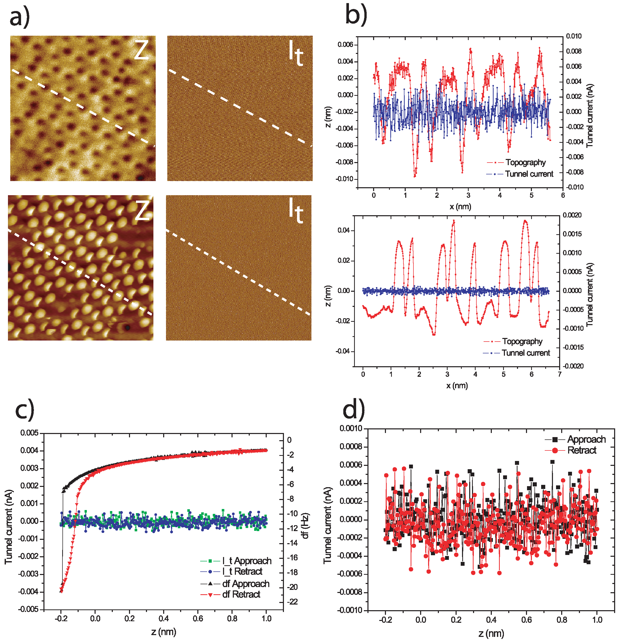
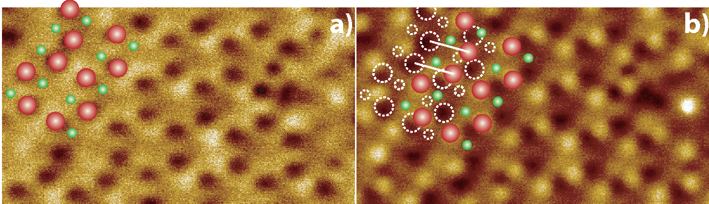
Supporting information is available highlighting that no tunnel current was measured during the experiments performed in this paper. We also discuss the assignment of atomic positions due to the double tip present in the high-setpoint “inverted” image presented in Figure 1.
| Supporting Information File 1: Complete additional experimental detail and figures | ||
| Format: PDF | Size: 884.2 KB | Download |
| Supporting Information File 2: Representative tunnel current data during zero bias imaging | ||
| Format: PNG | Size: 2.9 MB | Download |
| Supporting Information File 3: Atomic position assignment | ||
| Format: PNG | Size: 823.8 KB | Download |
Cite the Following Article
Effect of the tip state during qPlus noncontact atomic force microscopy of Si(100) at 5 K: Probing the probe
Adam Sweetman, Sam Jarvis, Rosanna Danza and Philip Moriarty
Beilstein J. Nanotechnol. 2012, 3, 25–32.
https://doi.org/10.3762/bjnano.3.3
How to Cite
Sweetman, A.; Jarvis, S.; Danza, R.; Moriarty, P. Beilstein J. Nanotechnol. 2012, 3, 25–32. doi:10.3762/bjnano.3.3
Download Citation
Citation data can be downloaded as file using the "Download" button or used for copy/paste from the text window
below.
Citation data in RIS format can be imported by all major citation management software, including EndNote,
ProCite, RefWorks, and Zotero.
Citations to This Article
Up to 20 of the most recent references are displayed here.
Scholarly Works
- Gordon, O. doi:10.1002/9783527834044.ch45
- Guo, H.; Zhang, J. Scanning probe microscopy of epitaxial oxide thin films. Epitaxial Growth of Complex Metal Oxides; Elsevier, 2022; pp 331–367. doi:10.1016/b978-0-08-102945-9.00011-3
- Gordon, O. M.; Moriarty, P. Machine learning at the (sub)atomic scale: next generation scanning probe microscopy. Machine Learning: Science and Technology 2020, 1, 023001. doi:10.1088/2632-2153/ab7d2f
- Katsube, D.; Ojima, S.; Inami, E.; Abe, M. Atomic-resolution imaging of rutile TiO2(110)-(1 × 2) reconstructed surface by non-contact atomic force microscopy. Beilstein journal of nanotechnology 2020, 11, 443–449. doi:10.3762/bjnano.11.35
- Gordon, O. M.; Junqueira, F. L. Q.; Moriarty, P. Embedding human heuristics in machine-learning-enabled probe microscopy. Machine Learning: Science and Technology 2020, 1, 015001. doi:10.1088/2632-2153/ab42ec
- Gordon, O. M.; D'Hondt, P.; Knijff, L.; Freeney, S. E.; Junqueira, F. L. Q.; Moriarty, P.; Swart, I. Scanning tunneling state recognition with multi-class neural network ensembles. Review of Scientific Instruments 2019, 90, 103704. doi:10.1063/1.5099590
- Li, M.; Zhuo, W.; Pang, H.; Lai, L. Improving the atomic-resolution AFM imaging of monolayer MoS2 for worn tips: a molecular dynamics study. Japanese Journal of Applied Physics 2019, 58, 055003. doi:10.7567/1347-4065/ab0646
- Giessibl, F. J. The qPlus sensor, a powerful core for the atomic force microscope. The Review of scientific instruments 2019, 90, 011101. doi:10.1063/1.5052264
- Sweetman, A.; Lekkas, I.; Moriarty, P. Mechano-chemical manipulation of Sn chains on Si(1 0 0) by NC-AFM. Journal of physics. Condensed matter : an Institute of Physics journal 2016, 29, 074003. doi:10.1088/1361-648x/29/7/074003
- Sweetman, A.; Jarvis, S.; Moriarty, P. Mechanochemistry at Silicon Surfaces. NanoScience and Technology; Springer International Publishing, 2015; pp 247–274. doi:10.1007/978-3-319-15588-3_13
- Barth, C. Manipulation of Metal Nanoparticles on Insulating Surfaces. Advances in Atom and Single Molecule Machines; Springer International Publishing, 2015; pp 93–110. doi:10.1007/978-3-319-17401-3_6
- Barth, C. Defects on Bulk MgO(001) Imaged by nc-AFM. Springer Series in Surface Sciences; Springer International Publishing, 2015; pp 215–239. doi:10.1007/978-3-319-14367-5_7
- Baykara, M. Z. Noncontact Atomic Force Microscopy for Atomic-Scale Characterization of Material Surfaces. Surface Science Tools for Nanomaterials Characterization; Springer Berlin Heidelberg, 2015; pp 273–316. doi:10.1007/978-3-662-44551-8_8
- Guo, H.; Zhang, J. Scanning probe microscopy (SPM) of epitaxial oxide thin films. Epitaxial Growth of Complex Metal Oxides; Elsevier, 2015; pp 295–328. doi:10.1016/b978-1-78242-245-7.00011-7
- Heyde, M.; Simon, G. H.; Lichtenstein, L. Surface and Interface Science; Wiley-VCH Verlag GmbH & Co. KGaA, 2014; pp 641–690. doi:10.1002/9783527680566.ch24
- Stirling, J.; Lekkas, I.; Sweetman, A.; Djuranovic, P.; Guo, Q.; Pauw, B. R.; Granwehr, J.; Lévy, R.; Moriarty, P. Critical assessment of the evidence for striped nanoparticles. PloS one 2014, 9, 108482. doi:10.1371/journal.pone.0108482
- Sweetman, A.; Rahe, P.; Moriarty, P. Unique determination of "subatomic" contrast by imaging covalent backbonding. Nano letters 2014, 14, 2265–2270. doi:10.1021/nl4041803
- Jarvis, S.; Kantorovich, L.; Moriarty, P. Structural development and energy dissipation in simulated silicon apices. Beilstein journal of nanotechnology 2013, 4, 941–948. doi:10.3762/bjnano.4.106
- Resta, A.; Leoni, T.; Barth, C.; Ranguis, A.; Becker, C.; Bruhn, T.; Vogt, P.; Le Lay, G. Atomic structures of silicene layers grown on Ag(111): scanning tunneling microscopy and noncontact atomic force microscopy observations. Scientific reports 2013, 3, 2399. doi:10.1038/srep02399
- Heyde, M.; Simon, G. H.; Lichtenstein, L. Resolving oxide surfaces – From point and line defects to complex network structures. physica status solidi (b) 2013, 250, 895–921. doi:10.1002/pssb.201248597