Guest Editor: H. Ihmels Beilstein J. Org. Chem.2019,15, 780–788.https://doi.org/10.3762/bjoc.15.74 Received 28 Jan 2019,
Accepted 14 Mar 2019,
Published 25 Mar 2019
The serotonin 5-hydroxytryptamine 3 receptor (5-HT3R) plays a unique role within the seven classes of the serotonin receptor family, as it represents the only ionotropic receptor, while the other six members are G protein-coupled receptors (GPCRs). The 5-HT3 receptor is related to chemo-/radiotherapy provoked emesis and dysfunction leads to neurodevelopmental disorders and psychopathologies. Since the development of the first serotonin receptor antagonist in the early 1990s, the range of highly selective and potent drugs expanded based on various chemical structures. Nevertheless, on-off-targeting of a pharmacophore’s activity with high spatiotemporal resolution as provided by photopharmacology remains an unsolved challenge bearing additionally the opportunity for detailed receptor examination. In the presented work, we summarize the synthesis, photochromic properties and in vitro characterization of azobenzene-based photochromic derivatives of published 5-HT3R antagonists. Despite reported proof of principle of direct azologization, only one of the investigated derivatives showed antagonistic activity lacking isomer specificity.
5-Hydroxytryptamine (5-HT), commonly known as serotonin [1,2] or enteramine [3,4], is a monoamine neurotransmitter and hormone which is produced in the brain and in intestines and regulates a large variety of physiological functions in the mammalian central and peripheral nervous system [1,5]. In the central nervous system (CNS), it modulates sleep–wake cycles, emesis, appetite, mood, memory, breathing, cognition and numerous other functions [6-9]. In the gastrointestinal (GI) tract, it causes peristalsis via either smooth muscle contraction or enteric nerve depolarization [10]. It is also found in the platelets, where it is presumably involved in blood coagulation and vasoconstriction. Furthermore, serotonin is one of the first neurotransmitters to appear during development [11] and may have an organizing function in the development of the mammalian CNS being involved in cell division, differentiation, survival, neuronal migration [12,13] and synaptogenesis [14]. Dysfunction of the 5-HT receptor (5-HTR) signalling during early developmental stages my lead to altered cognitive ability, neurodevelopmental disorders, and increased incidence of psychopathologies such as autism and schizophrenia [15,16].
Serotonin operates via seven classes of 5-HT receptors of which six are G protein-coupled receptors (GPCRs) and only one, the 5-HT3R, is a ligand-gated cation channel [5,6,17]. When this receptor was identified and cloned [18-20], it became clear that 5-HT3 takes a unique position as pentameric ligand-gated cation-selective ion channel belonging to the Cys-loop receptor subfamily. In vertebrates, this family also includes nicotinic acetylcholine receptors (nAChRs), γ-aminobutyric acid type A receptors (GABAARs), and glycine receptors (GlyRs). To date, five subunits of the 5-HT3 receptor are identified (5-HT3A–5-HT3E) [21]. Functional receptors are either constructed as 5-HT3A homopentamers or as heteropentamers containing 5-HT3A and 5-HT3B receptor subunits [22-24].
5-HT3 receptors are highly expressed in the brainstem, especially in areas involved in the vomiting reflex and in the dorsal horn of the spinal cord [25]. These receptors are also expressed presynaptically providing regulation of the neurotransmitters release [21,22].
Besides targeting of 5-HT3Rs for the treatment of psychiatric disorders, they are object to counteract postoperative nausea and chemo-/radiotherapy provoked emesis [26-29]. In the early 1990s, the first potent and selective 5-HT3 receptor antagonist ondansetron was initially developed [26]. Since then the development of 5-HT3R antagonists progressed. The first-generation antagonists are structurally categorized in three major classes: (I) carbazole derivatives (e.g., ondansetron), (II) indazoles (e.g., granisetron), and (III) indoles (e.g., dolasetron) [26,30]. Generally, 5-HT3R antagonists share a basic amine, a rigid (hetero-)aromatic system and a carbonyl group or isosteric equivalent which is coplanar to the aromatic system. Although the antagonists show a general structural motive, they differ in their binding affinities, dose responses, and side effects [22].
To improve prospective antagonists and obtain a systematic tool for receptor investigation, spatial and temporal restriction of ligand binding and concomitant activity regulation is desirable. Fuelled by light, the growing field of photopharmacology provides a noninvasive method to trigger a drug’s pharmacological response on demand [31-33]. To introduce photoresponsiveness into a biological system, different approaches are feasible, e.g., the use of caged ligands (CL) [34-37], photoswitchable tethered ligands (PTLs) [38-40], photoswitchable orthogonal remotely tethered ligands (PORTLs) [41] or photochromic ligands (PCLs) [31,42]. The latter ones represent small molecules, which can either be engineered via extension of the chemical structure of a known pharmacophore towards a photochromic moiety or via replacement of certain parts of the biomolecule to generate a photochromic hybrid biomolecule. In this context, various photochromic scaffolds including dithienylethenes, fulgi(mi)des, and azobenzenes are investigated [31,42]. The latter ones were already discovered in 1834 by E. Mitscherlich [43] but it took around another 100 years till G. S. Hartley [44] revealed their photo-induced trans–cis isomerization representing the time of birth of the azobenzene photoswitch. Benefiting of their accessible synthesis, large change in polarity and geometry upon switching, excellent photochromic properties and tuneability, azobenzenes are amongst the most widely used photochromic scaffolds [31,42,45-47]. Since their first use in a biological environment in the late 1960s for the photoregulation of the enzymatic activity of chymotrypsin [48], their applications in biology widely expanded towards receptor control [49-52] and fields as bacterial growth [53], vision restoration [53-55], the respiratory chain [56] and lipids [57,58]. Owing to the reported serotonin antagonists’ chemical structures, the use of azobenzene as photochromic scaffold in the presented work seemed axiomatic. Therefore, the primary design of our photochromic derivatives is based on the direct “azologization” [59] of reported non-photochromic antagonists [60,61] via replacement of the benzene-ring connecting amide bond and thioether, respectively, by an azo bridge.
Results and Discussion
Design and synthesis of azobenzene-based photochromic modulators
The reported [60,61] scaffolds of 5-HT3R antagonists are based on an aromatic system either connected to a purine/pyrimidine moiety via a thioether bridge or a quinoxaline moiety via an amide bond. Referring to this work performed by the groups of DiMauro [60] and Jensen [61], we envisioned that the replacement of the thioether or amide bond (Scheme 1) by an azo bridge would result in highly active photochromic serotonin 5-HTR antagonists controllable by irradiation with light. Based on the suggested receptor binding mode reported for one potent non-photochromic antagonist (lead structure of 16c) [61] we expected the extended trans-isomer as biologically active configuration whereas its bent cis-isomer should be inactive.
Scheme 1:
Approach of the direct azologization of reported [60,61] serotonin 5-HT3R antagonists via replacement of a thioether or amide bond by an azo bridge.
Scheme 1:
Approach of the direct azologization of reported [60,61] serotonin 5-HT3R antagonists via replacement of a...
The synthesis of the unsubstituted quinoxaline-based azobenzene derivatives 5a and 5b is based on a Baeyer [62]–Mills [63] reaction (Scheme 2). Therefore, nitrosoquinoxaline 3 was synthesized in a two-step procedure starting from 2-chloroquinoxaline (1), which was transformed into its oxime 2 using hydroxylammonium chloride [64]. The subsequent oxidation was performed using periodic acid as oxidant [65]. The subsequent reaction with differently substituted anilines in acetic acid [65] provided both quinoxaline azobenzene derivatives in good yields.
Scheme 2:
Synthesis of the differently substituted quinoxaline azobenzene derivatives 5a and 5b via Baeyer [62]–Mills [63] reaction [64,65].
Scheme 2:
Synthesis of the differently substituted quinoxaline azobenzene derivatives 5a and 5b via Baeyer [62]–M...
The methoxy-substituted quinoxaline azobenzene derivative 12a was synthesized via a different synthetic route depicted in Scheme 3. In a first step, p-toluidine (4a) was diazotized using sodium nitrite and subsequently reacted with the 2-chloroacetylacetone ester derivative 7 providing hydrazine 8[66]. Upon reaction of the chloro-ester 8 with phenylenediamine (9) in the presence of triethylamine the quinoxaline moiety was formed [67]. Oxidation of the hydrazine derivative 10 using hydrogen peroxide under an oxygen atmosphere afforded the quinoxaline azobenzene derivative 11[68]. Subsequent methylation using methyl iodide [69] mainly resulted in the formation of the N-methylated non-photochromic product 12b but in low yields also the desired photochromic methoxy-substituted quinoxaline azobenzene derivative 12a.
Scheme 3:
Synthesis of the methoxy-substituted quinoxaline derivative 12a via diazotization [66-69].
Scheme 3:
Synthesis of the methoxy-substituted quinoxaline derivative 12a via diazotization [66-69].
Synthesis of the purine and thienopyrimidine-based derivatives
Scheme 4 depicts the general procedure applied for the synthesis of differently substituted purine- and thienopyrimidine azobenzene derivatives. Differently substituted non-photochromic antagonists were chosen as lead structures delivering photochromic derivatives with varying electronic and thus photochromic properties. The respective arylamines 13a–c were converted into their corresponding hydrazines 14a–c via diazonium-salt formation using sodium nitrite and subsequent reduction using tin(II) chloride [70]. The following nucleophilic substitution at a chloro-substituted purine (15a,b) or thienopyrimidine (15c), respectively, and subsequent oxidation of the hydrazine moiety afforded the corresponding azobenzene derivatives 16a–d[71].
Scheme 4:
General procedure for the synthesis of purine- and thienopyrimidine-substituted arylazobenzenes and depiction of the corresponding structures [70,71].
Scheme 4:
General procedure for the synthesis of purine- and thienopyrimidine-substituted arylazobenzenes and...
Synthesis of azobenzene-extended thiopurine derivatives
To further tune the photochromism and compare the properties of direct azologization to azo-extension, two additional derivatives of the in vitro most promising naphthalene azopurine 16c were synthesized either by keeping the original thioether (Scheme 5) or replacing it by an amide bond (Scheme 6) known as common structural feature of 5-HT3R antagonists.
Scheme 5:
Synthesis of the thiomethyl-linked purine azobenzene 23[62,63,72-74].
Scheme 5:
Synthesis of the thiomethyl-linked purine azobenzene 23 [62,63,72-74].
Scheme 5 reflects the synthesis of the azo-extended thiomethylpurine 23 starting with the synthesis of hydroxymethylazobenzene 19[72] in a Baeyer [62]–Mills [63] reaction and subsequent nucleophilic substitution using cyanuric chloride (20) [73] providing chloromethyl azobenzene 21. The introduction of the thiopurine moiety in 23 was accomplished upon reaction of 21 with dihydropurinethione 22[74].
The amide-linked derivative of thiomethylpurine azobenzene 23 was synthesized via Baeyer [62]–Mills [63] formation of the carboxylated azobenzene 25 starting from aminobenzoic acid 24 and nitrosobenzene (18) [75]. Activation using thionyl chloride [76] afforded the acid chloride 26 and allowed amide-bond formation [77] for the generation of 28 (Scheme 6).
Photochromic properties
The investigation of the photochromic properties of the potential 5-HT3R antagonists 5a, 5b, 12a, 16a–d, 23, and 28 was performed in DMSO and depending on their solubility in phosphate buffer + 0.1% DMSO (16a–d) by UV–vis absorption spectroscopy. The compounds were dissolved at 50 µM in the respective solvent and irradiated with the indicated wavelengths to generate a substantial amount of their cis-isomer. This process can be followed by a decrease of the trans-absorption maximum at around 350–400 nm and an increase in absorption at around 450–500 nm in the UV–vis spectrum representing the cis-isomer (Figure 1, black arrows). The absorption bands of the trans and cis-isomers of compounds 12a, 16c, and 16d overlap to such an extent, that no new maximum representing the cis-isomer was observed and thus cis–trans isomerization only occurs thermally and is not triggerable by irradiation with visible light. Back-isomerization was triggered by irradiation with visible light (5a, 5b, 16a, 16b, 23, and 28) of the indicated wavelength or by thermal relaxation (5a, 5b, 12a, 16a–d, 23, and 28). The irradiation times were determined by following the UV–vis spectrum upon isomerization until no more changes in absorption were observed and the photostationary state (PSS) was reached. The points of intersection in the absorption spectrum upon switching (= isosbestic points) indicate a clear two-component switching between trans and cis-species without any degradation or formation of a side-product (Figure 1, dotted black arrows). The UV–vis absorption spectra of all compounds are depicted in Supporting Information File 1, Figures S1–S10 and the data are summarized in Table S1 and Table S2. A comparison of the differently substituted purine azobenzene derivatives revealed the beneficial effect of an o-chloro substitution on the photochromic properties of 16b compared to 16c as the electron density at the nitrogen-rich purine core is reduced. Further reduction of the electron density was achieved by using a thienopyrimidine (16a) instead of a purine core (16b–d). Nevertheless, the photochromic properties of those heterocyclic, especially purine-based azobenzenes, are rather poor. In addition to direct azologization, two azo-extended purine derivatives 23 and 28 were synthesized resulting in excellent photochromic properties. Figure 1 compares exemplarily the UV–vis absorption spectra of the naphthalene-azo-purine 16c (left) and its azo-extended azobenzene thioether purine 23 (right). The determination of the thermal half-lives (THL) of the cis-isomers of compounds 5a, 5b, 12a, 16a–d, 23, and 28 was accomplished by monitoring the increase in absorbance which corresponds to the evolution of the trans-isomer after irradiation and exposure to dark. In contrast to the heterocyclic compounds 5a, 5b, 12a, and 16a–d with a thermal half-life in the seconds to minutes range, the azo-extended compounds 23 and 28 showed only slow thermal back-isomerization (day range) at room temperature. Depending on the desired application, both properties may be of benefit. For thermally instable compounds, only one wavelength for switching is required. In case of thermally stable cis-isomers constant irradiation to maintain a substantial amount of the cis-isomer can be avoided.
Figure 1:
UV–vis absorption spectra measured at 50 µM in DMSO. Left: purine derivative 16c; right: azo-extended derivative 23.
Figure 1:
UV–vis absorption spectra measured at 50 µM in DMSO. Left: purine derivative 16c; right: azo-extend...
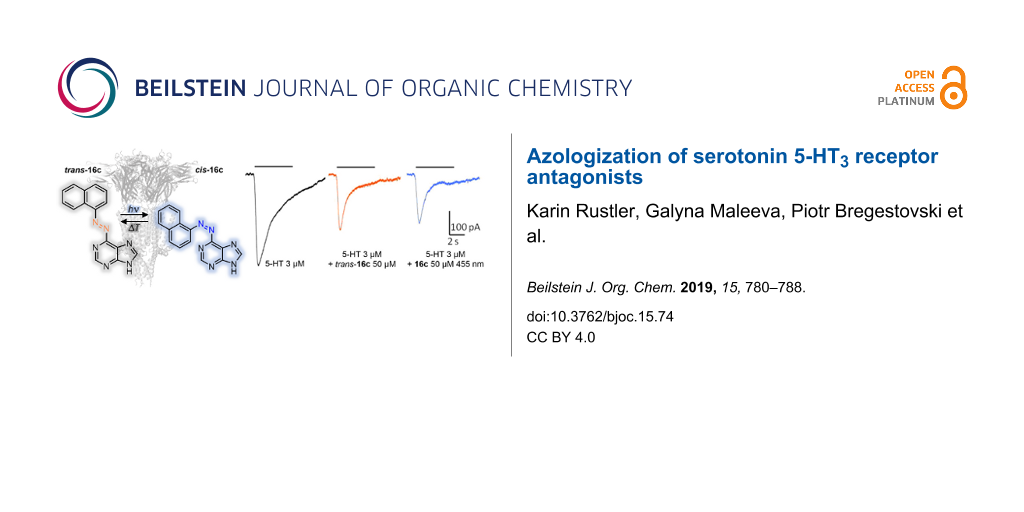
The synthesized azo antagonist derivatives 5a, 5b, 12a, 16a–d, 23, and 28 were tested for their inhibitory activity using the patch-clamp technique on heterologously expressed ionotropic homopentameric 5-HT3A receptors. Only upon addition of 16c the amplitude of the 5-HT3A mediated currents was decreased (Figure 2, left). Application of a 50 µM solution of trans-16c in its thermal equilibrium decreased the amplitude of 5-HT induced currents on 54 ± 3% (n = 4). However, irradiation-induced trans–cis isomerization with light of λ = 530 nm and 455 nm, respectively, had no significant effect on the amplitude of 5-HT3A-mediated currents (Figure 2, right).
Figure 2:
On the left panel representative traces of currents induced by the application of 3 µM 5HT (black trace), by 3 µM 5HT and 50 µM trans-16c (red trace), by 5HT and 16c under constant irradiation (455 nm, blue trace), and again by pure 5HT – wash-out of the studied compound (black trace) are shown. On the right panel, a graph representing the relative amplitudes of currents in control (black column), at application of trans-16c (red column), at application of 16c irradiated with blue light (blue column) and at wash-out (gray column) are shown. P > 0.05, paired t-test.
Figure 2:
On the left panel representative traces of currents induced by the application of 3 µM 5HT (black t...
In the presented work, we address the design, synthesis, photochromic characterization and in vitro investigation of in total nine azobenzene-based derivatives of reported 5-HT3R antagonists. Initially, seven photoligands (5a, 5b, 12a, and 16a–d) either based on quinoxaline (5a, 5b, and 12a) or purine derivatives (16a–d) with varying electronic and thus photochromic properties were synthesized by direct azologization of the respective leads. Especially the purine-based azobenzenes displayed high solubility in aqueous media. The beneficial effect of substituents reducing the overall electron density of the purine moiety (16a, 16b) resulted in higher photostationary states and better band separation compared to 16c and 16d. Still, only one compound (16c) showed antagonistic activity in patch-clamp studies. This might be explained by the fact that its corresponding non-photochromic lead is the inhibitory most active reported [61] antagonist among the investigated ones. The partial rigidization of the thioether via incorporation of an azo bridge might result in a vast loss of activity. Thereby, azologization of the less potent leads resulted in complete loss of inhibitory activity (5a, 5b, 12a, 16a, 16b, 16d) and only the originally most potent derivative 16c kept recordable antagonistic activity. The missing significant difference in activity upon irradiation-induced trans–cis isomerization of 16c is probably due to its moderate photochromic properties and slow trans–cis isomerization (Figure 1, left). During the patch-clamp analysis, the cells are continuously superfused with external solution resulting in a fast exchange of the surrounding media and co-applied tested compounds. Thus, the cis-PSS of 16c might not be reached by irradiation within the short time of compound application despite continuous irradiation. Therefore, two azobenzene-extended derivatives (23 and 28) with improved photochromic properties were synthesized but lost antagonistic activity probably due to their increased steric demand.
In ongoing studies, detailed molecular modelling is used to design potential photochromic antagonists fitting the requirements of the receptor’s binding pocket. Regarding the analysis method, compounds will be optimized towards either thermally stable cis-isomers to be tested separately upon prior irradiation or faster switching compounds.
Supporting Information
Supporting Information File 1:
Detailed photochromic characterization (UV–vis absorption spectra, cycle performances, thermal half-lives) and NMR spectra of all synthesized compounds are provided. The file contains crystal structures of compounds 12b and 16a and experimental procedures.
The authors are grateful for financial support to ERA SynBIO grant MODULIGHTOR (PCIN-2015-163-C02-01), the Bundesministerium für Bildung und Forschung (BMBF) and the Russian Science Foundation (grant number: 18-15-00313). We are also grateful to Dr. S. Lummis for kind providing with cDNA of 5HT3A subunit. KR thanks Josef Kiermaier and Wolfgang Söllner for mass spectrometry analysis. KR thanks the X-ray department for crystal structure analysis.
References
Barnes, N. M.; Neumaier, J. F. Tocris Sci. Rev. Ser.2015,34, 1–16.
Return to citation in text:
[1]
[2]
Rapport, M. M. J. Biol. Chem.1949,180, 961–969.
Return to citation in text:
[1]
Erspamer, V.; Asero, B. Nature1952,169, 800–801. doi:10.1038/169800b0
Return to citation in text:
[1]
Erspamer, V. Naunyn-Schmiedebergs Arch. Exp. Pathol. Pharmakol.1940,196, 343–365. doi:10.1007/bf01861121
Return to citation in text:
[1]
Davis, J. M.; Alderson, N. L.; Welsh, R. S. Am. J. Clin. Nutr.2000,72, 573S–578S. doi:10.1093/ajcn/72.2.573s
Return to citation in text:
[1]
Ray, R. S.; Corcoran, A. E.; Brust, R. D.; Kim, J. C.; Richerson, G. B.; Nattie, E.; Dymecki, S. M. Science2011,333, 637–642. doi:10.1126/science.1205295
Return to citation in text:
[1]
Vitalis, T.; Cases, O.; Passemard, S.; Callebert, J.; Parnavelas, J. G. Eur. J. Neurosci.2007,26, 331–344. doi:10.1111/j.1460-9568.2007.05661.x
Return to citation in text:
[1]
Hoyer, D.; Clarke, D. E.; Fozard, J. R.; Hartig, P. R.; Martin, G. R.; Mylecharane, E. J.; Saxena, P. R.; Humphrey, P. P. A. Pharmacol. Rev.1994,46, 157–203.
Return to citation in text:
[1]
Derkach, V.; Surprenant, A.; North, R. A. Nature1989,339, 706–709. doi:10.1038/339706a0
Return to citation in text:
[1]
Maricq, A. V.; Peterson, A. S.; Brake, A. J.; Myers, R. M.; Julius, D. Science1991,254, 432–437. doi:10.1126/science.1718042
Return to citation in text:
[1]
Kilpatrick, G. J.; Jones, B. J.; Tyers, M. B. Nature1987,330, 746–748. doi:10.1038/330746a0
Return to citation in text:
[1]
Lummis, S. C. R. J. Biol. Chem.2012,287, 40239–40245. doi:10.1074/jbc.r112.406496
Return to citation in text:
[1]
[2]
Thompson, A. J.; Lummis, S. C. R. Curr. Pharm. Des.2006,12, 3615–3630. doi:10.2174/138161206778522029
Return to citation in text:
[1]
[2]
[3]
Davies, P. A.; Pistis, M.; Hanna, M. C.; Peters, J. A.; Lambert, J. J.; Hales, T. G.; Kirkness, E. F. Nature1999,397, 359–363. doi:10.1038/16941
Return to citation in text:
[1]
Dubin, A. E.; Huvar, R.; D'Andrea, M. R.; Pyati, J.; Zhu, J. Y.; Joy, K. C.; Wilson, S. J.; Galindo, J. E.; Glass, C. A.; Luo, L.; Jackson, M. R.; Lovenberg, T. W.; Erlander, M. G. J. Biol. Chem.1999,274, 30799–30810. doi:10.1074/jbc.274.43.30799
Return to citation in text:
[1]
Miquel, M.-C.; Emerit, M. B.; Nosjean, A.; Simon, A.; Rumajogee, P.; Brisorgueil, M.-J.; Doucet, E.; Hamon, M.; Vergé, D. Eur. J. Neurosci.2002,15, 449–457. doi:10.1046/j.0953-816x.2001.01872.x
Return to citation in text:
[1]
Miller, P. S.; Smart, T. G. Trends Pharmacol. Sci.2010,31, 161–174. doi:10.1016/j.tips.2009.12.005
Return to citation in text:
[1]
Barnes, N. M.; Hales, T. G.; Lummis, S. C. R.; Peters, J. A. Neuropharmacology2009,56, 273–284. doi:10.1016/j.neuropharm.2008.08.003
Return to citation in text:
[1]
Szymański, W.; Beierle, J. M.; Kistemaker, H. A. V.; Velema, W. A.; Feringa, B. L. Chem. Rev.2013,113, 6114–6178. doi:10.1021/cr300179f
Return to citation in text:
[1]
[2]
[3]
[4]
Lerch, M. M.; Hansen, M. J.; van Dam, G. M.; Szymanski, W.; Feringa, B. L. Angew. Chem., Int. Ed.2016,55, 10978–10999. doi:10.1002/anie.201601931
Return to citation in text:
[1]
Broichhagen, J.; Frank, J. A.; Trauner, D. Acc. Chem. Res.2015,48, 1947–1960. doi:10.1021/acs.accounts.5b00129
Return to citation in text:
[1]
Mayer, G.; Heckel, A. Angew. Chem., Int. Ed.2006,45, 4900–4921. doi:10.1002/anie.200600387
Return to citation in text:
[1]
Reiner, A.; Isacoff, E. Y. Photoswitching of Cell Surface Receptors Using Tethered Ligands. In Photoswitching Proteins; Cambridge, S., Ed.; Methods in Molecular Biology, Vol. 1148; Humana Press: New York, USA; pp 45–68. doi:10.1007/978-1-4939-0470-9_4
Return to citation in text:
[1]
Donthamsetti, P. C.; Winter, N.; Schönberger, M.; Levitz, J.; Stanley, C.; Javitch, J. A.; Isacoff, E. Y.; Trauner, D. J. Am. Chem. Soc.2017,139, 18522–18535. doi:10.1021/jacs.7b07659
Return to citation in text:
[1]
Lin, W.-C.; Tsai, M.-C.; Rajappa, R.; Kramer, R. H. J. Am. Chem. Soc.2018,140, 7445–7448. doi:10.1021/jacs.8b03942
Return to citation in text:
[1]
Broichhagen, J.; Damijonaitis, A.; Levitz, J.; Sokol, K. R.; Leippe, P.; Konrad, D.; Isacoff, E. Y.; Trauner, D. ACS Cent. Sci.2015,1, 383–393. doi:10.1021/acscentsci.5b00260
Return to citation in text:
[1]
Brieke, C.; Rohrbach, F.; Gottschalk, A.; Mayer, G.; Heckel, A. Angew. Chem., Int. Ed.2012,51, 8446–8476. doi:10.1002/anie.201202134
Return to citation in text:
[1]
[2]
[3]
Mitscherlich, E. Ann. Pharm. (Lemgo, Ger.)1834,12, 311–314. doi:10.1002/jlac.18340120282|
Return to citation in text:
[1]
Hartley, G. S. Nature1937,140, 281–282. doi:10.1038/140281a0
Return to citation in text:
[1]
Dong, M.; Babalhavaeji, A.; Samanta, S.; Beharry, A. A.; Woolley, G. A. Acc. Chem. Res.2015,48, 2662–2670. doi:10.1021/acs.accounts.5b00270
Return to citation in text:
[1]
Beharry, A. A.; Sadovski, O.; Woolley, G. A. J. Am. Chem. Soc.2011,133, 19684–19687. doi:10.1021/ja209239m
Return to citation in text:
[1]
Weston, C. E.; Richardson, R. D.; Haycock, P. R.; White, A. J. P.; Fuchter, M. J. J. Am. Chem. Soc.2014,136, 11878–11881. doi:10.1021/ja505444d
Return to citation in text:
[1]
Kaufman, H.; Vratsanos, S. M.; Erlanger, B. F. Science1968,162, 1487–1489. doi:10.1126/science.162.3861.1487
Return to citation in text:
[1]
Deal, W. J.; Erlanger, B. F.; Nachmansohn, D. Proc. Natl. Acad. Sci. U. S. A.1969,64, 1230–1234. doi:10.1073/pnas.64.4.1230
Return to citation in text:
[1]
Volgraf, M.; Gorostiza, P.; Numano, R.; Kramer, R. H.; Isacoff, E. Y.; Trauner, D. Nat. Chem. Biol.2006,2, 47–52. doi:10.1038/nchembio756
Return to citation in text:
[1]
Volgraf, M.; Gorostiza, P.; Szobota, S.; Helix, M. R.; Isacoff, E. Y.; Trauner, D. J. Am. Chem. Soc.2007,129, 260–261. doi:10.1021/ja067269o
Return to citation in text:
[1]
Hauwert, N. J.; Mocking, T. A. M.; Da Costa Pereira, D.; Kooistra, A. J.; Wijnen, L. M.; Vreeker, G. C. M.; Verweij, E. W. E.; De Boer, A. H.; Smit, M. J.; De Graaf, C.; Vischer, H. F.; de Esch, I. J. P.; Wijtmans, M.; Leurs, R. J. Am. Chem. Soc.2018,140, 4232–4243. doi:10.1021/jacs.7b11422
Return to citation in text:
[1]
Wegener, M.; Hansen, M. J.; Driessen, A. J. M.; Szymanski, W.; Feringa, B. L. J. Am. Chem. Soc.2017,139, 17979–17986. doi:10.1021/jacs.7b09281
Return to citation in text:
[1]
[2]
Polosukhina, A.; Litt, J.; Tochitsky, I.; Nemargut, J.; Sychev, Y.; De Kouchkovsky, I.; Huang, T.; Borges, K.; Trauner, D.; Van Gelder, R. N.; Kramer, R. H. Neuron2012,75, 271–282. doi:10.1016/j.neuron.2012.05.022
Return to citation in text:
[1]
Tochitsky, I.; Polosukhina, A.; Degtyar, V. E.; Gallerani, N.; Smith, C. M.; Friedman, A.; Van Gelder, R. N.; Trauner, D.; Kaufer, D.; Kramer, R. H. Neuron2014,81, 800–813. doi:10.1016/j.neuron.2014.01.003
Return to citation in text:
[1]
Simeth, N. A.; Kneuttinger, A. C.; Sterner, R.; König, B. Chem. Sci.2017,8, 6474–6483. doi:10.1039/c7sc00781g
Return to citation in text:
[1]
Frank, J. A.; Franquelim, H. G.; Schwille, P.; Trauner, D. J. Am. Chem. Soc.2016,138, 12981–12986. doi:10.1021/jacs.6b07278
Return to citation in text:
[1]
Pernpeintner, C.; Frank, J. A.; Urban, P.; Roeske, C. R.; Pritzl, S. D.; Trauner, D.; Lohmüller, T. Langmuir2017,33, 4083–4089. doi:10.1021/acs.langmuir.7b01020
Return to citation in text:
[1]
Schoenberger, M.; Damijonaitis, A.; Zhang, Z.; Nagel, D.; Trauner, D. ACS Chem. Neurosci.2014,5, 514–518. doi:10.1021/cn500070w
Return to citation in text:
[1]
Sparling, B. A.; DiMauro, E. F. Bioorg. Med. Chem. Lett.2017,27, 3207–3218. doi:10.1016/j.bmcl.2017.04.073
Return to citation in text:
[1]
[2]
[3]
[4]
Trattnig, S. M.; Harpsøe, K.; Thygesen, S. B.; Rahr, L. M.; Ahring, P. K.; Balle, T.; Jensen, A. A. J. Biol. Chem.2012,287, 25241–25254. doi:10.1074/jbc.m112.360370
Return to citation in text:
[1]
[2]
[3]
[4]
[5]
[6]
Mills, C. J. Chem. Soc., Trans.1895,67, 925–933. doi:10.1039/ct8956700925
Return to citation in text:
[1]
[2]
[3]
[4]
[5]
[6]
von Wantoch Rekowski, M.; Pyriochou, A.; Papapetropoulos, N.; Stößel, A.; Papapetropoulos, A.; Giannis, A. Bioorg. Med. Chem.2010,18, 1288–1296. doi:10.1016/j.bmc.2009.12.027
Return to citation in text:
[1]
[2]
Harsányi, K.; Gönczi, C.; Korbonits, D. Justus Liebigs Ann. Chem.1973, 190–194. doi:10.1002/jlac.197319730203
Return to citation in text:
[1]
[2]
[3]
Murarka, S.; Martín-Gago, P.; Schultz-Fademrecht, C.; Al Saabi, A.; Baumann, M.; Fansa, E. K.; Ismail, S.; Nussbaumer, P.; Wittinghofer, A.; Waldmann, H. Chem. – Eur. J.2017,23, 6083–6093. doi:10.1002/chem.201603222
Return to citation in text:
[1]
[2]
Colotta, V.; Catarzi, D.; Varano, F.; Cecchi, L.; Filacchioni, G.; Galli, A.; Costagli, C. Arch. Pharm. (Weinheim, Ger.)1997,330, 387–391. doi:10.1002/ardp.19973301206
Return to citation in text:
[1]
[2]
Párkányi, C.; Abdelhamid, A. O.; Shawali, A. S. J. Heterocycl. Chem.1984,21, 521–524. doi:10.1002/jhet.5570210251
Return to citation in text:
[1]
[2]
Pierau, S.; Dale, G. Novel Compounds having an anti-bacterial Activity. WO Pat. Appl. WO2006021448 A1, March 2, 2006.
Return to citation in text:
[1]
[2]
Wang, X.; Chen, Y.-F.; Yan, W.; Cao, L.-L.; Ye, Y.-H. Molecules2016,21, No. 1574. doi:10.3390/molecules21111574
Return to citation in text:
[1]
[2]
Kolarski, D.; Szymanski, W.; Feringa, B. L. Org. Lett.2017,19, 5090–5093. doi:10.1021/acs.orglett.7b02361
Return to citation in text:
[1]
[2]
Stawski, P.; Sumser, M.; Trauner, D. Angew. Chem., Int. Ed.2012,51, 5748–5751. doi:10.1002/anie.201109265
Return to citation in text:
[1]
[2]
del Barrio, J.; Horton, P. N.; Lairez, D.; Lloyd, G. O.; Toprakcioglu, C.; Scherman, O. A. J. Am. Chem. Soc.2013,135, 11760–11763. doi:10.1021/ja406556h
Return to citation in text:
[1]
[2]
Laufer, S. A.; Domeyer, D. M.; Scior, T. R. F.; Albrecht, W.; Hauser, D. R. J. J. Med. Chem.2005,48, 710–722. doi:10.1021/jm0408767
Return to citation in text:
[1]
[2]
Coleman, G. H.; Nichols, G.; McCloskey, C. M.; Anspon, H. D. Org. Synth.1945,25, 87–89. doi:10.15227/orgsyn.025.0087
Return to citation in text:
[1]
[2]
Liu, C. F.; Zeng, Y.; Lu, X. W. Peptide Nucleic Acid Monomers and Oligomers. WO Pat. Appl. WO2010027326 A1, March 2, 2006.
Return to citation in text:
[1]
[2]
References 31,42,45-47
31.
Szymański, W.; Beierle, J. M.; Kistemaker, H. A. V.; Velema, W. A.; Feringa, B. L. Chem. Rev.2013,113, 6114–6178. doi:10.1021/cr300179f
Deal, W. J.; Erlanger, B. F.; Nachmansohn, D. Proc. Natl. Acad. Sci. U. S. A.1969,64, 1230–1234. doi:10.1073/pnas.64.4.1230
50.
Volgraf, M.; Gorostiza, P.; Numano, R.; Kramer, R. H.; Isacoff, E. Y.; Trauner, D. Nat. Chem. Biol.2006,2, 47–52. doi:10.1038/nchembio756
51.
Volgraf, M.; Gorostiza, P.; Szobota, S.; Helix, M. R.; Isacoff, E. Y.; Trauner, D. J. Am. Chem. Soc.2007,129, 260–261. doi:10.1021/ja067269o
52.
Hauwert, N. J.; Mocking, T. A. M.; Da Costa Pereira, D.; Kooistra, A. J.; Wijnen, L. M.; Vreeker, G. C. M.; Verweij, E. W. E.; De Boer, A. H.; Smit, M. J.; De Graaf, C.; Vischer, H. F.; de Esch, I. J. P.; Wijtmans, M.; Leurs, R. J. Am. Chem. Soc.2018,140, 4232–4243. doi:10.1021/jacs.7b11422
Trattnig, S. M.; Harpsøe, K.; Thygesen, S. B.; Rahr, L. M.; Ahring, P. K.; Balle, T.; Jensen, A. A. J. Biol. Chem.2012,287, 25241–25254. doi:10.1074/jbc.m112.360370
Trattnig, S. M.; Harpsøe, K.; Thygesen, S. B.; Rahr, L. M.; Ahring, P. K.; Balle, T.; Jensen, A. A. J. Biol. Chem.2012,287, 25241–25254. doi:10.1074/jbc.m112.360370
Trattnig, S. M.; Harpsøe, K.; Thygesen, S. B.; Rahr, L. M.; Ahring, P. K.; Balle, T.; Jensen, A. A. J. Biol. Chem.2012,287, 25241–25254. doi:10.1074/jbc.m112.360370
Frank, J. A.; Franquelim, H. G.; Schwille, P.; Trauner, D. J. Am. Chem. Soc.2016,138, 12981–12986. doi:10.1021/jacs.6b07278
58.
Pernpeintner, C.; Frank, J. A.; Urban, P.; Roeske, C. R.; Pritzl, S. D.; Trauner, D.; Lohmüller, T. Langmuir2017,33, 4083–4089. doi:10.1021/acs.langmuir.7b01020
Wegener, M.; Hansen, M. J.; Driessen, A. J. M.; Szymanski, W.; Feringa, B. L. J. Am. Chem. Soc.2017,139, 17979–17986. doi:10.1021/jacs.7b09281
54.
Polosukhina, A.; Litt, J.; Tochitsky, I.; Nemargut, J.; Sychev, Y.; De Kouchkovsky, I.; Huang, T.; Borges, K.; Trauner, D.; Van Gelder, R. N.; Kramer, R. H. Neuron2012,75, 271–282. doi:10.1016/j.neuron.2012.05.022
55.
Tochitsky, I.; Polosukhina, A.; Degtyar, V. E.; Gallerani, N.; Smith, C. M.; Friedman, A.; Van Gelder, R. N.; Trauner, D.; Kaufer, D.; Kramer, R. H. Neuron2014,81, 800–813. doi:10.1016/j.neuron.2014.01.003
Trattnig, S. M.; Harpsøe, K.; Thygesen, S. B.; Rahr, L. M.; Ahring, P. K.; Balle, T.; Jensen, A. A. J. Biol. Chem.2012,287, 25241–25254. doi:10.1074/jbc.m112.360370
Trattnig, S. M.; Harpsøe, K.; Thygesen, S. B.; Rahr, L. M.; Ahring, P. K.; Balle, T.; Jensen, A. A. J. Biol. Chem.2012,287, 25241–25254. doi:10.1074/jbc.m112.360370
Trattnig, S. M.; Harpsøe, K.; Thygesen, S. B.; Rahr, L. M.; Ahring, P. K.; Balle, T.; Jensen, A. A. J. Biol. Chem.2012,287, 25241–25254. doi:10.1074/jbc.m112.360370
Davis, J. M.; Alderson, N. L.; Welsh, R. S. Am. J. Clin. Nutr.2000,72, 573S–578S. doi:10.1093/ajcn/72.2.573s
8.
Ray, R. S.; Corcoran, A. E.; Brust, R. D.; Kim, J. C.; Richerson, G. B.; Nattie, E.; Dymecki, S. M. Science2011,333, 637–642. doi:10.1126/science.1205295
Davies, P. A.; Pistis, M.; Hanna, M. C.; Peters, J. A.; Lambert, J. J.; Hales, T. G.; Kirkness, E. F. Nature1999,397, 359–363. doi:10.1038/16941
24.
Dubin, A. E.; Huvar, R.; D'Andrea, M. R.; Pyati, J.; Zhu, J. Y.; Joy, K. C.; Wilson, S. J.; Galindo, J. E.; Glass, C. A.; Luo, L.; Jackson, M. R.; Lovenberg, T. W.; Erlander, M. G. J. Biol. Chem.1999,274, 30799–30810. doi:10.1074/jbc.274.43.30799
Stawski, P.; Sumser, M.; Trauner, D. Angew. Chem., Int. Ed.2012,51, 5748–5751. doi:10.1002/anie.201109265
73.
del Barrio, J.; Horton, P. N.; Lairez, D.; Lloyd, G. O.; Toprakcioglu, C.; Scherman, O. A. J. Am. Chem. Soc.2013,135, 11760–11763. doi:10.1021/ja406556h
74.
Laufer, S. A.; Domeyer, D. M.; Scior, T. R. F.; Albrecht, W.; Hauser, D. R. J. J. Med. Chem.2005,48, 710–722. doi:10.1021/jm0408767
Hoyer, D.; Clarke, D. E.; Fozard, J. R.; Hartig, P. R.; Martin, G. R.; Mylecharane, E. J.; Saxena, P. R.; Humphrey, P. P. A. Pharmacol. Rev.1994,46, 157–203.
del Barrio, J.; Horton, P. N.; Lairez, D.; Lloyd, G. O.; Toprakcioglu, C.; Scherman, O. A. J. Am. Chem. Soc.2013,135, 11760–11763. doi:10.1021/ja406556h
Reiner, A.; Isacoff, E. Y. Photoswitching of Cell Surface Receptors Using Tethered Ligands. In Photoswitching Proteins; Cambridge, S., Ed.; Methods in Molecular Biology, Vol. 1148; Humana Press: New York, USA; pp 45–68. doi:10.1007/978-1-4939-0470-9_4
39.
Donthamsetti, P. C.; Winter, N.; Schönberger, M.; Levitz, J.; Stanley, C.; Javitch, J. A.; Isacoff, E. Y.; Trauner, D. J. Am. Chem. Soc.2017,139, 18522–18535. doi:10.1021/jacs.7b07659
40.
Lin, W.-C.; Tsai, M.-C.; Rajappa, R.; Kramer, R. H. J. Am. Chem. Soc.2018,140, 7445–7448. doi:10.1021/jacs.8b03942


![[1860-5397-15-74-i1]](/bjoc/content/inline/1860-5397-15-74-i1.svg?scale=2.0&max-width=1024&background=FFFFFF)
![[1860-5397-15-74-i2]](/bjoc/content/inline/1860-5397-15-74-i2.svg?scale=2.0&max-width=1024&background=FFFFFF)
![[1860-5397-15-74-i3]](/bjoc/content/inline/1860-5397-15-74-i3.svg?scale=2.0&max-width=1024&background=FFFFFF)
![[1860-5397-15-74-i4]](/bjoc/content/inline/1860-5397-15-74-i4.svg?scale=2.0&max-width=1024&background=FFFFFF)
![[1860-5397-15-74-i5]](/bjoc/content/inline/1860-5397-15-74-i5.svg?scale=2.0&max-width=1024&background=FFFFFF)
![[1860-5397-15-74-i6]](/bjoc/content/inline/1860-5397-15-74-i6.svg?scale=2.0&max-width=1024&background=FFFFFF)
![[1860-5397-15-74-1]](/bjoc/content/figures/1860-5397-15-74-1.png?scale=2.0&max-width=1024&background=FFFFFF)
![[1860-5397-15-74-2]](/bjoc/content/figures/1860-5397-15-74-2.png?scale=2.0&max-width=1024&background=FFFFFF)