Abstract
4-(2-Fluorophenyl)-7-methoxycoumarin (6) was synthesized by Pechmann reaction under mild conditions via a three-step reaction. The solution-state 1H NMR spectra of 6 showed a strong intramolecular interaction between F and H5 (JFH = 2.6 Hz) and 13C NMR suggested that this C–F···H–C coupling is a through-space interaction. The 2D 19F-{1H} HOESY and 1H-{19F} 1D experiments were done to confirm this F···H interaction. The single crystal X-ray structure and the DFT-optimized structure showed that the fluorinated phenyl ring favors the orientation with the fluorine atom closer to H5 than H3. The X-ray structure also showed the existence of the intermolecular C–F···H–C interaction.

Graphical Abstract
Introduction
Coumarins constitute one of the big classes of naturally occurring compounds. The first coumarin was isolated from the tonka bean (Dipteryx odorata) in 1820 and, to date, more than 1300 coumarins have been identified from natural sources [1,2]. Coumarins have been reported to play a vital role as food and cosmetics constituents, cigarette additives, and dye-sensitized solar cells [3,4]. In addition, coumarins possess some biological activities such as anti-inflammatory [5], antitumor [6], anti-oxidant [7], antibacterial [8], hepatoprotective, anticoagulant, antiviral and antithrombotic activities [9]. The variety of uses of these compounds resulted in an increase in demand for large quantities of coumarins. Due to an insufficient natural supply to meet this demand for these compounds, numerous methods for the synthesis of these compounds have been developed, examples are the Pechmann condensation [10,11], Stille coupling reaction [12], Knoevenagel condensation [13], Heck coupling reaction [14], Kostanecki reaction, Baylis–Hillman reaction [15], Michael reaction [16], Suzuki–Miyaura cross-coupling reaction [17], Negishi cross-coupling reaction [18] and Wittig reaction [17]. The concept of the incorporation of fluorine into organic molecules has gained much interest since Fried and Sabo reported the improvement of the therapeutic index of cortisol by the incorporation of a fluorine atom in the 9α position of the structure [19]. Since then, the fluorine-containing drugs have come onto the market and they are amongst the best-selling pharmaceutical drugs, including Lipitor®, Prevacid®, Advair Discus® and Lexapro® [20-22]. The incorporation of fluorine may improve the activity of biologically active compounds as it imparts a variety of properties such as enhanced binding interaction, metabolic stability, and reaction selectivity by changing physical and chemical properties [23-26].
Hydrogen bonds (HBs) are associated with highly electronegative atoms (oxygen, nitrogen, fluorine) and have been observed to govern the conformational structure of some molecules as well as the alignment of the molecules within a crystal structure [27-29]. Moreover, HBs have been reported to play a vital role in a ligand–receptor interaction that determines the biological activity of a molecule. Oxygen and nitrogen have been proven to be good hydrogen-bond acceptors which form strong intermolecular and intramolecular hydrogen bonds, however, fluorine is still denied hydrogen-bond acceptor status by some scientists.
There is evidence of the existence of C–F···H interaction in organic molecules [30,31]. Early reports by Glusker and co-workers in 1983 and 1994 showed C–F···H interactions in structures found in the Cambridge Crystallographic Data Centre database [32]. Similar evidence was reported by Howard, O’Hagan, Desiraju and their co-workers where the C–F···H interaction was observed, although the conclusions of the two groups were different – O’Hagan et al. concluded that fluorine is not a good hydrogen-bond acceptor, whereas Desiraju et al. concluded that the interaction has genuine hydrogen-bond character [24,33-36].
The C–F···H–C interaction is amongst the weakest of hydrogen bonding phenomena because a carbon acid (C–H) is weak, therefore is a weak donor, and the acceptor is non-polarizable, therefore is a poor acceptor [34-36]. Wang and co-workers reported the existence of the C–F···H–C intramolecular hydrogen bond in the structure of aromatic triazole foldmers [37]. In their study, using crystallographic and DFT data, they concluded that their folded conformers are held by C–F···H–C hydrogen bonds. To further these studies, we have synthesized a fluorine-containing phenylcoumarin in order to study the fluorine-hydrogen bond. The crystal structure and solution-state NMR data of the coumarin 6 were studied to examine any C–F···H–C hydrogen bond interactions. DFT calculations were performed to determine the preferred conformations of the structure that might exhibit a C–F···H–C hydrogen bond.
Results and Discussion
Synthesis of 2-fluorophenylcoumarin 6
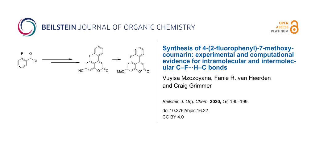
4-(2-Fluorophenyl)-7-methoxycoumarin (6) was synthesized under mild conditions via a three-step reaction (Scheme 1) and the first step was the synthesis of the fluorinated β-keto ester 3. Methyl acetoacetate (2) was treated with MgCl2, Et3N and n-BuLi in DCM and then with 2-fluorobenzoyl chloride (1) to yield methyl 2-fluorobenzoylacetate (3) [38,39]. These reactions are very rare in the literature, however, there are similar reactions for the synthesis of β-keto esters as reported by Sijbesma et al. [40] and Anwar [41]. The second step of the synthesis was the Pechmann reaction, commonly used for the synthesis of coumarins [42,43]. Methyl 2-fluorobenzoylacetate (3) was reacted with resorcinol (4) in the presence of H2SO4 at 35 °C, and 7-hydroxy-4-(2-fluorophenyl)coumarin (5) [39,44] was obtained as a light yellow solid. The last step of the synthesis was the methylation of the hydroxy group of coumarin 5 with dimethyl sulfate, to form 4-(2-fluorophenyl)-7-methoxycoumarin (6).
![[1860-5397-16-22-i1]](/bjoc/content/inline/1860-5397-16-22-i1.svg?scale=2.0&max-width=1024&background=FFFFFF)
Scheme 1: Synthesis of 4-(2-fluorophenyl)-7-methoxycoumarin (6).
Scheme 1: Synthesis of 4-(2-fluorophenyl)-7-methoxycoumarin (6).
Discussion
During the synthesis of coumarin 6, solution-state NMR spectroscopy was used to characterize compounds 3, 5, and 6 (1H and 13C spectra are available in Supporting Information File 1). The 1H spectrum of coumarin 6 showed H···F interactions for H3′, H4′, H5′ and H6′ which is typical through-bond (TB) coupling. However, the peaks that caught our particular attention were the singlet peak at 6.25 ppm and a doublet-of-doublets (dd) peak at 7.16 ppm assigned to H3 and H5, respectively (Figure 1). The H5 signal was expected to be a doublet (not a dd) due to 3J coupling to H6, since an H,H-COSY experiment does not show coupling between H5 and H8 (Figure S9, Supporting Information File 1). It became clear that the splitting of the signal from H5 was due to coupling with the 19F atom by comparing the spectra from the 1H and 1H-{19F} experiments (Figure 1) which showed the H5 peak as a doublet with 19F decoupling. While the doublet-of-doublets signal for H5 collapses into a doublet with 19F decoupling, there are no significant changes in the line-shape for the signal of H3 with 19F decoupling (Figure 1).
To determine whether the observation of 19F–H5 coupling for coumarin 6 was a solvent dependent phenomenon, a comparison was made between the 1H and 1H-{19F} spectra in CDCl3 and acetone-d6. Splitting of the H5 signal was observed in both solvents (Figure 1) suggesting that the 19F–H5 coupling for coumarin 6 is solvent independent. A literature report of the NMR characterization of a structurally similar 7-hydroxycoumarin performed in DMSO-d6 has the signal for H5 reported as a singlet [44].
![[1860-5397-16-22-1]](/bjoc/content/figures/1860-5397-16-22-1.png?scale=2.0&max-width=1024&background=FFFFFF)
Figure 1: 1H NMR spectra for the “aromatic” region of coumarin 6; comparison of 1H spectrum and 1H-{19F} spectrum in CDCl3 and 1H spectrum and 1H-{19F} spectrum in acetone-d6.
Figure 1: 1H NMR spectra for the “aromatic” region of coumarin 6; comparison of 1H spectrum and 1H-{19F} spec...
Since coumarins 6 and 5 have similar structure, the only difference is at position seven, 6 has a methoxy group while 5 has a hydroxy group, a similar study was carried out for coumarin 5 (Figure S10b, Supporting Information File 1).
The question posed at this point was “is this a through-bond (TB) or through-space (TS) effect”?
To answer this question, we analysed a 13C-{1H} spectrum of coumarin 6 dissolved in CDCl3 and the signal corresponding to C5 was found to be a doublet (J = 1.4 Hz) but the signals corresponding to C4 and C4a were singlets, and this indicates that this coupling is not a TB effect, because if it were a TB effect, the signals for C4 and C4a would also likely be split. Similar splitting of C5 was observed in coumarin 5, also in different solvents as shown in Figure 2 and there was no splitting of the signals corresponding to C4 and C4a as shown in Figures S22 and S21 (Supporting Information File 1). These observations were not found in the similar derivatives of coumarin 5 (7-hydroxy-4-(3-fluorophenyl)coumarin and 7-hydroxy-4-(4-fluorophenyl)coumarin) found in the literature where the fluorine atom is in the third (C3') and the fourth (C4') position, respectively, of the phenyl ring [39]. The spectra for these derivatives showed an H5 signal as a doublet (not doublet-of-doublets as observed in coumarin 5 and 6) and C5 as a singlet, indicating that they do not possess the through-space F···H5 or/and F···C5 coupling since the fluorine atom is a bit further away from H5 and C5.
![[1860-5397-16-22-2]](/bjoc/content/figures/1860-5397-16-22-2.png?scale=2.0&max-width=1024&background=FFFFFF)
Figure 2: 13C NMR spectra for coumarin 5 and 6; showing the splitting of the signal corresponding to C5.
Figure 2: 13C NMR spectra for coumarin 5 and 6; showing the splitting of the signal corresponding to C5.
To confirm our findings, we further ran a 19F,1H-HOESY experiment and it showed clear H5···19F and H3···19F coupling (Figure 3). Evidence of a HOESY interaction between H5···19F and H3···19F indicates that neither the H3···19F nor the H5···19F interaction limits the C4–C1′ bond rotation.
![[1860-5397-16-22-3]](/bjoc/content/figures/1860-5397-16-22-3.png?scale=2.0&max-width=1024&background=FFFFFF)
Figure 3: 19F,1H-HOESY NMR spectrum for coumarin 6 illustrating two through-space interactions.
Figure 3: 19F,1H-HOESY NMR spectrum for coumarin 6 illustrating two through-space interactions.
The geometry of coumarin 6 (single molecule, gas phase) was optimized using the B3LYP functional and the 6-311G basis set, as implemented in Gaussian-09W (Rev. C.01) [45]. The superposition of the single-crystal X-ray structure (red) and the optimized structure (green) is shown in Figure 4.
![[1860-5397-16-22-4]](/bjoc/content/figures/1860-5397-16-22-4.png?scale=2.0&max-width=1024&background=FFFFFF)
Figure 4: Superposition of single-crystal X-ray structure (red) and DFT-optimized structure (green); RMSD 0.3 Å (hydrogen atoms omitted for clarity).
Figure 4: Superposition of single-crystal X-ray structure (red) and DFT-optimized structure (green); RMSD 0.3...
The DFT optimized geometric structure is shown in Figure 5 and has a dihedral angle, Φ (C2'–C1'–C4–C4a) of 65.3°. Following the optimization, the dihedral angle Φ was varied through a 360° rotation to examine the effect of changing the relative position of the fluorinated ring and the energy profile for this variation is shown in Figure 6. When Φ = 5°, the F···H5 distance is at its shortest (dF···H5 = 2.0 Å) and the fluorinated ring is almost coplanar with the coumarin ring, and the molecule is at its least stable conformation due to the electron–electron (e–e) repulsion of H5 and fluorine. The second least stable conformation is found at Φ = 185°, with the fluorine atom and H3 in close proximity (dF···H3 = 2.0 Å).
![[1860-5397-16-22-5]](/bjoc/content/figures/1860-5397-16-22-5.png?scale=1.7&max-width=1024&background=FFFFFF)
Figure 5: DFT-optimized structure for coumarin (6).
Figure 5: DFT-optimized structure for coumarin (6).
![[1860-5397-16-22-6]](/bjoc/content/figures/1860-5397-16-22-6.png?scale=2.0&max-width=1024&background=FFFFFF)
Figure 6: Plots of relative energy (black trace, no units), interatomic distance F–H5 (red trace, Å), interatomic distance F–H3 (blue trace, Å) as a function of dihedral angle Φ C2’–C1’–C4–C4a (°).
Figure 6: Plots of relative energy (black trace, no units), interatomic distance F–H5 (red trace, Å), interat...
Examples of through-space coupling between fluorine and hydrogen atoms in organic molecules are reported in the chemical literature [46,47], with magnitudes as large as 7 Hz for 7JHF [46]
One report of TS-coupling between F and H atoms comments that “...it appears that significant coupling only occurs when the proton–fluorine closest approach distance is within the sum of the van der Waals radii of hydrogen and fluorine (c. 2.55 Å)...” [47]. During rotation of the fluorophenyl ring (Figure 6), the F–H distance (for both F–H5 and F–H3) varies between 2 and 5 Å, so for some part of the rotation, both the distances F–H5 and F–H3 fall inside the limit of ≈2.55 Å, although not simultaneously. For a fixed geometry like that of the crystal structure, this would suggest that through-space F–H coupling would be observed between F and H5 but not between F and H3, as in Figure 1, where there is an obvious change to the shape of the signal corresponding to H5 but a negligible change to the shape of the signal corresponding to H3, with the application of {19F} decoupling. However, the F–H HOESY spectrum in Figure 3 shows coupling between the fluorine atom and both H5 and H3. The authors interpretation of these observations (Figure 1 and Figure 3) is that, in solution, rotation of the fluorophenyl ring (about C4–C1’) is permitted but that the average geometry of coumarin 6 has the fluorine atom closer to H5 than H3.
The theoretical NMR data for twenty-four conformations of coumarin 6 were obtained from Gaussian 09W (Rev C.01) at the B3LYP/6-311G level. Geometry optimization and calculation of NMR parameters for TMS and CCl3F at the same level provided reference chemical shifts for 1H, 13C, and 19F. The chemical shifts for the lowest energy structure (Φ = 65.3°) and the most unstable conformer (Φ = 5°) are used as examples (Table 1). The theoretical chemical shifts for the carbons appeared to be shifted downfield relative to the experimental carbon peaks (for both stable and unstable conformers) as shown by ‘change’ (Δ = −ve, experimental − theoretical) in Table 1.
Table 1: Experimental and theoretical (gas phase) 1H and 13C chemical shift (δ) for atoms within six bonds from fluorine for coumarin 6 and RSMD values.
| Atom | Exp | Φ = 65.3° a | Φ = 5° b | ||
| C/H | δ (ppm) | δ (ppm) | Δ(ppm)c | δ (ppm) | Δ(ppm)c |
| C2 | 160.9 | 169.1 | −8.2 | 170.1 | −9.2 |
| C3 | 112.5 | 118.4 | −5.9 | 117.7 | −5.2 |
| H3 | 6.25 | 6.00 | 0.25 | 6.75 | −0.50 |
| C4 | 150.5 | 158.0 | −7.5 | 152.6 | −2.1 |
| C4a | 112.4 | 120.8 | −8.4 | 120.2 | −7.8 |
| C5 | 127.8 | 135.0 | −7.2 | 139.2 | −11.4 |
| H5 | 7.16 | 6.97 | 0.19 | 8.16 | −1.00 |
| C6 | 113.5 | 121.4 | −7.9 | 119.7 | −6.2 |
| C1' | 123.2 | 135.2 | −12.0 | 130.6 | −7.4 |
| C2' | 159.1 | 172.8 | −13.7 | 173.7 | −14.6 |
| C3' | 116.3 | 123.0 | −6.7 | 126.8 | −10.5 |
| H3' | 7.29 | 7.20 | 0.09 | 7.30 | −0.01 |
| C4' | 131.5 | 137.5 | −6.0 | 138.6 | −7.1 |
| H4' | 7.50 | 7.52 | −0.02 | 7.46 | 0.04 |
| C5' | 130.5 | 132.6 | −2.1 | 132.7 | −2.2 |
| H5' | 7.35 | 7.37 | −0.02 | 7.39 | −0.04 |
| C6' | 124.7 | 138.0 | −13.3 | 136.9 | −12.2 |
| H6' | 7.23 | 7.31 | −0.08 | 8.06 | −0.83 |
| RMSD values |
13C NMR = 8.84 ppm
1H NMR = 0.138 ppm |
13C NMR = 8.79 ppm
1H NMR = 0.569 ppm |
|||
aConformer with Φ = 65 °. bConformer with Φ = 5 °. cExperimental – theoretical, e.g., C2: 160.9 − 169.1 = −8.2 ppm.
Comparing the experimental and the calculated 13C NMR chemical shifts for both the optimized and least-stable DFT-generated conformations, the RMSD values were found to be 8.84 ppm and 8.79 ppm, respectively. The RMSD value for the calculated 1H NMR chemical shifts of the optimized conformer was found to be substantially smaller (RMSD = 0.14 ppm) than that for the least-stable conformer (RMSD = 0.57 ppm).
The theoretical coupling constants at the same 24 geometries from geometry scan for coumarin 6 were obtained using the same functional and level of theory (B3LYP/6-311G). The coupling constants (F···C or F···H) for selected nuclei of interest were obtained using a scaling factor calculated from the observed and calculated 1JFC value for CFCl3 [48]. Selected graphs of the plots of nJFH and nJFC as a function of rotation of the fluorophenyl ring are included in Supporting Information File 1.
Figures S18 and S19 show the magnitude of 5JFH and 6JFH between the fluorine atom and H3 and H5, respectively. It can be seen that, at the B3LYP/6-311G) level, when coupling between F and either H3 or H5 is large enough to be observed in an NMR spectrum (magnitude ≈1–11 Hz for H5, ≈1–5 Hz for H3), coupling to the other nucleus is near zero and may not necessarily be observed. This is consistent with the idea of an average angle in solution that places the fluorine atom closer to H5 than H3, since F–H5 coupling is obvious in the experimental NMR spectrum (Figure 1), while F–H3 coupling is not. A similar prediction is made for coupling between F–C5 and F–C3 (Figures S14 and S17 in Supporting Information File 1). At angles that would manifest in an experimental F–H5 coupling of ≈2.5 Hz, with near-zero F–H3 coupling, Gaussian predicts small F–C coupling to both C5 and C3. This is consistent with the experimental observation of 5JFC ≈ 1.4 Hz between F and C5 and no observed coupling between F and C3, in the 13C-{1H} spectrum. Gaussian also predicts F–C coupling between the fluorine atom and carbons C4 and C4a, magnitude <1 Hz and 1–1.5 Hz, respectively. While the <1 Hz F–C4 coupling might not be noticeable in the 13C-{1H} spectrum, it should be possible to see F–C4a coupling of 1–1.5 Hz, since the F–C5 coupling is observable at a similar magnitude. The model (B3LYP/6-311G) seems to work reasonably well for coupling between F and H3, H5, C3, C4, and C5 but not for C4a.
The single crystal X-ray analysis of coumarin 6 was carried out as it has not been reported previously [CCDC No.: 1868146]. The crystals of 6 were obtained by slow evaporation of methanol/dichloromethane and were found to be of the monoclinic crystal system with space group C2/c (Figure 7).
The crystal structure shows that the fluorinated phenyl ring is at a torsion angle (Φ, C2'–C1'–C4–C4a angle) of 54.44° to the coumarin moiety. The F···H5 TS-distance of 2.547 Å is small enough to induce some rotational constraint on the C4 and C1' bond, as the constraint was observed at an F···H distance of 2.9 Å [37]. The short contact interactions show that there are C–F···H–C intermolecular interactions to the neighboring molecules (F–H6 and F–H5; different molecules) that play a crucial role in crystal packing (Figure 7).
![[1860-5397-16-22-7]](/bjoc/content/figures/1860-5397-16-22-7.png?scale=2.0&max-width=1024&background=FFFFFF)
Figure 7: Short contacts within the single-crystal X-ray structure of coumarin 6.
Figure 7: Short contacts within the single-crystal X-ray structure of coumarin 6.
As mentioned above, the structure of coumarin 6 was optimized and the dihedral angle, Φ (C2'–C1'–C4–C4a) was found to be 65.3° (Figure 5), which is comparible close to that found in the crystal structure (Φ = 54.4°). The TS distance between F and H5 for the optimized structure is 2.807 Å which is relatively close to that of the crystal structure (2.547 Å). Selected comparisons are shown in Table 2.
Table 2: Comparison of some features of the X-ray crystal structure and DFT optimized structure of 6: Through-space (TS) and dihedral angle (Φ).
| Run | TS distance (Å) | Dihedral angle (°) | ||
| F···H5 | F···C5 | H6···H3 | Φ | |
| 6 (exp) | 2.547 | 2.934 | 2.535 | 54.44 |
| 6 (DFT) | 2.807 | 3.174 | 2.874 | 65.27 |
| difference | 0.260 | 0.24 | 0.339 | 10.83 |
Conclusion
The synthesis of 4-(2-fluorophenyl)-7-methoxycoumarin (6) via the Pechmann reaction was successful. The solution-state 1H and 13C NMR spectra of 6 showed that there is a strong intramolecular interaction between F and H5 (JFH = 2.6 Hz) and suggest that this interaction is through-space C–F···H–C coupling, since C5 is coupled to F (JFC = 1.4 Hz) whereas C4 and C4a are not. The 2D HOESY spectrum shows the F···H5 coupling and also F···H3 coupling, seemingly weaker than F···H5 since splitting of the H3 signal is not observed in the 1H and 1H-{19F} 1D spectra. The single crystal X-ray structure showed that the fluorinated phenyl ring is orientated in a manner that brings the fluorine atom closer to H5 than H3. The same orientation was observed in the DFT-optimized (B3LYP/6-311G) structure. The X-ray data also showed the intermolecular C–F···H–C interactions which, together with other interactions, are resposible for the crystal packing.
Experimental
General
All reagents (including solvents) were purchased from the chemical suppliers Aldrich, Fluka and Merck. For all moisture-sensitive reactions, the glassware was thoroughly dried in an oven at ca. 140 °C for 12 h prior to use, and anhydrous solvents were used under inert conditions. Qualitative thin-layer chromatography (TLC, silica gel 60254, aluminum backed) was used to monitor the reactions. Visualization of the TLC plates was achieved using an iodine tank and/or fluorescence on exposure to short wavelength ultraviolet light (254 nm). For purification, column chromatography (silica gel 60, 0.040–0.063 mm) or centrifugal chromatography conducted on a Harrison Research Chromatotron model 7924T (glass plates coated with silica gel 60 PF254 containing gypsum, 2 and 4 mm thick layer) was used.
Nuclear magnetic resonance (NMR) spectra were recorded on a Bruker Avance 400 spectrometer equipped with a 5 mm BBOZ probe at frequencies of 400 MHz, 100 MHz, and 376 MHz for 1H, 13C, and 19F, respectively. High-resolution mass spectrometry (HRMS) was performed on a Waters LCT Premier time-of-flight mass spectrometer.
Synthesis of methyl 2-fluorobenzoylacetate (3): To a stirred mixture of MgCl2 (2.0 g, 21 mmol) and Et3N (2.1 g, 21 mmol) in dry DCM (15 mL) at room temperature, methyl acetoacetate (2, 2.0 g, 17 mmol) was added slowly. The mixture was stirred for 30 min before the temperature was reduced to 0 °C. n-BuLi (20 mL of a 1.6 M in hexane, 32 mmol) was added slowly into the mixture and the mixture was stirred for a further 30 min. 2-Fluorobenzoyl chloride (1, 2.7 g, 17 mmol) was added dropwise into the mixture and the mixture was stirred for 15 min. The reaction mixture was allowed to reach room temperature and was stirred overnight. To the reaction, was added 5 M HCl (8 mL) and distilled water (10 mL) and the mixture was extracted with DCM (3 × 30 mL). The organic layer was dried over anhydrous MgSO4 and the solvent was removed in vacuo. The resulting yellow product was purified by silica gel column chromatography with 10% EtOAc in hexane as eluent and 3 was obtained as a light orange viscous liquid (2.7 g, 81%), TLC Rf 0.50 (hexane/EtOAc, 9:1). 1H NMR (400 MHz, CDCl3) 3.76 (s, 3H, H4), 4.01 (d, 1hJH,F = 3.4 Hz, 2H, H2), 7.15 (ddd, 3JH,F = 12.1 Hz, 3JH,H = 8.5 Hz, 4JH,H = 1.0 Hz, 1H, H3'), 7.26 (t, 3JH,H = 7.6 Hz, 1H, H5'), 7.57 (m, 1H, H4'), 7.95 (ddd, 3JH,H = 7.6 Hz, 4JH,F = 6.2 Hz, 4JH,H = 1.9 Hz, 1H, H6'); 13C NMR (100 MHz, CDCl3) 49.6 (d, 2hJC,F = 8.1 Hz, C2), 52.3 (C4), 116.7 (d, 2JC,F = 24.1 Hz, C3'), 124.7 (d, 3JC,F = 2.9 Hz, C6'), 129.3 (d, 2JC,F = 21.7 Hz, C1'), 131.0 (d, 4JC,F = 2.3 Hz, C5'), 135.5 (d, 3JC,F = 9.6 Hz, C4'), 162.2 (d, 1JC,F = 254.3 Hz, C2’), 167.8 (d, 3hJC,F = 3.0 Hz, C3), 190.1 (d, TSJC,F = 3.7 Hz, C1).
Synthesis of 7-hydroxy-4-(2-fluorophenyl)coumarin (5): To a mixture of resorcinol (2.0 g, 18 mmol) and methyl 2-fluorobenzoylacetate (3.5 g, 18 mmol) was added H2SO4 (8 mL, 75%). The temperature of a stirred mixture was increased to 35 °C. After stirring for 5 h, the mixture was poured into crushed ice and neutralized with a NaOH solution. The mixture was filtered under vacuum and the residue was washed with plenty of water. The resulting product was purified by silica gel column chromatography with 60% EtOAc in hexane as eluent and 5 was obtained as a light yellow solid (4.2 g, 91%), mp 204–207 °C, TLC Rf 0.45 (hexane/EtOAc, 2:3). 1H NMR (400 MHz, DMSO-d6) 6.24 (s, 1H, H3), 6.77 (dd, 3JH,H = 8.6 Hz, 4JH,H = 2.4 Hz, 1H, H6), 6.81 (d, 4JH,H = 2.4 Hz, 1H, H8), 7.03 (dd, 3JH,H = 8.6 Hz, 1hJH,F = 2.6 Hz, 1H, H5), 7.37–7.45 (m, 2H, H3', H6'), 7.50 (td, 3JH,H = 7.5 Hz, 4JH,H = 1.8 Hz, 1H, H5'), 7.61 (m, 1H, H4'), 10.67 (s, 1H, OH); 13C NMR (100 MHz, DMSO-d6) 102.6 (C8), 110.7 (C4a), 112.1 (C3), 113.4 (C6), 116.1 (d, 2JC,F = 21.3 Hz, C3'), 122.7 (d, 2JC,F = 15.3 Hz, C1'), 125.2 (d, 3JC,F = 3.6 Hz, C6'), 127.9 (d, 2hJC,F = 1.6 Hz, C5), 130.8 (d, 4JC,F = 2.9 Hz, C5'), 132.0 (d, 3JC,F = 8.2 Hz, C4'), 150.3 (C4), 155.2 (C8a), 158.6 (d, 1JC,F = 248.6 Hz, C2'), 160.0 (C2), 161.6 (C7); HRMS–ESI+ (m/z): [M + Na]+ calcd for C15H9O3FNa, 279.0433; found, 279.0437,
Synthesis of 4-(2-fluorophenyl)-7-methoxycoumarin (6): A mixture of 7-hydroxy-4-(2-fluorophenyl)coumarin (5 ,0.77 g, 3.0 mmol), dimethyl sulfate (0.76 g, 6.0 mmol) and K2CO3 (0.83 g, 6.0 mmol) was refluxed in acetone (20 mL) for 4 h. The reaction mixture was cooled to room temperature and and brine (50 mL) was added then extracted with ethyl acetate (3 × 40 mL). The organic layer was dried over anhydrous MgSO4 and the solvent was removed in vacuo. The resulting light yellow product was purified by silica gel column chromatography with 60% EtOAc in hexane as eluent and 6 was obtained as a yellow crystalline solid (0.78 g, 2.9 mmol, 97%), mp 167–170 ºC, TLC Rf 0.54 (hexanes/EtOAc, 3:2). 1H NMR (400 MHz, CDCl3) 3.88 (s, 3H, OMe), 6.25 (s, 1H, H3), 6.79 (dd, 3JH,H = 8.9 Hz, 4JH,H = 2.5 Hz, 1H, H6), 6.89 (d, 4JH,H = 2.5 Hz, 1H, H8), 7.16 (dd, 3JH,H = 8.9 Hz, 1hJH,F = 2.5 Hz, 1H, H5), 7.23 (dd, 3JH,H = 7.8 Hz, 4JH,F = 4.5 Hz, 1H, H6'), 7.29 (td, 3JH,H = 3JH,F = 7.8 Hz, 4JH,H = 1.8 Hz, 1H, H3'), 7.35 (td 3JH,H = 7.8 Hz, 4JH,H = 1.8 Hz, 1H, H5'), 7.50 (m, 1H, H4'); 13C NMR (100 MHz, CDCl3) 55.8 (OMe), 101.0 (C8), 112.4 (C4a), 112.5 (C3), 113.5 (C6), 116.3 (d, 2JC,F = 21.9 Hz, C3'), 123.2 (d, 2JC,F = 15.4 Hz, C1'), 124.7 (d, 3JC,F = 3.7 Hz, C6'), 127.8 (d, 2hJC,F = 1.4 Hz, C5), 130.5 (d, 4JC,F = 3.1 Hz, C5'), 131.5 (d, 3JC,F = 7.9 Hz, C4'), 150.5 (C4), 155.7 (C8a), 159.1 (d, 1JC,F = 250.0 Hz, C2'), 160.9 (C2), 163.0 (C7). HRMS–ESI+ (m/z): [M + Na]+ calcd for C16H11O3FNa, 293.0590; found, 293.0587.
Supporting Information
| Supporting Information File 1: Copies of NMR spectra for compound 3, 5 and 6, single crystal X-ray data for compound 6, Gaussian calculation data of J-values for compound 6 and HRMS for compound 6. | ||
| Format: PDF | Size: 1.3 MB | Download |
Acknowledgements
The authors would like to acknowledge the Ph.D. thesis by Vuyisa Mzozoyana (University of KwaZulu-Natal, South Africa) as the main source of this work. The authors would like to thank Dr Rainner Kerssebaum of Bruker-DE for his kind assistance with the programming of a gradient HOESY experiment compatible with Topspin 2.1.
References
-
Lin, M.-H.; Chou, Y.-S.; Tsai, Y.-J.; Chou, D.-S. J. Exp. Clin. Med. 2011, 3, 126–131. doi:10.1016/j.jecm.2011.04.006
Return to citation in text: [1] -
Hoult, J. R. S.; Payá, M. Gen. Pharmacol.: Vasc. Syst. 1996, 27, 713–722. doi:10.1016/0306-3623(95)02112-4
Return to citation in text: [1] -
Hara, K.; Sayama, K.; Ohga, Y.; Shinpo, A.; Suga, S.; Arakawa, H. Chem. Commun. 2001, 569–570. doi:10.1039/b010058g
Return to citation in text: [1] -
Givel, M. Tob. Control 2003, 12, 401–405. doi:10.1136/tc.12.4.401
Return to citation in text: [1] -
Emmanuel-Giota, A. A.; Fylaktakidou, K. C.; Litinas, K. E.; Nicolaides, D. N.; Hadjipavlou-Litina, D. J. J. Heterocycl. Chem. 2001, 38, 717–722. doi:10.1002/jhet.5570380329
Return to citation in text: [1] -
Marchenko, M. M.; Kopyl’chuk, G. P.; Shmarakov, I. A.; Ketsa, O. V.; Kushnir, V. N. Pharm. Chem. J. 2006, 40, 296–297. doi:10.1007/s11094-006-0113-8
Return to citation in text: [1] -
Vukovic, N.; Sukdolak, S.; Solujic, S.; Niciforovic, N. Arch. Pharmacal Res. 2010, 33, 5–15. doi:10.1007/s12272-010-2220-z
Return to citation in text: [1] -
Basanagouda, M.; Kulkarni, M. V.; Sharma, D.; Gupta, V. K.; Pranesha; Sandhyarani, P.; Rasal, V. P. J. Chem. Sci. 2009, 121, 485–495. doi:10.1007/s12039-009-0058-z
Return to citation in text: [1] -
Jung, K.; Park, Y.-J.; Ryu, J.-S. Synth. Commun. 2008, 38, 4395–4406. doi:10.1080/00397910802369513
Return to citation in text: [1] -
Vilar, S.; Quezada, E.; Santana, L.; Uriarte, E.; Yánez, M.; Fraiz, N.; Alcaide, C.; Cano, E.; Orallo, F. Bioorg. Med. Chem. Lett. 2006, 16, 257–261. doi:10.1016/j.bmcl.2005.10.013
Return to citation in text: [1] -
Maheswara, M.; Siddaiah, V.; Damu, G. L. V.; Rao, Y. K.; Rao, C. V. J. Mol. Catal. A: Chem. 2006, 255, 49–52. doi:10.1016/j.molcata.2006.03.051
Return to citation in text: [1] -
Cherry, K.; Parrain, J.-L.; Thibonnet, J.; Duchêne, A.; Abarbri, M. J. Org. Chem. 2005, 70, 6669–6675. doi:10.1021/jo050638z
Return to citation in text: [1] -
Heravi, M. M.; Hekmatshoar, R.; Emamgholizadeh, M. Phosphorus, Sulfur Silicon Relat. Elem. 2004, 179, 1893–1896. doi:10.1080/10426500490466814
Return to citation in text: [1] -
Barancelli, D. A.; Salles, A. G., Jr.; Taylor, J. G.; Correia, C. R. D. Org. Lett. 2012, 14, 6036–6039. doi:10.1021/ol302923f
Return to citation in text: [1] -
Kaye, P. T.; Musa, M. A. Synthesis 2002, 2701–2706. doi:10.1055/s-2002-35984
Return to citation in text: [1] -
Ray, S. K.; Singh, P. K.; Molleti, N.; Singh, V. K. J. Org. Chem. 2012, 77, 8802–8808. doi:10.1021/jo301513x
Return to citation in text: [1] -
Jung, J.-W.; Kim, N.-J.; Yun, H.; Han, Y. T. Molecules 2018, 23, 2417. doi:10.3390/molecules23102417
Return to citation in text: [1] [2] -
Wu, J.; Liao, Y.; Yang, Z. J. Org. Chem. 2001, 66, 3642–3645. doi:10.1021/jo0102157
Return to citation in text: [1] -
Fried, J.; Sabo, E. F. J. Am. Chem. Soc. 1954, 76, 1455–1456. doi:10.1021/ja01634a101
Return to citation in text: [1] -
Hagmann, W. K. J. Med. Chem. 2008, 51, 4359–4369. doi:10.1021/jm800219f
Return to citation in text: [1] -
Barradell, L. B.; Faulds, D.; McTavish, D. Drugs 1992, 44, 225–250. doi:10.2165/00003495-199244020-00007
Return to citation in text: [1] -
Gether, U.; Andersen, P. H.; Larsson, O. M.; Schousboe, A. Trends Pharmacol. Sci. 2006, 27, 375–383. doi:10.1016/j.tips.2006.05.003
Return to citation in text: [1] -
Purser, S.; Moore, P. R.; Swallow, S.; Gouverneur, V. Chem. Soc. Rev. 2008, 37, 320–330. doi:10.1039/b610213c
Return to citation in text: [1] -
O'Hagan, D. Chem. Soc. Rev. 2008, 37, 308–319. doi:10.1039/b711844a
Return to citation in text: [1] [2] -
Isanbor, C.; O’Hagan, D. J. Fluorine Chem. 2006, 127, 303–319. doi:10.1016/j.jfluchem.2006.01.011
Return to citation in text: [1] -
Kirk, K. L. J. Fluorine Chem. 2006, 127, 1013–1029. doi:10.1016/j.jfluchem.2006.06.007
Return to citation in text: [1] -
Beijer, F. H.; Kooijman, H.; Spek, A. L.; Sijbesma, R. P.; Meijer, E. W. Angew. Chem., Int. Ed. 1998, 37, 75–78. doi:10.1002/(sici)1521-3773(19980202)37:1/2<75::aid-anie75>3.0.co;2-r
Return to citation in text: [1] -
Duarte, C. J.; Freitas, M. P. J. Mol. Struct. 2009, 930, 135–139. doi:10.1016/j.molstruc.2009.05.004
Return to citation in text: [1] -
Fonseca, T. A. O.; Freitas, M. P.; Cormanich, R. A.; Ramalho, T. C.; Tormena, C. F.; Rittner, R. Beilstein J. Org. Chem. 2012, 8, 112–117. doi:10.3762/bjoc.8.12
Return to citation in text: [1] -
Nath, B.; Baruah, J. B. Cryst. Growth Des. 2013, 13, 5146–5155. doi:10.1021/cg401220x
Return to citation in text: [1] -
Ridout, J.; Probert, M. R. Cryst. Growth Des. 2013, 13, 1943–1948. doi:10.1021/cg301773h
Return to citation in text: [1] -
Murray-Rust, P.; Stallings, W. C.; Monti, C. T.; Preston, R. K.; Glusker, J. P. J. Am. Chem. Soc. 1983, 105, 3206–3214. doi:10.1021/ja00348a041
Return to citation in text: [1] -
Desiraju, G. R. Acc. Chem. Res. 2002, 35, 565–573. doi:10.1021/ar010054t
Return to citation in text: [1] -
Howard, J. A. K.; Hoy, V. J.; O'Hagan, D.; Smith, G. T. Tetrahedron 1996, 52, 12613–12622. doi:10.1016/0040-4020(96)00749-1
Return to citation in text: [1] [2] -
Pedireddi, V. R.; Desiraju, G. R. J. Chem. Soc., Chem. Commun. 1992, 988–990. doi:10.1039/c39920000988
Return to citation in text: [1] [2] -
Thakur, T. S.; Kirchner, M. T.; Bläser, D.; Boese, R.; Desiraju, G. R. CrystEngComm 2010, 12, 2079–2085. doi:10.1039/b925082d
Return to citation in text: [1] [2] -
Shang, J.; Gallagher, N. M.; Bie, F.; Li, Q.; Che, Y.; Wang, Y.; Jiang, H. J. Org. Chem. 2014, 79, 5134–5144. doi:10.1021/jo500582c
Return to citation in text: [1] [2] -
Jeon, M.-K.; Kim, K. Tetrahedron 1999, 55, 9651–9667. doi:10.1016/s0040-4020(99)00585-2
Return to citation in text: [1] -
Mzozoyana, V. Synthesis of fluorinated benzophenones and phenylcoumarins. Ph.D. Thesis, UKZN, 2015.
Return to citation in text: [1] [2] [3] -
Hirschberg, J. H. K. K.; Koevoets, R. A.; Sijbesma, R. P.; Meijer, E. W. Chem. – Eur. J. 2003, 9, 4222–4231. doi:10.1002/chem.200204495
Return to citation in text: [1] -
Anwar, H. F. Synlett 2009, 2711–2712. doi:10.1055/s-0029-1218016
Return to citation in text: [1] -
Panetta, J. A.; Rapoport, H. J. Org. Chem. 1982, 47, 946–950. doi:10.1021/jo00345a009
Return to citation in text: [1] -
Raad, I.; Terreux, R.; Richomme, P.; Matera, E.-L.; Dumontet, C.; Raynaud, J.; Guilet, D. Bioorg. Med. Chem. 2006, 14, 6979–6987. doi:10.1016/j.bmc.2006.06.026
Return to citation in text: [1] -
Timonen, J. M.; Nieminen, R. M.; Sareila, O.; Goulas, A.; Moilanen, L. J.; Haukka, M.; Vainiotalo, P.; Moilanen, E.; Aulaskari, P. H. Eur. J. Med. Chem. 2011, 46, 3845–3850. doi:10.1016/j.ejmech.2011.05.052
Return to citation in text: [1] [2] -
Gaussian 16, Revision C.01; Gaussian, Inc.: Wallingford, CT, 2016.
Return to citation in text: [1] -
Agranat, I.; Rabinovitz, M.; Gosnay, I.; Weitzen-Dagan, A. J. Am. Chem. Soc. 1972, 94, 2889–2891. doi:10.1021/ja00763a074
Return to citation in text: [1] [2] -
Adcock, W.; Rizvi, S. Aust. J. Chem. 1973, 26, 2659–2663. doi:10.1071/ch9732659
Return to citation in text: [1] [2] -
Malkina, O. L.; Salahub, D. R.; Malkin, V. G. J. Chem. Phys. 1996, 105, 8793–8800. doi:10.1063/1.472658
Return to citation in text: [1]
| 42. | Panetta, J. A.; Rapoport, H. J. Org. Chem. 1982, 47, 946–950. doi:10.1021/jo00345a009 |
| 43. | Raad, I.; Terreux, R.; Richomme, P.; Matera, E.-L.; Dumontet, C.; Raynaud, J.; Guilet, D. Bioorg. Med. Chem. 2006, 14, 6979–6987. doi:10.1016/j.bmc.2006.06.026 |
| 39. | Mzozoyana, V. Synthesis of fluorinated benzophenones and phenylcoumarins. Ph.D. Thesis, UKZN, 2015. |
| 44. | Timonen, J. M.; Nieminen, R. M.; Sareila, O.; Goulas, A.; Moilanen, L. J.; Haukka, M.; Vainiotalo, P.; Moilanen, E.; Aulaskari, P. H. Eur. J. Med. Chem. 2011, 46, 3845–3850. doi:10.1016/j.ejmech.2011.05.052 |
| 1. | Lin, M.-H.; Chou, Y.-S.; Tsai, Y.-J.; Chou, D.-S. J. Exp. Clin. Med. 2011, 3, 126–131. doi:10.1016/j.jecm.2011.04.006 |
| 2. | Hoult, J. R. S.; Payá, M. Gen. Pharmacol.: Vasc. Syst. 1996, 27, 713–722. doi:10.1016/0306-3623(95)02112-4 |
| 7. | Vukovic, N.; Sukdolak, S.; Solujic, S.; Niciforovic, N. Arch. Pharmacal Res. 2010, 33, 5–15. doi:10.1007/s12272-010-2220-z |
| 18. | Wu, J.; Liao, Y.; Yang, Z. J. Org. Chem. 2001, 66, 3642–3645. doi:10.1021/jo0102157 |
| 48. | Malkina, O. L.; Salahub, D. R.; Malkin, V. G. J. Chem. Phys. 1996, 105, 8793–8800. doi:10.1063/1.472658 |
| 6. | Marchenko, M. M.; Kopyl’chuk, G. P.; Shmarakov, I. A.; Ketsa, O. V.; Kushnir, V. N. Pharm. Chem. J. 2006, 40, 296–297. doi:10.1007/s11094-006-0113-8 |
| 17. | Jung, J.-W.; Kim, N.-J.; Yun, H.; Han, Y. T. Molecules 2018, 23, 2417. doi:10.3390/molecules23102417 |
| 37. | Shang, J.; Gallagher, N. M.; Bie, F.; Li, Q.; Che, Y.; Wang, Y.; Jiang, H. J. Org. Chem. 2014, 79, 5134–5144. doi:10.1021/jo500582c |
| 5. | Emmanuel-Giota, A. A.; Fylaktakidou, K. C.; Litinas, K. E.; Nicolaides, D. N.; Hadjipavlou-Litina, D. J. J. Heterocycl. Chem. 2001, 38, 717–722. doi:10.1002/jhet.5570380329 |
| 16. | Ray, S. K.; Singh, P. K.; Molleti, N.; Singh, V. K. J. Org. Chem. 2012, 77, 8802–8808. doi:10.1021/jo301513x |
| 46. | Agranat, I.; Rabinovitz, M.; Gosnay, I.; Weitzen-Dagan, A. J. Am. Chem. Soc. 1972, 94, 2889–2891. doi:10.1021/ja00763a074 |
| 3. | Hara, K.; Sayama, K.; Ohga, Y.; Shinpo, A.; Suga, S.; Arakawa, H. Chem. Commun. 2001, 569–570. doi:10.1039/b010058g |
| 4. | Givel, M. Tob. Control 2003, 12, 401–405. doi:10.1136/tc.12.4.401 |
| 17. | Jung, J.-W.; Kim, N.-J.; Yun, H.; Han, Y. T. Molecules 2018, 23, 2417. doi:10.3390/molecules23102417 |
| 47. | Adcock, W.; Rizvi, S. Aust. J. Chem. 1973, 26, 2659–2663. doi:10.1071/ch9732659 |
| 12. | Cherry, K.; Parrain, J.-L.; Thibonnet, J.; Duchêne, A.; Abarbri, M. J. Org. Chem. 2005, 70, 6669–6675. doi:10.1021/jo050638z |
| 14. | Barancelli, D. A.; Salles, A. G., Jr.; Taylor, J. G.; Correia, C. R. D. Org. Lett. 2012, 14, 6036–6039. doi:10.1021/ol302923f |
| 10. | Vilar, S.; Quezada, E.; Santana, L.; Uriarte, E.; Yánez, M.; Fraiz, N.; Alcaide, C.; Cano, E.; Orallo, F. Bioorg. Med. Chem. Lett. 2006, 16, 257–261. doi:10.1016/j.bmcl.2005.10.013 |
| 11. | Maheswara, M.; Siddaiah, V.; Damu, G. L. V.; Rao, Y. K.; Rao, C. V. J. Mol. Catal. A: Chem. 2006, 255, 49–52. doi:10.1016/j.molcata.2006.03.051 |
| 46. | Agranat, I.; Rabinovitz, M.; Gosnay, I.; Weitzen-Dagan, A. J. Am. Chem. Soc. 1972, 94, 2889–2891. doi:10.1021/ja00763a074 |
| 47. | Adcock, W.; Rizvi, S. Aust. J. Chem. 1973, 26, 2659–2663. doi:10.1071/ch9732659 |
| 9. | Jung, K.; Park, Y.-J.; Ryu, J.-S. Synth. Commun. 2008, 38, 4395–4406. doi:10.1080/00397910802369513 |
| 44. | Timonen, J. M.; Nieminen, R. M.; Sareila, O.; Goulas, A.; Moilanen, L. J.; Haukka, M.; Vainiotalo, P.; Moilanen, E.; Aulaskari, P. H. Eur. J. Med. Chem. 2011, 46, 3845–3850. doi:10.1016/j.ejmech.2011.05.052 |
| 8. | Basanagouda, M.; Kulkarni, M. V.; Sharma, D.; Gupta, V. K.; Pranesha; Sandhyarani, P.; Rasal, V. P. J. Chem. Sci. 2009, 121, 485–495. doi:10.1007/s12039-009-0058-z |
| 13. | Heravi, M. M.; Hekmatshoar, R.; Emamgholizadeh, M. Phosphorus, Sulfur Silicon Relat. Elem. 2004, 179, 1893–1896. doi:10.1080/10426500490466814 |
| 39. | Mzozoyana, V. Synthesis of fluorinated benzophenones and phenylcoumarins. Ph.D. Thesis, UKZN, 2015. |
| 23. | Purser, S.; Moore, P. R.; Swallow, S.; Gouverneur, V. Chem. Soc. Rev. 2008, 37, 320–330. doi:10.1039/b610213c |
| 24. | O'Hagan, D. Chem. Soc. Rev. 2008, 37, 308–319. doi:10.1039/b711844a |
| 25. | Isanbor, C.; O’Hagan, D. J. Fluorine Chem. 2006, 127, 303–319. doi:10.1016/j.jfluchem.2006.01.011 |
| 26. | Kirk, K. L. J. Fluorine Chem. 2006, 127, 1013–1029. doi:10.1016/j.jfluchem.2006.06.007 |
| 19. | Fried, J.; Sabo, E. F. J. Am. Chem. Soc. 1954, 76, 1455–1456. doi:10.1021/ja01634a101 |
| 20. | Hagmann, W. K. J. Med. Chem. 2008, 51, 4359–4369. doi:10.1021/jm800219f |
| 21. | Barradell, L. B.; Faulds, D.; McTavish, D. Drugs 1992, 44, 225–250. doi:10.2165/00003495-199244020-00007 |
| 22. | Gether, U.; Andersen, P. H.; Larsson, O. M.; Schousboe, A. Trends Pharmacol. Sci. 2006, 27, 375–383. doi:10.1016/j.tips.2006.05.003 |
| 38. | Jeon, M.-K.; Kim, K. Tetrahedron 1999, 55, 9651–9667. doi:10.1016/s0040-4020(99)00585-2 |
| 39. | Mzozoyana, V. Synthesis of fluorinated benzophenones and phenylcoumarins. Ph.D. Thesis, UKZN, 2015. |
| 40. | Hirschberg, J. H. K. K.; Koevoets, R. A.; Sijbesma, R. P.; Meijer, E. W. Chem. – Eur. J. 2003, 9, 4222–4231. doi:10.1002/chem.200204495 |
| 34. | Howard, J. A. K.; Hoy, V. J.; O'Hagan, D.; Smith, G. T. Tetrahedron 1996, 52, 12613–12622. doi:10.1016/0040-4020(96)00749-1 |
| 35. | Pedireddi, V. R.; Desiraju, G. R. J. Chem. Soc., Chem. Commun. 1992, 988–990. doi:10.1039/c39920000988 |
| 36. | Thakur, T. S.; Kirchner, M. T.; Bläser, D.; Boese, R.; Desiraju, G. R. CrystEngComm 2010, 12, 2079–2085. doi:10.1039/b925082d |
| 37. | Shang, J.; Gallagher, N. M.; Bie, F.; Li, Q.; Che, Y.; Wang, Y.; Jiang, H. J. Org. Chem. 2014, 79, 5134–5144. doi:10.1021/jo500582c |
| 32. | Murray-Rust, P.; Stallings, W. C.; Monti, C. T.; Preston, R. K.; Glusker, J. P. J. Am. Chem. Soc. 1983, 105, 3206–3214. doi:10.1021/ja00348a041 |
| 24. | O'Hagan, D. Chem. Soc. Rev. 2008, 37, 308–319. doi:10.1039/b711844a |
| 33. | Desiraju, G. R. Acc. Chem. Res. 2002, 35, 565–573. doi:10.1021/ar010054t |
| 34. | Howard, J. A. K.; Hoy, V. J.; O'Hagan, D.; Smith, G. T. Tetrahedron 1996, 52, 12613–12622. doi:10.1016/0040-4020(96)00749-1 |
| 35. | Pedireddi, V. R.; Desiraju, G. R. J. Chem. Soc., Chem. Commun. 1992, 988–990. doi:10.1039/c39920000988 |
| 36. | Thakur, T. S.; Kirchner, M. T.; Bläser, D.; Boese, R.; Desiraju, G. R. CrystEngComm 2010, 12, 2079–2085. doi:10.1039/b925082d |
| 27. | Beijer, F. H.; Kooijman, H.; Spek, A. L.; Sijbesma, R. P.; Meijer, E. W. Angew. Chem., Int. Ed. 1998, 37, 75–78. doi:10.1002/(sici)1521-3773(19980202)37:1/2<75::aid-anie75>3.0.co;2-r |
| 28. | Duarte, C. J.; Freitas, M. P. J. Mol. Struct. 2009, 930, 135–139. doi:10.1016/j.molstruc.2009.05.004 |
| 29. | Fonseca, T. A. O.; Freitas, M. P.; Cormanich, R. A.; Ramalho, T. C.; Tormena, C. F.; Rittner, R. Beilstein J. Org. Chem. 2012, 8, 112–117. doi:10.3762/bjoc.8.12 |
| 30. | Nath, B.; Baruah, J. B. Cryst. Growth Des. 2013, 13, 5146–5155. doi:10.1021/cg401220x |
| 31. | Ridout, J.; Probert, M. R. Cryst. Growth Des. 2013, 13, 1943–1948. doi:10.1021/cg301773h |
© 2020 Mzozoyana et al.; licensee Beilstein-Institut.
This is an Open Access article under the terms of the Creative Commons Attribution License (https://creativecommons.org/licenses/by/4.0). Please note that the reuse, redistribution and reproduction in particular requires that the authors and source are credited.
The license is subject to the Beilstein Journal of Organic Chemistry terms and conditions: (https://www.beilstein-journals.org/bjoc)