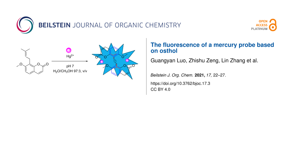
Abstract
The ability of osthol (OST) to recognize mercury ions in aqueous solution was studied using fluorescence, UV–vis spectrophotometry, mass spectrometry, and 1H NMR spectroscopy, and the recognition mechanism is discussed. The results showed that OST and Hg2+ can form a complex with a stoichiometric ratio of 1:1. The binding constant was 1.552 × 10⁵ L∙mol−1, having a highly efficient and specific selectivity for Hg2+. The fluorescence intensity of OST showed a good linear correlation with the Hg2+ concentration (6.0 × 10⁻⁵ to 24.0 × 10⁻⁵ mol∙L−1, R2 = 0.9954), and the detection limit of the probe was 5.04 × 10⁻⁸ mol∙L−1, which can be used for the determination of Hg2+ traces.

Graphical Abstract
Introduction
Mercury is a dangerous heavy-metal pollutant. Inorganic mercury (Hg2+) can be transformed into methyl mercury (MeHg+) by sulfate-reducing bacteria [1-3]. MeHg+ can accumulate in organisms through the food chain, resulting in serious and irreversible nerve damage. Therefore, it is very important to develop a highly sensitive and selective method for mercury detection. At present, the detection of mercury mainly includes atomic absorption and atomic emission spectrometry [4], inductively coupled plasma mass spectrometry [5,6], and capillary electrophoresis [7]. However, the application of these analytical methods in mercury detection is limited due to the complex sample pretreatment procedures, expensive instruments, and other factors [8,9]. Fluorescent-probe instruments have been widely used due to the advantages of the simple operation, low cost, and high sensitivity [10-12]. In recent years, many fluorescent molecular probes have been reported. Because Hg2+ has a very strong quenching effect on fluorescence, most of the fluorescent molecular Hg2+ probes are of the fluorescence-quenching type and are easily interfered with by other quenching processes [13-17]. Fluorescence-enhanced probes have received wide attention because of their enhanced fluorescence signal, which can better exclude the influence of instrument noise and other factors, reduce measurement errors, and thus have a higher sensitivity. Osthol (OST) is a coumarin compound extracted from the fruit of Cnidium monnieri (L.) cuss [18,19]. Modern pharmacological studies have shown that OST has antihypertension, anti-epilepsy [20], anti-arrhythmia, anti-fatty-liver-disease, antitumor [21], antiosteoporosis, and other effects [22]. As a new type of fluorescence probe [23], OST as a probe for the detection of mercury has not been reported. In this paper, OST was used as a fluorescent probe since the fluorescence intensity of OST at 406 nm increased significantly when Hg2+ was added. The probe had a high selectivity and sensitivity for Hg2+ recognition and can be used for the quantitative detection and monitoring of mercury ions in the environment.
Results and Discussion
OST fluorescence probe for Hg2+ identification
Selectivity of the fluorescent OST probe to metal ions
The specificity of a probe for metal ions is the key factor to evaluate the performance of fluorescent probes. As shown in Figure 1A, the selectivity of OST to common metal ions is monitored by the fluorescence spectrum (λex = 325 nm). OST exhibits a weak fluorescence intensity at λex = 406 nm. When adding different metal ions (Hg2+, Ca2+, Na+, Mg2+, Al3+, Cd2+, Cu2+, Pb2+, Ni2+, Co2+, Fe3+, Ag+, Cr3+, and other metal ions) in 10 times the equivalent concentration to the OST probe, only Hg2+ produced an obvious fluorescence enhancement (the intensity increased sharply by about 27 times), and the other metal ions had little effect on the fluorescence intensity of OST, which indicates that the probe had a high selectivity for Hg2+. At the same time, under irradiation with 365 nm ultraviolet light, it was found that the OST solution was colorless and transparent when metal ions other than mercury were added. Only after Hg2+ was added, it showed a bright blue fluorescence (Figure 1B). These obvious changes in the color and fluorescence intensity indicated that OST had a good Hg2+ recognition effect and achieved the visual detection of Hg2+.
![[1860-5397-17-3-1]](/bjoc/content/figures/1860-5397-17-3-1.png?scale=2.0&max-width=1024&background=FFFFFF)
Figure 1: (A) Fluorescence spectra of OST (c = 3.0 × 10⁻⁵ mol∙L−1) upon the addition of various metal ions. (B) Fluorescent color change under ultraviolet light at 365 nm upon adding Hg2+ ions. 1: OST + Hg2+ solution, 2: OST + other ions, and 3: OST solution.
Figure 1: (A) Fluorescence spectra of OST (c = 3.0 × 10⁻⁵ mol∙L−1) upon the addition of various metal ions. (...
Influence of coexisting ions on the detection of Hg2+
To investigate the interference ability of coexisting ions with the determination of Hg2+, a coexisting ion experiment was carried out (Figure 2) [24]. The fluorescence intensity was measured by adding 1.2 × 10⁻⁴ mol∙L−1 of various metal ions to the OST–Hg2+ (c = 3.0 × 10⁻⁵ mol∙L−1) probe system. It is found that Fe3+ had a certain fluorescence quenching effect on the system due to the paramagnetism [25], but the fluorescence quenching effect was not enough to affect the recognition of mercury ions by the probe, and the presence of other coexisting metal ions did not affect the fluorescence spectrum change of the OST–Hg2+ system.
![[1860-5397-17-3-2]](/bjoc/content/figures/1860-5397-17-3-2.png?scale=2.0&max-width=1024&background=FFFFFF)
Figure 2: Metal ion selectivity of OST–Hg2+ (c = 3.0 × 10⁻⁵ mol∙L−1) in the presence of 1.2 × 10⁻⁴ mol∙L−1 of various metal ions (H2O/CH3OH 97:3, v/v, λex = 325 nm).
Figure 2: Metal ion selectivity of OST–Hg2+ (c = 3.0 × 10⁻⁵ mol∙L−1) in the presence of 1.2 × 10⁻⁴ mol∙L−1 of...
Drawing the standard curve [26]
Figure 3A shows the fluorescence spectrum (λex = 325 nm) of the OST probe following interaction with Hg2+ at different concentrations. With the increase of the Hg2+ concentration, the fluorescence intensity at 406 nm showed an increase of the Hg2+ dependence. The fluorescence intensity reached the maximum when 1.0 equivalent of Hg2+ was added. When the molar Hg2+/OST ratio was 1:1, the fluorescence intensity increased by about 27 times, and a further increase of the Hg2+ concentration did not cause further changes in the fluorescence intensity (Figure 3B). The linear relationship between the magnitude of the increase in the fluorescence intensity and the concentration of the mercury ions was in the range of 6.0 × 10⁻⁵ to 24.0 × 10⁻⁵ mol∙L−1 (Figure 4). The linear regression equation was y = 664.91 ∙ x − 94.92, and the coefficient of determination was R2 = 0.9954. According to the formula for the limit of detection (LOD) = 3 ∙ σ/K (n = 7, where σ is the standard deviation and K is the slope of the calibration curve) [27], the detection limit of the OST probe for Hg2+ was 5.04 × 10⁻⁸ mol∙L−1.
![[1860-5397-17-3-3]](/bjoc/content/figures/1860-5397-17-3-3.png?scale=2.0&max-width=1024&background=FFFFFF)
Figure 3: Fluorescence spectra of OST (c = 3 × 10⁻⁵ mol∙L−1) in H2O/CH3OH 97:3, v/v in the presence of an increasing concentration of Hg2+ ions (λex = 325 nm). (A) n(Hg2+)/n(OST) = 0, 0.1, 0.2, 0.3, 0.4, 0.5, 0.6, 0.7, 0.8, 0.9, 1.0, 1.1, 1.2, 1.3, 1.4, 1.5, 1.6, 1.7, 1.8, 1.9, 2.0 (a→t). (B) Plot of the fluorescence intensity of OST at 406 nm.
Figure 3: Fluorescence spectra of OST (c = 3 × 10⁻⁵ mol∙L−1) in H2O/CH3OH 97:3, v/v in the presence of an inc...
![[1860-5397-17-3-4]](/bjoc/content/figures/1860-5397-17-3-4.png?scale=2.0&max-width=1024&background=FFFFFF)
Figure 4: The standard Job curve of mercury ions to OST (c = 3.0 × 10⁻⁵ mol∙L−1) at 406 nm (pH 7.0, H2O/CH3OH 97:3, v/v, λex = 325 nm).
Figure 4: The standard Job curve of mercury ions to OST (c = 3.0 × 10⁻⁵ mol∙L−1) at 406 nm (pH 7.0, H2O/CH3OH...
Impact of the pH value of the solution
The effects of the pH value on the fluorescence intensity of the system are shown in Figure S1, Supporting Information File 1. The fluorescence intensity of the OST probe was weak in the pH value range of 2–10. When Hg2+ was added, the fluorescence intensity of OST–Hg2+ was strong, and stable in the pH value range of 4–10. Especially at pH 4–8.0, the fluorescence intensity reached the maximum value. Therefore, we chose pH 4–8.0 as the best measurement conditions.
Effect of the solvent
Figure S2 in Supporting Information File 1 shows the effects of different solvents on the fluorescence intensity of OST and OST–Hg2+. The results showed that the fluorescence intensity of OST was relatively weak in the studied solvents, but the fluorescence signal of OST was significantly enhanced after adding Hg2+, and the fluorescence intensity in different solvents was not very different. Because Hg2+ is often detected in water systems, pH 7.0 and H2O/CH3OH 97:3, v/v was selected as the solvent system for the detection.
Study on the mechanism of the OST as a fluorescent Hg2+ probe
The binding ratio of the OST–Hg2+ complex
The stoichiometric ratio between OST and Hg2+ was determined using the Job method (constant mole variation), and the results are shown in Figure S3, Supporting Information File 1. The maximum UV absorption intensity was achieved at n(Hg2+)/n((OST) + (Hg2+)) = 0.5 at 325 nm, indicating that the binding ratio of the OST probe to Hg2+ was 1:1. Combined with mass spectrometry data (Figure 5), the fragment peak of ESIMS at m/z 892.5422 corresponded to [OST2–Hg2]+ (calculated as 892.5429), indicating that OST and Hg2+ formed a 2:2 complex.
![[1860-5397-17-3-5]](/bjoc/content/figures/1860-5397-17-3-5.png?scale=2.0&max-width=1024&background=FFFFFF)
Figure 5: Mass spectrum of the probe with Hg2+.
Figure 5: Mass spectrum of the probe with Hg2+.
1H NMR titration analysis of the mode of interaction between the probe and Hg2+
The chemical shift of the resonance peak between the OST probe and Hg2+ in a 1H NMR titration experiment was used to infer the binding mode of the probe and Hg2+. Figure 6 shows a 1H NMR titration of OST in the presence of Hg2+ at different concentrations. With the addition of Hg2+, the OST proton resonances shift. At n(Hg2+)/n(OST) = 1, the chemical shift of the proton signals did not change, indicating that the interaction ratio of OST and Hg2+ was 1:1. At this time, the signals of the protons Ha, Hb, Hc, Hd, and He shifted downfield by 0.02, 0.03, 0.06, 0.04, and 0.04 ppm, respectively. This was due to the formation of the OST–Hg2+ complex, and the oxygen atoms on the aromatic ring were complexed by Hg2+, and thus enhancing the electron absorption effect on the aromatic ring, reducing the electron cloud density, leading to the proton signal of the aromatic ring moving to a lower field. The signals of the protons Hf, Hh, and Hi were shifted to a higher field by 0.02, 0.35, and 0.18 ppm, respectively. The formation of the OST–Hg2+ complex decreased the distance between the two aromatic rings so that the protons Hf, Hh, and Hi on the branched chain were in the shielding region of the benzene ring and their signals shifted to a higher field. When the protons Hh and Hi of the two methyl groups were in the shielding region of the aromatic ring, the chemical shifts moved to a higher field. Because Hh is located in the central area of the shield and Hi is at the edge of the shield, the Δδ value of Hh is relatively large, and finally both moved to δ 1.35.
![[1860-5397-17-3-6]](/bjoc/content/figures/1860-5397-17-3-6.svg?scale=2.0&max-width=1024&background=FFFFFF)
Figure 6: 1H NMR titration spectra recorded for OST (c = 5 × 10⁻⁴ mol∙L−1) during the addition of different molar equivalents of Hg2+ (400 MHz, D2O, n(Hg2+)/n(OST) = 0 (A), 0.2 (B), 0.4 (C), 0.8 (D, E), 1.0 (F), 1.2 (G), pH 7.0, D2O/CH3OH 97:3, v/v).
Figure 6: 1H NMR titration spectra recorded for OST (c = 5 × 10⁻⁴ mol∙L−1) during the addition of different m...
Based on the 1H NMR and mass spectrometry data, combined with the analysis of the UV–vis spectrum, the interaction mode between OST and Hg2+ is suggested as shown in Figure 7. Hg2+ and the oxygen atoms of the lactone ring and the methoxy group formed a stable complex, resulting in the enhanced fluorescence.
![[1860-5397-17-3-7]](/bjoc/content/figures/1860-5397-17-3-7.svg?scale=2.0&max-width=1024&background=FFFFFF)
Figure 7: The binding mode of OST and Hg2+.
Figure 7: The binding mode of OST and Hg2+.
Isothermal titration calorimetry analysis
A Hg2+ solution (2 × 10⁻³ mol∙L−1, 25 μL) was added to the 1 × 10⁻⁴ mol∙L−1 OST probe solution at 25 °C to record the exothermic binding isotherms [28], and the measurement results obtained are shown in Figure S4 and Table S1, Supporting Information File 1. By the formula ΔG = −R ⋅ T ⋅ ln(Ka) = ΔH − T ⋅ ΔS, with ΔH < 0 and T ⋅ ΔS < 0, ΔG was determined to be <0, and therefore the thermodynamic parameters show that the binding process between OST and Hg2+ was mainly determined by ΔH, with an action ratio of 1:1 and a K value of 1.552 × 10⁵ L ∙ mol−1.
Measurement of the fluorescence quantum yield
The fluorescence quantum yield of ʟ-tryptophan at an excitation wavelength of 293 nm was 0.14, which was taken as the standard (Figure S5, Supporting Information File 1) [29,30]. The integration range of ʟ-tryptophan was 280–540 nm and that of OST–Hg2+ was 300–540 nm, and the fluorescence quantum yield of the aqueous probe solution was measured to be 0.08. Although this quantum yield is not high, the selectivity and sensitivity for the fluorescence analysis are good. This can be better used to detect Hg2+ ions and has a certain analytical value.
Determination of Hg2+ in a water sample
Tap water was directly used for the determination of Hg2+ using the standard addition method, and the recovery rate of the sample was determined. The results are listed in Table 1.
Table 1: Determination of Hg2+ in tap water.a
|
Hg2+
(μmol∙L−1) |
detected (mean ± SD, μmol∙L−1) |
recovery rate
(%) |
RSD
(n = 3, %) |
| 0 | – | – | – |
| 12 | 11.95 | 99.58 | 2.63 |
| 16 | 15.86 | 99.13 | 0.31 |
| 18 | 17.67 | 98.17 | 0.65 |
aSD = standard deviation, RSD = relative standard deviation.
As shown in Table 1, the probe can not only identify Hg2+ but also effectively detect Hg2+ in tap water, which further proves that the applicational value of the probe is considerable.
Conclusion
In this paper, OST was used as the fluorescent probe to establish a new method for the determination of Hg2+. Within the range of 6.0 × 10⁻⁵ to 24.0 × 10⁻⁵ mol∙L−1, the change in the fluorescence intensity of the system had a good linear relationship with the concentration of mercury ions. The linear equation was y = 664.91 ∙ x − 94.92, the detection limit was 5.04 × 10⁻⁸ mol∙L−1, and the recognition process was the result of a chelation fluorescence enhancement mechanism. The method is simple to be executed, has a high sensitivity and good selectivity, and can be used to quantitatively detect and monitor mercury ions in the environment.
Supporting Information
| Supporting Information File 1: General information and descriptions of the methods. | ||
| Format: PDF | Size: 1.1 MB | Download |
References
-
Ullrich, S. M.; Tanton, T. W.; Abdrashitova, S. A. Crit. Rev. Environ. Sci. Technol. 2001, 31, 241–293. doi:10.1080/20016491089226
Return to citation in text: [1] -
Regnell, O.; Watras, C. J. Environ. Sci. Technol. 2019, 53, 4–19. doi:10.1021/acs.est.8b02709
Return to citation in text: [1] -
Li, Y.; Zhao, J.; Zhong, H.; Wang, Y.; Li, H.; Li, Y.-F.; Liem-Nguyen, V.; Jiang, T.; Zhang, Z.; Gao, Y.; Chai, Z. Environ. Sci. Technol. 2019, 53, 1844–1852. doi:10.1021/acs.est.8b03511
Return to citation in text: [1] -
Fong, B. M. W.; Siu, T. S.; Lee, J. S. K.; Tam, S. J. Anal. Toxicol. 2007, 31, 281–287. doi:10.1093/jat/31.5.281
Return to citation in text: [1] -
Yuan, H.; Gao, S.; Liu, X.; Li, H.; Günther, D.; Wu, F. Geostand. Geoanal. Res. 2004, 28, 353–370. doi:10.1111/j.1751-908x.2004.tb00755.x
Return to citation in text: [1] -
Rastogi, L.; Dash, K.; Arunachalam, J. J. Pharm. Anal. 2013, 3, 180–186. doi:10.1016/j.jpha.2013.02.004
Return to citation in text: [1] -
Macka, M.; Haddad, P. R. Electrophoresis 1997, 18, 2482–2501. doi:10.1002/elps.1150181237
Return to citation in text: [1] -
Du, J.; Hu, M.; Fan, J.; Peng, X. Chem. Soc. Rev. 2012, 41, 4511–4535. doi:10.1039/c2cs00004k
Return to citation in text: [1] -
Formica, M.; Fusi, V.; Giorgi, L.; Micheloni, M. Coord. Chem. Rev. 2012, 256, 170–192. doi:10.1016/j.ccr.2011.09.010
Return to citation in text: [1] -
Chen, X.; Pradhan, T.; Wang, F.; Kim, J. S.; Yoon, J. Chem. Rev. 2012, 112, 1910–1956. doi:10.1021/cr200201z
Return to citation in text: [1] -
Quang, D. T.; Kim, J. S. Chem. Rev. 2010, 110, 6280–6301. doi:10.1021/cr100154p
Return to citation in text: [1] -
Wang, X.; Ma, X.; Wen, J.; Geng, Z.; Wang, Z. Talanta 2020, 207, 120311. doi:10.1016/j.talanta.2019.120311
Return to citation in text: [1] -
YouMing, Z.; BingBing, S.; Peng, Z. Sci. China: Chem. 2013, 56, 612–618.
Return to citation in text: [1] -
Xu, Y.; Jiang, Z.; Xiao, Y.; Zhang, T.-T.; Miao, J.-Y.; Zhao, B.-X. Anal. Chim. Acta 2014, 807, 126–134. doi:10.1016/j.aca.2013.11.042
Return to citation in text: [1] -
Hennrich, G.; Walther, W.; Resch-Genger, U.; Sonnenschein, H. Inorg. Chem. 2001, 40, 641–644. doi:10.1021/ic000827u
Return to citation in text: [1] -
Yang, M.-H.; Thirupathi, P.; Lee, K.-H. Org. Lett. 2011, 13, 5028–5031. doi:10.1021/ol201683t
Return to citation in text: [1] -
Jing, S.; Zheng, C.; Pu, S.; Fan, C.; Liu, G. Dyes Pigm. 2014, 107, 38–44. doi:10.1016/j.dyepig.2014.03.023
Return to citation in text: [1] -
Pan, Z.; Fang, Z.; Lu, W.; Liu, X.; Zhang, Y. J. Ethnopharmacol. 2015, 175, 456–462. doi:10.1016/j.jep.2015.10.009
Return to citation in text: [1] -
Lian, Q. Zhongyaocai 2003, 26, 141–144.
Return to citation in text: [1] -
Luszczki, J. J.; Wojda, E.; Andres-Mach, M.; Cisowski, W.; Glensk, M.; Glowniak, K.; Czuczwar, S. J. Epilepsy Res. 2009, 85, 293–299. doi:10.1016/j.eplepsyres.2009.03.027
Return to citation in text: [1] -
Liang, H.-J.; Suk, F.-M.; Wang, C.-K.; Hung, L.-F.; Liu, D.-Z.; Chen, N.-Q.; Chen, Y.-C.; Chang, C.-C.; Liang, Y.-C. Chem.-Biol. Interact. 2009, 181, 309–315. doi:10.1016/j.cbi.2009.08.003
Return to citation in text: [1] -
Zhang, H.; Zhang, H.; Qu, C.; Bai, L.; Ding, L. Spectrochim. Acta, Part A 2007, 68, 474–477. doi:10.1016/j.saa.2006.12.013
Return to citation in text: [1] -
Zhao, H.; Song, F.; Zhang, J.; Wang, F.; Liu, J.; Liu, Y. J. Opt. Soc. Am. B 2013, 30, 2387–2392. doi:10.1364/josab.30.002387
Return to citation in text: [1] -
Lin, Q.; Fu, Y.-P.; Chen, P.; Wei, T.-B.; Zhang, Y.-M. Dyes Pigm. 2013, 96, 1–6. doi:10.1016/j.dyepig.2012.06.023
Return to citation in text: [1] -
Wang, H.-H.; Xue, L.; Yu, C.-L.; Qian, Y.-Y.; Jiang, H. Dyes Pigm. 2011, 91, 350–355. doi:10.1016/j.dyepig.2011.04.007
Return to citation in text: [1] -
Wen, J.; Geng, Z.; Yin, Y.; Zhang, Z.; Wang, Z. Dalton Trans. 2011, 40, 1984–1989. doi:10.1039/c0dt01262a
Return to citation in text: [1] -
Ding, S.-Y.; Dong, M.; Wang, Y.-W.; Chen, Y.-T.; Wang, H.-Z.; Su, C.-Y.; Wang, W. J. Am. Chem. Soc. 2016, 138, 3031–3037. doi:10.1021/jacs.5b10754
Return to citation in text: [1] -
O’Neill, M. A. A.; Gaisford, S. Int. J. Pharm. 2011, 417, 83–93. doi:10.1016/j.ijpharm.2011.01.038
Return to citation in text: [1] -
de Lucas, N. C.; Santos, G. L. C.; Gaspar, C. S.; Garden, S. J.; Netto-Ferreira, J. C. J. Photochem. Photobiol., A 2014, 294, 121–129. doi:10.1016/j.jphotochem.2014.08.009
Return to citation in text: [1] -
Wohlgemuth, M.; Bonačić-Koutecký, V.; Mitrić, R. J. Chem. Phys. 2011, 135, 054105. doi:10.1063/1.3622563
Return to citation in text: [1]
| 28. | O’Neill, M. A. A.; Gaisford, S. Int. J. Pharm. 2011, 417, 83–93. doi:10.1016/j.ijpharm.2011.01.038 |
| 29. | de Lucas, N. C.; Santos, G. L. C.; Gaspar, C. S.; Garden, S. J.; Netto-Ferreira, J. C. J. Photochem. Photobiol., A 2014, 294, 121–129. doi:10.1016/j.jphotochem.2014.08.009 |
| 30. | Wohlgemuth, M.; Bonačić-Koutecký, V.; Mitrić, R. J. Chem. Phys. 2011, 135, 054105. doi:10.1063/1.3622563 |
| 1. | Ullrich, S. M.; Tanton, T. W.; Abdrashitova, S. A. Crit. Rev. Environ. Sci. Technol. 2001, 31, 241–293. doi:10.1080/20016491089226 |
| 2. | Regnell, O.; Watras, C. J. Environ. Sci. Technol. 2019, 53, 4–19. doi:10.1021/acs.est.8b02709 |
| 3. | Li, Y.; Zhao, J.; Zhong, H.; Wang, Y.; Li, H.; Li, Y.-F.; Liem-Nguyen, V.; Jiang, T.; Zhang, Z.; Gao, Y.; Chai, Z. Environ. Sci. Technol. 2019, 53, 1844–1852. doi:10.1021/acs.est.8b03511 |
| 8. | Du, J.; Hu, M.; Fan, J.; Peng, X. Chem. Soc. Rev. 2012, 41, 4511–4535. doi:10.1039/c2cs00004k |
| 9. | Formica, M.; Fusi, V.; Giorgi, L.; Micheloni, M. Coord. Chem. Rev. 2012, 256, 170–192. doi:10.1016/j.ccr.2011.09.010 |
| 26. | Wen, J.; Geng, Z.; Yin, Y.; Zhang, Z.; Wang, Z. Dalton Trans. 2011, 40, 1984–1989. doi:10.1039/c0dt01262a |
| 7. | Macka, M.; Haddad, P. R. Electrophoresis 1997, 18, 2482–2501. doi:10.1002/elps.1150181237 |
| 27. | Ding, S.-Y.; Dong, M.; Wang, Y.-W.; Chen, Y.-T.; Wang, H.-Z.; Su, C.-Y.; Wang, W. J. Am. Chem. Soc. 2016, 138, 3031–3037. doi:10.1021/jacs.5b10754 |
| 5. | Yuan, H.; Gao, S.; Liu, X.; Li, H.; Günther, D.; Wu, F. Geostand. Geoanal. Res. 2004, 28, 353–370. doi:10.1111/j.1751-908x.2004.tb00755.x |
| 6. | Rastogi, L.; Dash, K.; Arunachalam, J. J. Pharm. Anal. 2013, 3, 180–186. doi:10.1016/j.jpha.2013.02.004 |
| 24. | Lin, Q.; Fu, Y.-P.; Chen, P.; Wei, T.-B.; Zhang, Y.-M. Dyes Pigm. 2013, 96, 1–6. doi:10.1016/j.dyepig.2012.06.023 |
| 4. | Fong, B. M. W.; Siu, T. S.; Lee, J. S. K.; Tam, S. J. Anal. Toxicol. 2007, 31, 281–287. doi:10.1093/jat/31.5.281 |
| 25. | Wang, H.-H.; Xue, L.; Yu, C.-L.; Qian, Y.-Y.; Jiang, H. Dyes Pigm. 2011, 91, 350–355. doi:10.1016/j.dyepig.2011.04.007 |
| 20. | Luszczki, J. J.; Wojda, E.; Andres-Mach, M.; Cisowski, W.; Glensk, M.; Glowniak, K.; Czuczwar, S. J. Epilepsy Res. 2009, 85, 293–299. doi:10.1016/j.eplepsyres.2009.03.027 |
| 22. | Zhang, H.; Zhang, H.; Qu, C.; Bai, L.; Ding, L. Spectrochim. Acta, Part A 2007, 68, 474–477. doi:10.1016/j.saa.2006.12.013 |
| 18. | Pan, Z.; Fang, Z.; Lu, W.; Liu, X.; Zhang, Y. J. Ethnopharmacol. 2015, 175, 456–462. doi:10.1016/j.jep.2015.10.009 |
| 19. | Lian, Q. Zhongyaocai 2003, 26, 141–144. |
| 23. | Zhao, H.; Song, F.; Zhang, J.; Wang, F.; Liu, J.; Liu, Y. J. Opt. Soc. Am. B 2013, 30, 2387–2392. doi:10.1364/josab.30.002387 |
| 13. | YouMing, Z.; BingBing, S.; Peng, Z. Sci. China: Chem. 2013, 56, 612–618. |
| 14. | Xu, Y.; Jiang, Z.; Xiao, Y.; Zhang, T.-T.; Miao, J.-Y.; Zhao, B.-X. Anal. Chim. Acta 2014, 807, 126–134. doi:10.1016/j.aca.2013.11.042 |
| 15. | Hennrich, G.; Walther, W.; Resch-Genger, U.; Sonnenschein, H. Inorg. Chem. 2001, 40, 641–644. doi:10.1021/ic000827u |
| 16. | Yang, M.-H.; Thirupathi, P.; Lee, K.-H. Org. Lett. 2011, 13, 5028–5031. doi:10.1021/ol201683t |
| 17. | Jing, S.; Zheng, C.; Pu, S.; Fan, C.; Liu, G. Dyes Pigm. 2014, 107, 38–44. doi:10.1016/j.dyepig.2014.03.023 |
| 10. | Chen, X.; Pradhan, T.; Wang, F.; Kim, J. S.; Yoon, J. Chem. Rev. 2012, 112, 1910–1956. doi:10.1021/cr200201z |
| 11. | Quang, D. T.; Kim, J. S. Chem. Rev. 2010, 110, 6280–6301. doi:10.1021/cr100154p |
| 12. | Wang, X.; Ma, X.; Wen, J.; Geng, Z.; Wang, Z. Talanta 2020, 207, 120311. doi:10.1016/j.talanta.2019.120311 |
| 21. | Liang, H.-J.; Suk, F.-M.; Wang, C.-K.; Hung, L.-F.; Liu, D.-Z.; Chen, N.-Q.; Chen, Y.-C.; Chang, C.-C.; Liang, Y.-C. Chem.-Biol. Interact. 2009, 181, 309–315. doi:10.1016/j.cbi.2009.08.003 |
© 2021 Luo et al.; licensee Beilstein-Institut.
This is an Open Access article under the terms of the Creative Commons Attribution License (https://creativecommons.org/licenses/by/4.0). Please note that the reuse, redistribution and reproduction in particular requires that the author(s) and source are credited and that individual graphics may be subject to special legal provisions.
The license is subject to the Beilstein Journal of Organic Chemistry terms and conditions: (https://www.beilstein-journals.org/bjoc/terms)