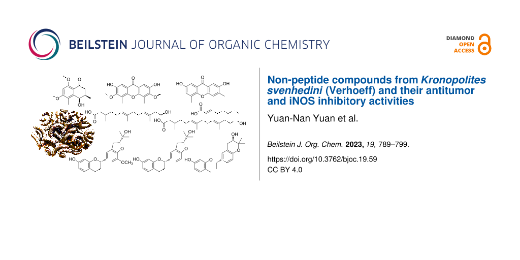
Abstract
Six new compounds, including a tetralone 1, two xanthones 2 and 3, a flavan derivative 4, and two nor-diterpenoids 7 and 8, accompanied by two known flavan derivatives 5 and 6 and a known olefine acid (9) were isolated from whole bodies of Kronopolites svenhedini (Verhoeff). The structures of the new compounds were determined by 1D and 2D nuclear magnetic resonance (NMR) and other spectroscopic methods, as well as computational methods. Selected compounds were evaluated for their biological properties against a mouse pancreatic cancer cell line and inhibitory effects on iNOS and COX-2 in RAW264.7 cells.

Graphical Abstract
Introduction
The millipede (class Diplopoda) is a pervasive arthropod in nature, functioning as a decomposer in forest ecosystems [1]. Most current research on millipedes centers around biological sciences and environmental sciences, such as community changes [2-4]. Limited studies on millipede chemical composition and biological activity have revealed the presence of antimicrobial peptides [5], defensive alkaloids [6], and defensive long chain alcohol acetates [7]. Historically, in China, numerous records documented the utilization of animals like arthropods for medicinal purposes. Millipedes hereby represent a traditional Chinese medicine (TCM) with anti-inflammatory, analgesic, stomach-soothing, and fatigue-relieving effects [8]. K. svenhedini (Diplopoda, Strongylosomidae) is a millipede species first described by Verhoeff in 1934 [9]. Based on literature [10], we know that K. svenhedini is the animal source of the TCM millipedes. In our studies of arthropods over the years, we have found that non-peptide small molecules play a significant role in chemical structures and biological activities [11-16].
In examining the chemical constituents of the millipede K. svenhedini, the focus was directed toward non-peptide small molecules, leading to the isolation of six new and three known compounds (Figure 1) from its extract. These structures were determined by 1D and 2D NMR spectra and the experimental and calculated electronic circular dichroism (ECD) spectra. The six new compounds have been named kronopoone A (1), kronopoiols A (2) and B (3), 5-O-methyldaphnegiralin C1 (4), and kronoponoids A (7) and B (8). Biological activity experiments were conducted with the isolated compounds, revealing that compounds 3–5 exhibited the expression of iNOS in a dose-dependent manner.
![[1860-5397-19-59-1]](/bjoc/content/figures/1860-5397-19-59-1.svg?scale=2.0&max-width=1024&background=FFFFFF)
Results and Discussion
Structural identification
Compound 1, a yellow gum, possesses the molecular formula C14H18O4 (six degrees of unsaturation), as deduced from its HRESIMS [M + H]+ ion peak at m/z 251.1274 (calcd for C14H19O4, 251.1278). The 1H NMR data (Table 1 and Figure S1 in Supporting Information File 1) display one aromatic proton [δH 7.02 (s, 1H, H-7)], two methoxy signals [δH 3.96 (s, 3H, H3-12) and 3.73 (s, 3H, H3-11)], and two methyl signals [δH 2.51 (s, 3H, H3-10) and 1.09 (d, J = 6.8 Hz, 3H, H3-9)]. The 13C NMR and DEPT spectra of compound 1 (Table 1 and Figure S2 in Supporting Information File 1) exhibit 14 resonances attributable to two methyl groups, two methoxy carbons, one methylene, three methines (one sp2 and one of them oxygenated), one ketone, and five sp2 carbons (two of them oxygenated). Some of these signals resemble those of 8-O-methylteratosphaerone B [17], suggesting compound 1 being an analogue, but with an additional methyl group on the benzene ring. The HMBC correlations (Figure 2 and Figure S5 in Supporting Information File 1) of H3-10/C-4 (δC 72.9, weak), C-4a (δC 136.1), C-5 (δC 124.5), C-6 (δC 148.5) disclosed that C-10 is connected to C-5 in compound 1. The coupling constant was used to determine the relative configuration of the cyclohexanone segment in 1. The small coupling constant (J3,4 = 3.0 Hz) indicated that H-3 and H-4 are on the same side of the ring, corroborated by the literature [18]. The absolute configuration of compound 1 was identified as 3R,4R in accordance with the experimental and calculated ECD spectra (Figure 3 and Figure S7 in Supporting Information File 1). As a result, the structure of 1 was defined and designated as kronopoone A.
Table 1: 1H (600 MHz) and 13C NMR (150 MHz) data of compound 1 (δ in ppm, J in Hz, methanol-d4).
| No. | δH (mult, J, amount) | δC mult | No. | δH (mult, J, amount) | δC mult |
| C-1 | 201.3 C | C-7 | 7.02 (s, 1H) | 110.8 CH | |
| C-2 |
2.64 (dd, J = 17.2, 10.3, 1H)
2.48 (dd, J = 17.2, 4.7, 1H) |
43.7 CH2 | C-8 | 158.0 C | |
| C-3 | 2.36 (m, 1H) | 35.8 CH | C-8a | 145.6 C | |
| C-4 | 4.69 (d, J = 3.0, 1H) | 72.9 CH | C-9 | 1.09 (d, J = 6.8, 3H) | 16.3 CH3 |
| C-4a | 136.1 C | C-10 | 2.51 (s, 3H) | 14.1 CH3 | |
| C-5 | 124.5 C | C-11 | 3.73 (s, 3H) | 60.7 CH3 | |
| C-6 | 148.5 C | C-12 | 3.96 (s, 3H) | 56.3 CH3 | |
![[1860-5397-19-59-2]](/bjoc/content/figures/1860-5397-19-59-2.svg?scale=2.0&max-width=1024&background=FFFFFF)
Figure 2: Key 1H–1H COSY, HMBC, and ROESY correlations of compounds 1–4, 7, and 8.
Figure 2: Key 1H–1H COSY, HMBC, and ROESY correlations of compounds 1–4, 7, and 8.
![[1860-5397-19-59-3]](/bjoc/content/figures/1860-5397-19-59-3.png?scale=2.0&max-width=1024&background=FFFFFF)
Figure 3: Calculated and experimental ECD spectra of compounds 1 and 4.
Figure 3: Calculated and experimental ECD spectra of compounds 1 and 4.
Compound 2, isolated as a brown solid, was showed to have the molecular formula C17H16O6 (ten degrees of unsaturation) on the basis of its HRESIMS [M + H]+ ion peak at m/z 317.1008 (calcd for C17H17O6, 317.1020). The 1H NMR data (Table 2 and Figure S8 in Supporting Information File 1) reveal an aromatic signal [δH 7.47 (s, 2H, H-1, H-8)], a methoxy signal [δH 3.95 (s, 6H, H3-11, H3-14)], and a methyl signal [δH 2.48 (s, 6H, H3-12, H3-13)]. The 13C NMR and DEPT spectra (Table 2 and Figure S9 in Supporting Information File 1) show that compound 2 is comprised of 9 carbons, including one methyl, one methoxy carbon, one sp2 methine, one ketone, and five sp2 carbons (three of them oxygenated). The 1H and 13C NMR data and the molecular formula indicated that the compound possesses the same two pentasubstituted benzene rings, suggesting an axially symmetric structure. The methoxy group is situated at C-3, as determined by the HMBC correlation (Figure 2 and Figure S12 in Supporting Information File 1) of H3-11/C-3 (δC 154.1). The hydroxy group and the methyl group are positioned at C-2 and C-4, respectively, as deduced by comparing the HMBC correlations of H-1/C-2 (δC 148.7), C-9a (δC 118.1), C-9 (δC 178.5), and H3-12/C-3, C-4 (δC 121.6), C-4a (δC 151.0), C-11 (δC 61.0, weak) with those in the literature [18-21]. These results suggest that the second benzene ring shares the same structure. By comparing the 1H and 13C chemical shifts with similar compounds [19-22], the NMR data implied the presence of C-8a–C-9–C-9a and C-4a–O–C-10a bonds in the structure of 2. Consequently, the structure of compound 2 was identified and named kronopoiol A.
Table 2: 1H (600 MHz) and 13C NMR (150 MHz) data of compounds 2 and 3 (δ in ppm, J in Hz, methanol-d4).
| No. | 2 | 3 | |||
| δH (mult, J, amount) | δC mult | δH (mult, J, amount) | δC mult | ||
| C-1 | 7.47 (s, 1H, overlap) | 108.3 CH | 7.47 (s, 1H) | 108.2 CH | |
| C-2 | 148.7 C | 153.8 C | |||
| C-3 | 154.1 C | 137.5 C | |||
| C-4 | 121.6 C | 7.39 (d, J = 1.0, 1H) | 120.5 CH | ||
| C-4a | 151.0 C | 151.6 C | |||
| C-5 | 121.6 C | 130.2 C | |||
| C-6 | 154.1 C | 7.16 (dd, J = 3.1, 1.0, 1H) | 126.1 CH | ||
| C-7 | 148.7 C | 154.4 C | |||
| C-8 | 7.47 (s, 1H, overlap) | 108.3 CH | 7.38 (d, J = 3.1, 1H) | 107.2 CH | |
| C-8a | 118.1 C | 122.5 C | |||
| C-9 | 178.5 C | 179.1 C | |||
| C-9a | 118.1 C | 120.5 CH | |||
| C-10a | 151.0 C | 150.1 C | |||
| C-11 | 3.95 (s, 3H, overlap) | 61.0 CH3 | 2.37 (s, 3H) | 17.1 CH3 | |
| C-12 | 2.48 (s, 3H, overlap) | 9.2 CH3 | 2.52 (s, 3H) | 15.8 CH3 | |
| C-13 | 2.48 (s, 3H, overlap) | 9.2 CH3 | |||
| C-14 | 3.95 (s, 3H, overlap) | 61.0 CH3 | |||
Compound 3, a brown solid, was assigned the molecular formula C15H12O4 (ten degrees of unsaturation), by analysis of its HRESIMS [M + H]+ ion peak at m/z 257.0802 (calcd for C15H13O4, 257.0808). The 1H NMR data (Table 2 and Figure S14 in Supporting Information File 1) show a typical AB spin system [δH 7.47 (s, 1H, H-1), 7.39 (d, J = 1.0 Hz, 1H, H-4)], which corresponds to a 1,2,4,5-tetrasubstituted benzene substructure. Additional aromatic proton signals [δH 7.38 (d, J = 3.1 Hz, 1H, H-8) and δH 7.16 (dd, J = 3.4, 1.0 Hz, 1H, H-6)] suggest the presence of a 1,2,3,5-tetrasubstituted benzene substructure. The 13C NMR and DEPT spectra of 3 (Table 2 and Figure S15 in Supporting Information File 1) exhibit 15 resonances classified into two methyls, four sp2 methines, one ketone, and eight sp2 carbons (four of them oxygenated). The two methyl groups are positioned at C-3 and C-5, as deduced by the HMBC correlations (Figure 2 and Figure S18 in Supporting Information File 1) of H3-11/C-2 (δC 153.8), C-3 (δC 137.5), C-4 (δC 120.5) and H3-12/C-5 (δC 130.2), C-6 (δC 126.1), C-10a (δC 150.1), C-8a (δC 61.0, weak). By comparing the HMBC correlations of H-1/C-2, C-9a (δC 120.5), C-9 (δC 179.1), and H-8/C-7 (δC 154.4), C-9 (δC 179.1) with those in the literature [18-22], it was concluded that the hydroxy groups are positioned at C-2 and C-7, respectively. A comparison between compounds 2 and 3 revealed that both possess C-8a–C-9–C-9a and C-4a–O–C-10a bonds. As a result, the structure of 3 is defined and named as kronopoiol B.
Compound 4 was obtained as a brown solid. The molecular formula of it was determined to be C21H24O5 (ten degrees of unsaturation) deduced from its HRESIMS [M + H]+ ion peak at m/z 357.1680 (calcd for C21H25O5, 357.1697). The 1H NMR data (Table 3 and Figure S20 in Supporting Information File 1) show three typical aromatic signals [δH 6.86 (m, 1H, H-5, overlap), 6.31 (dd, J = 8.2, 2.4 Hz, 1H, H-6), and 6.26 (d, J = 2.4 Hz, 1H, H-8)], suggesting the presence of a 1,2,4-trisubstituted benzene substructure. Additionally, two aromatic signals at δH 6.86 (m, 1H, H-2', overlap) and δH 6.84 (s, 1H, H-6') are observed in the 1H NMR spectrum, indicating the presence of a 1,3,4,5-tetrasubstituted benzene substructure. The 13C NMR and DEPT spectra of 4 (Table 3 and Figure S21 in Supporting Information File 1) contain 21 resonances ascribed to two methyls, one methoxy carbon, three methylenes, seven methines (five sp2 and two of them oxygenated), one oxygenated carbon, and seven sp2 carbons (four of them oxygenated). Based on this information, compound 4 was deduced to be similar to daphnegiralin C1 [23], with both sharing the same 7-hydroxyflavan skeleton. The distinction in compound 4 is an additional methoxy group, which is connected to C-5' as supported by the HMBC correlation (Figure 2 and Figure S24 in Supporting Information File 1) of 5'-OCH3 (δH 3.85)/C-5' (δC 136.1). Two asymmetric carbon centers are present at C-2 and C-2″ in compound 4. According to the literature [22], the absolute configuration at C-2 for 4 was assigned as S, from the Cotton effects in its ECD curve (Figure S26 in Supporting Information File 1) [283 nm (Δε −0.71)]. The absolute configuration of 4 was determined as 2S,2″R based on the comparison of the experimental and ECD spectra (Figure 3 and Figure S26 in Supporting Information File 1). Consequently, the structure of 4 is defined and named 5-O-methyldaphnegiralin C1.
Table 3: 1H (500 MHz) and 13C NMR (150 MHz) data of 4 (δ in ppm, J in Hz, methanol-d4).
| No. | δH (mult, J, amount) | δC mult | No. | δH (mult, J, amount) | δC mult |
| C-2 | 4.92 (m, 1H) | 79.2 CH | C-3' | 129.8 C | |
| C-3 |
2.12 (m, 1H)
2.00 (m, 1H) |
31.6 CH2 | C-4' | 149.2 C | |
| C-4 |
2.86 (ddd, J = 16.5, 11.3, 5.7, 1H)
2.67 (m, 1H) |
25.5 CH3 | C-5' | 145.2 CH | |
| C-5 | 6.86 (m, 1H, overlap) | 131.0 CH | C-6' | 6.84 (s, 1H) | 111.4 CH |
| C-6 | 6.31 (dd, J = 8.2, 2.4, 1H) | 109.1 CH | C-1'' | 3.19 (m, 2H) | 32.0 CH2 |
| C-7 | 157.6 C | C-2'' | 4.63 (t, J = 9.0, 1H) | 91.1 CH | |
| C-8 | 6.26 (d, J = 2.4, 1H) | 104.1 CH | C-3'' | 72.5 C | |
| C-9 | 157.1 C | C-4'' | 1.24 (s, 3H) | 25.5 CH3 | |
| C-10 | 114.3 C | C-5'' | 1.26 (s, 3H) | 25.0 CH3 | |
| C-1' | 136.5 C | 5'-OCH3 | 3.85 (s, 3H) | 56.8 CH3 | |
| C-2' | 6.86 (m, 1H, overlap) | 116.2 CH | |||
Compound 7, a light yellow gum, has the molecular formula C16H28O3 (three degrees of unsaturation) as determined by its HRESIMS [M + H]+ ion peak at m/z 269.2116 (calcd for C16H29O3, 269.2111). The 1H NMR spectrum (Table 4 and Figure S27 in Supporting Information File 1) displays two olefinic protons [δH 5.18 (t, J = 7.0 Hz, 1H, H-10) and 5.12 (t, J = 7.0 Hz, 1H,H-6)], one oxymethylene group [δH 3.59 (t, J = 7.1 Hz, 2H, H2-13)], and three methyl signals [δH 1.63 (s, 3H, H3-16), 1.61 (s, 3H, H3-15), and 0.96 (s, 3H, H3-14)]. The 13C NMR and DEPT spectra of compound 7 (Table 4 and Figure S28 in Supporting Information File 1) contain 16 resonances attributable to three methyls, seven methylenes (one of them oxygenated), three methines (two sp2), one carbonyl carbon, and two sp2 quaternary carbons. The 1H–1H COSY spectrum (Figure 2 and Figure S29 in Supporting Information File 1) of compound 7 disclosed the existence of correlations of H2-2 (δ 2.29, 2.08)/H-3 (δH 1.91, 1.93)/H2-4 (δH 1.38, 1.24)/H2-5 (δH 2.03, 2H)/H-6 (δH 5.12), H-3 (δH 1.93)/H2-15 (δH 0.96), H2-8 (δH 2.03, 2H)/H2-9 (δH 2.11, 2H)/H-10 (δH 5.18), and H2-12 (δH 2.20, 2H)/H2-13 (δH 3.59, 2H), which revealed three partial structures a (C-2 to C-6), b (C-8 to C-10), and c (C-12 to C-13). The partial structures a and b were connected to C-7 by the correlations of H3-16 (δH 1.61)/C-6 (δC 125.6), C-7 (δC 136.0), C-8 (δC 40.7), and H-8/C-6, C-7 in the HMBC spectrum (Figure 2 and Figure S31 in Supporting Information File 1). The partial structures b and c were connected to C-11, as confirmed by the HMBC correlations of H3-17 (δH 1.63)/C-10 (δC 127.4), C-11 (δC 132.9), C-12 (δC 43.8), and H-12/C-10, C-11. The presence of a conjugated carboxylic acid was verified by the HMBC correlation of H2-2 to C-1 (δC 177.1). Concerning the geometry of 7, the ROESY correlation (Figure 2 and Figure S32 in Supporting Information File 1) of H-10/H2-12 disclosed that the ∆10,11 configuration was E. However, the overlapping signals of H2-5 and H2-8 made it difficult to determine the geometry of the ∆6,7 in the same manner. By comparing the 1H and 13C chemical shifts with those of similar compounds [24-28], the geometry of the ∆6,7 was determined to be E. The configuration at C-3 remains undetermined due to its long carbon chain. As a consequence, the structure of compound 7, named kronoponoid A, was determined to be as showed in Figure 1.
Table 4: 1H (500 MHz) and 13C NMR (150 MHz) data of compounds 7 and 8 (δ in ppm, J in Hz, methanol-d4).
| No. | 7 | 8 | |||
| δH (mult, J, amount) | δC mult | δH (mult, J, amount) | δC mult | ||
| C-1 | 177.1 C | 182.5 C | |||
| C-2 |
2.29 (dd, J = 14.8, 6.0, 1H)
2.08 (m, 1H) |
42.6 CH2 |
2.19 (m, 1H)
1.94 (m, 1H, overlap) |
47.3 CH2 | |
| C-3 | 1.93 (m, 1H) | 31.1 CH | 1.94 (m, 1H, overlap) | 32.2 CH | |
| C-4 |
1.38 (m, 1H)
1.24 (m, 1H) |
37.8 CH2 |
1.38 (m, 1H)
1.19 (m, 1H) |
38.5 CH2 | |
| C-5 | 2.03 (m, 2H, overlap) | 26.3 CH2 |
2.02 (m, 1H)
1.98 (m, 1H, overlap) |
26.6 CH2 | |
| C-6 | 5.12 (t, J = 7.0, 1H) | 125.6 C | 5.16 (m, 1H, overlap) | 126.0 CH | |
| C-7 | 136.0 C | 135.6 C | |||
| C-8 | 2.03 (m, 2H, overlap) | 40.7 CH2 | 1.98 (m, 2H, overlap) | 40.9 CH2 | |
| C-9 | 2.11 (m, 2H, overlap) | 27.6 CH2 | 2.08 (m, 2H, overlap) | 27.9 CH2 | |
| C-10 | 5.18 (t, J = 7.0, 1H) | 127.4 C | 5.16 (m, 1H, overlap) | 126.0 CH | |
| C-11 | 132.9 C | 135.2 C | |||
| C-12 | 2.20 (m, 2H, overlap) | 43.8 CH2 | 2.08 (m, 2H, overlap) | 36.8 CH2 | |
| C-13 | 3.59 (t, J = 7.1, 2H) | 61.9 CH2 | 1.75 (m, 2H) | 29.0 CH2 | |
| C-14 | 3.97 (t, J = 6.6, 2H) | 68.9 CH2 | |||
| C-15 | 0.96 (s, 3H) | 20.0 CH3 | 0.94 (d, J = 6.2, 3H) | 20.3 CH3 | |
| C-16 | 1.61 (s, 3H) | 16.0 CH3 | 1.61 (s, 3H, overlap) | 16.1 CH3 | |
| C-17 | 1.63 (s, 3H) | 16.3 CH3 | 1.61 (s, 3H, overlap) | 16.0 CH3 | |
Compound 8, a light yellow gum, possesses the molecular formula C17H30O3 (three degrees of unsaturation) deduced from its HRESIMS [M + H]+ ion peak at m/z 283.2268 (calcd for C17H31O3, 283.2268). The 1H NMR data (Table 4 and Figure S34 in Supporting Information File 1) display two olefinic protons [δH 5.16 (m, 2H, H-6, H-10)], one oxymethylene group [δH 3.97 (t, J = 6.6 Hz, 2H, H2-14)], and three methyl signals [δH 1.61 (s, 6H, H3-16, H3-17), and 0.94 (d, J = 6.2 Hz, 3H, H3-15)]. The 13C NMR and DEPT spectra (Table 4 and Figure S35 in Supporting Information File 1) show that this substance contains 17 resonances, including three methyls, eight methylenes (one of them oxygenated), three methines (two sp2), one carbonyl carbon, and two sp2 quaternary carbons. A comparison of the NMR data of compound 8 (Table 4) with that of compound 7, indicated that both compounds possess the same general skeleton structure, with the only difference being an additional methine group in compound 8. The 1H–1H COSY correlations (Figure 2 and Figure S36 in Supporting Information File 1) of H2-12 (δH 1.98, 2H)/H2-13 (δH 2.08, 2H)/H2-14 (δH 3.97, 2H) and the HMBC correlations (Figure 2 and Figure S38 in Supporting Information File 1) of H-12/C-10 (δC 126.0), C-11 (δC 135.2), and H-13/C-11 revealed the structure of C-12 to C-14, which differs from 7. Owing to the overlapping signals between H2-5 and H2-8, H2-9 and H2-12, the geometry of the double bonds was determined as 6E,10E through a comparison of the 1H and 13C chemical shifts with similar compounds [24-28]. The configuration at C-3 remains undetermined due to its long carbon chain. Thus far, the structure of compound 8 was identified as shown in Figure 1 and named as kronoponoid B.
Of note, the structures of compounds 2–4 are common in plants but rare in animals. Whether these compounds originate from plants or animals so far remains unknown.
In addition to the above mentioned compounds, three known compounds were identified as daphnegiralin C1 (5) [23], daphnegiranol C1 (6) [29], and (E)-oct-2-enoic acid (9) [30] by comparing their spectroscopic data with those in the literature.
Biological evaluation
To explore the bioactive potential of the isolated compounds, cytotoxic and anti-inflammatory properties were evaluated. In particular, a mouse pancreatic cancer cell line (Panc02-h7-GP-GFP) was used to determine cytotoxicity. Additionally, LPS-induced pro-inflammatory expression of iNOS and COX-2 in RAW264.7 cells was evaluated. Antitumor activity of compounds 1–5, 7, and 8 was assessed via a cell proliferation assay using Panc02-h7-GP-GFP cells. Unfortunately, none of the compounds did inhibit the proliferation of Panc02-h7-GP-GFP cells at a concentration of 20 μM (Figure S41 in Supporting Information File 1). On the other hand, an enhancement of CD8+ T cells was investigated at corresponding concentrations of compounds 2–5. Regrettably, no enhancement of CD8+ T cells was observed (Figure S43 in Supporting Information File 1). To examine the toxicity of compounds 1–5, 7, and 8, the CCK-8 assay was employed to detect the viability of RAW264.7 cells. The results indicate that the compounds did not exhibit significant toxicity toward RAW264.7 cells at the utilized concentrations (Figure 4A and B). Meanwhile, compounds 1‒5, 7, and 8 were evaluated for their anti-inflammatory activity against pro-inflammatory expression of iNOS and COX-2. The results demonstrated that compounds 3–5 exhibited inhibitory effects on LPS-induced iNOS in RAW264.7 cells in a dose-dependent manner (Figure 4C, D and E). However, all tested compounds were inactive against LPS-induced COX-2 in RAW264.7 cells.
![[1860-5397-19-59-4]](/bjoc/content/figures/1860-5397-19-59-4.png?scale=2.0&max-width=1024&background=FFFFFF)
Figure 4: Biological evaluation. Compounds 3‒5 inhibited the expression of LPS-induced pro-inflammatory expression of iNOS in RAW264.7 cells. Cells were treated with corresponding concentrations of compounds or DMSO for 2 h before being exposed to 1 μg/mL LPS for 12 h. (A) and (B), the proliferation of RAW264.7 cells in response to compounds at 20 and 40 μM was assessed by the CCK-8 assay. Data represent mean ± SEM values of three experiments. *p < 0.05, ** p < 0.01, and *** p < 0.001 compared with the CON group (DMSO alone). (C)‒(E), Western blotting was used to determine the protein levels of iNOS and COX-2, using GAPDH as a control and dexamethasone (DEX) as a positive reference drug.
Figure 4: Biological evaluation. Compounds 3‒5 inhibited the expression of LPS-induced pro-inflammatory expre...
Conclusion
Six new and three known non-peptide small molecules were isolated from the millipede Kronopolites svenhedini (Verhoeff), and their structures were characterized using spectroscopic and ECD calculation methods. Biological evaluation of compounds 3‒5 showed inhibitory activities against LPS-induced iNOS in RAW264.7 cells. These findings contribute new insights into the chemistry and biological activity of arthropod-derived non-peptide small molecules.
Experimental
General
1D and 2D NMR spectra were acquired using Bruker AV-500 and AV-600 spectrometers (Bruker), with tetramethylsilane (TMS) used as an internal standard. HRESIMS data were obtained using a Shimazu LC-20 CE AB SCIEX QTOF X500R MS spectrometer (Shimadzu Corporation, Tokyo, Japan). Optical rotations (ORD) were collected with a Horiba SEPA-300 polarimeter. Ultraviolet (UV) and circular dichroism (CD) spectra were carried out on a Jasco J-815 CD spectrometer (JASCO). Column chromatography (CC) was performed using macroporous adsorbent resin Amberlite TM XAD 16N (particle size 20–60 mesh, Rohm and Haas Company), MCI gel CHP 20P (particle size 75–150 μm, Mitsubishi Chemical Industries, Japan), RP-18 (particle size 40–60 μm; Daiso Co.), C-18 silica gel (particle size 40–60 μm; Daiso Co., Japan), Sephadex LH-20 (Amersham Biosciences), and YMC gel ODS-A-HG (particle size 40–60 μm; YMC Co. Japan). A Saipuruisi chromatograph with a semi-preparative high-pressure infusion pump (SP-5030) and a semi-preparative UV–vis dual wavelength detector (UV200) were utilized for RP–HPLC. A YMC-Pack ODS-A column (250 mm × 20 mm, i.d., S-5 μm) was employed for preparative HPLC, while three columns (a YMC-Pack ODS-A column (250 mm × 10 mm, i.d., 5 μm), a Stabllity 100 C30 column (25 mm × 10 mm, i.d., 5 μm), and an Inetex-Biphenyl 100A column (250 mm × 10 mm, i.d., 5 μm)) were used for semi-preparative HPLC.
Arthropod material
The dried arthropod bodies of Kronopolites svenhedini (Verhoeff) were purchased from Qunkang Pharmaceutical Co. in Anhui Province, PR China, in July 2021. The voucher specimen of this material (CHYX-0674) has been deposited at the School of Pharmaceutical Sciences, Health Science Center, Shenzhen University, PR China.
Extraction and isolation
The dried and powdered bodies of Kronopolites svenhedini (Verhoeff) (49 kg) were extracted using 50% EtOH (4 × 120 L, 24 h each time) to yield a crude extract. This extract (5.2 kg) was partitioned into six fractions (Fr. A–Fr. F) utilizing a macroporous adsorbent resin column eluted with gradient aqueous MeOH (0–100%). Fr. E (180 g) was separated into ten parts (Fr. E1–Fr. E9, Fr. EA) using an MCI gel CHP 20P column (MeOH/H2O, 60–100%). Fr. E2 (2.0 g) was further divided into six portions (Fr. E21–Fr. E26) by Sephadex LH-20 (MeOH/H2O, 70%). Fr. E25 (248.6 mg) was subjected to preparative HPLC (MeOH/H2O (0.04% TFA), 50–100%, flow rate: 10 mL min−1) to obtain six fractions (Fr. E251–Fr. E256). Fr. E254 was concentrated under reduced pressure to yield compound 9 (20.00 mg). Additionally, Fr. E255 (51.8 mg) was further purified using semi-preparative HPLC (C30, MeOH/H2O, 25%, flow rate: 3 mL min−1) to yield compound 8 (16.23 mg, tR = 25.04 min). Fr. E5 (9.0 g) was separated into eleven parts (Fr. E51–Fr. E59, Fr. E5A–B) by Sephadex LH-20 (MeOH/H2O, 70%). Fr. E53 (4.26 g) was divided into eleven portions (Fr. E531–Fr. E539, Fr. E53A, Fr. E53B) by using an MCI gel CHP 20P column (MeOH/H2O, 10–100%). Subsequently, Fr. E537 (354.8 mg) was divided into three parts (Fr. E5371–Fr. E5373) by Sephadex LH-20 (MeOH). Fr. E5373 (216.6 mg) was further fractionated into eight parts by a silica gel column (PE/EtOAc 2:1–1:1, to DCM/MeOH 20:1–1:1), and Fr. E53732 (63.8 mg) was further purified using semi-preparative HPLC (ODS-A, MeCN/H2O (0.04% TFA), 55%, flow rate: 3 mL min−1) to afford compound 7 (15.70 mg, tR = 14.05 min). Fr. E54 (415.3 mg) was separated into twelve fractions (Fr. E541–Fr.E549, Fr. E54A–Fr.E54C) using an MCI gel CHP 20P column (MeOH/H2O, 30–100%), followed by semi-preparative HPLC (ODS-A, MECN/H2O (0.04% TFA), 28%, flow rate: 3 mL min−1) to obtain compound 1 (1.32 mg, tR = 27.89 min). Fr. E8 (13.1 g) was partitioned into five portions (Fr. E81–Fr. E85) by Sephadex LH-20 (MeOH). Fr. E84 (207.3 mg) was divided into thirteen portions (Fr. E841–Fr. E849, Fr. E84A–Fr. E84D) using an ODS-A-HG column (MeOH/H2O, 30–100%). Fr. E846 (21.4 mg) was purified via semi-preparative HPLC (C30, MeCN/H2O (0.04% TFA), 42%, flow rate: 3 mL min−1) to afford compound 4 (0.97 mg, tR = 51.45 min). Fr. E847 (21.4 mg) was purified through semi-preparative HPLC (ODS-A, MeCN/H2O (0.04% TFA), 40%, flow rate: 3 mL min−1) to afford compound 5 (1.21 mg, tR = 56.80 min) and compound 6 (1.16 mg, tR = 61.32 min). Fr. E85 (686.7 mg) was separated into ten fractions (Fr. E851–Fr. E859, Fr. E85A) using an RP-18 column (MeOH/H2O, 30–100%). Fr. E856 (31.3 mg) was purified using semi-preparative HPLC (Inetex-Biphenyl, MECN/H2O (0.04% TFA), 38%, flow rate: 3 mL min−1) to afford compounds 3 (0.99 mg, tR = 25.33 min) and 2 (0.76 mg, tR = 30.38 min).
Compound characterization
Kronopoone A (1): yellow gum; [α]D25 +9.38 (c 0.32, MeOH); UV (MeOH) λmax, nm (log ε): 208 (2.92), 229 (2.88), 275 (2.78); ECD (MeOH) λ, nm (Δε): 210 (−1.88), 231 (+0.34), 251 (+0.02), 273 (−0.41), 327 (+0.39); HRESIMS (m/z): [M + H]+ calcd for C14H19O4, 251.1278; found, 251.1274; 1H and 13C NMR data, see Table 1.
Kronopoiol A (2): brown solid; UV (MeOH) λmax, nm (log ε): 202 (3.56), 242 (3.58), 280 (3.13), 324 (3.15), 367 (2.89); HRESIMS (m/z): [M + H]+ calcd for C17H17O6, 317.1020; found 317.1008 ; 1H and 13C NMR data, see Table 2.
Kronopoiol B (3): brown solid; UV (MeOH) λmax, nm (log ε): 201 (3.18), 241 (3.44), 260 (3.22), 324 (2.73), 378 (2.69); HRESIMS (m/z): [M + H]+ calcd for C15H13O4, 257.0808; found, 257.0802 ; 1H and 13C NMR data, see Table 2.
5-O-Methyldaphnegiralin C1 (4): brown solid; [α]D25 +30.77 (c 0.26, MeOH); UV (MeOH) λmax, nm (log ε): 207 (3.92), 280 (3.14); ECD (MeOH) λ, nm (Δε): 208 (+15.19), 237 (−1.90), 250 (+0.23), 283 (−0.71); HRESIMS (m/z): [M + H]+ calcd for C21H25O5, 357.1697; found, 357.1680; 1H and 13C NMR data, see Table 3.
Kronoponoid A (7): light yellow gum; [α]D25 +7.50 (c 0.40, MeOH); UV (MeOH) λmax, nm (log ε): 202 (3.68); HRESIMS (m/z): [M + H]+ calcd for C16H29O3, 269.2111; found, 269.2116 ; 1H and 13C NMR data, see Table 4.
Kronoponoid B (8): light yellow gum; [α]D25 +2.50 (c 0.40, MeOH); UV (MeOH) λmax, nm (log ε): 202 (3.81); HRESIMS (m/z): [M + H]+ calcd for C17H31O3, 283.2268; found, 283.2268 ; 1H and 13C NMR data, see Table 4.
Computational methods
The CONFLEX 7 searches, considering the Molecular Merck force field (MMFF94) and DFT/TDDFT calculations, were performed for model compounds (3R,4R)-1, (3S,4S)-1, (2S,2″R)-4, (2S,2″S)-4, (2R,2″S)-4, and (2R,2″R)-4 using the Spartan'14 software package and the Gaussian 09 program package. The ECD calculations of the predominant conformers (80%) were conducted using DFT calculations at the B3LYP/6-311G(d,p) level of theory. The program SpecDis 1.62 was used to generate the CD spectra [31].
Biological evaluation
Antitumor assay
Panc02-h7-GP-GFP cells (derived from the transformation of mouse pancreatic cancer cell line Panc02-h7) were maintained at 37 °C in a 5% CO2 atmosphere using high-glucose DMEM (GIBCO, USA) containing 10% fetal bovine serum (FBS, GIBCO, USA), 100 U/mL penicillin, 10 μg/mL streptomycin, and 10 μg/mL puromycin. Cells were seeded at a density of 5000 cells/well in 96-well plates under the same incubation conditions. Following overnight culture, cells were pretreated with compounds at a 20 μM concentration or DMSO for 48 h, with gemcitabine as a positive reference drug. Subsequently, Cell Count Kit-8 (CCK-8, MCE, USA) was added to each well at a 10 μL concentration for 2 h. The absorbance at 450 nm was measured using a microplate reader (TECAN, Switzerland).
Antitumor activity assay of CD8+ T cells in vitro
This assay, along with the associated experimental procedures, received approval from the Institutional Animal Care and Use Committee of Shenzhen University Health Science Center and the Animal Experimentation Ethics Committee of Shenzhen University Health Science Center (AEWC-202300026). All animal housing and handling adhered to the research ethics guidelines set forth by the Institutional Animal Care and Use Committee of Shenzhen University Health Science Center and the Animal Experimentation Ethics Committee of Shenzhen University Health Science Center.
Panc02-h7-GP-GFP cells were treated with trypsin (0.25%, Sigma) and resuspended in PBS (GIBCO, USA) following centrifugation. The cell suspension was then injected into the pancreas of mice (1 × 10⁶ cells per mouse). After 14 days, tumors were excised, digested with a digestion solution, ground, and centrifuged to produce a cell suspension. Lymphocytes were isolated from the cell suspension using the Percoll method and were further enriched (via negative selection) to obtain naive CD8+ T cells. The enrichment effect and phenotype of the CD8+ cells were detected by flow cytometry (BD, USA). CD8+ T cells and Panc02-h7-GP-GFP cells were co-cultured with the corresponding concentrations of compounds or DMSO for 18 h. Fluorescence intensity was measured using a microplate reader (emission at 476 nm, excitation at 514 nm).
Anti-inflammatory assay
RAW264.7 (a mouse macrophage cell line) cells were incubated in high-glucose DMEM (GIBCO, USA) containing 10% fetal bovine serum (FBS, GIBCO, USA), 100 U/mL penicillin, and 100 μg/mL streptomycin. Cells were maintained in an atmosphere of 5% CO2 at 37 °C and subsequently plated in 96-well plates at a concentration of 2 × 10⁴ cells/mL under the same incubation conditions. Following overnight incubation, the cells were treated with the corresponding concentration of the compound or DMSO for 24 h. CCK-8 (Beyotime, China) solution was added and incubated for 1 h. The absorbance of the solution in the 96-well plate was measured using a microplate reader (450 nm, BioTek, USA), and the cell survival rate was calculated.
Western blot analysis was used to detect protein levels in cells. RAW264.7 cells were pre-treated with the corresponding concentration of the compound or DMSO for 2 h and then stimulated with lipopolysaccharide (LPS, 1 μg/mL) for 12 h. Subsequently, radioimmunoprecipitation assay (RIPA) buffer (Beyotime, PR China) containing a protease inhibitor (Roche, Germany) was used to extract total protein from cells. Protein content samples was determined by the BCA assay (Thermo, USA).
Equivalent protein extracts were separated by 10% SDS-PAGE and transferred onto PVDF membranes. The membranes were blocked with 5% BSA and incubated with the indicated antibodies overnight at 4 °C. Following this, the membranes were incubated with horseradish peroxidase (HRP)-conjugated secondary antibody at room temperature. The ECL kit (Pierce, USA) and analysis system (Bio-Rad, CA, USA) were used to visualize and detect the bands. Immunoblot densitometric analysis results were processed using ImageJ software (NIH, USA).
Supporting Information
| Supporting Information File 1: NMR, HRESIMS, and CD spectra for new compounds and the figures of antitumor activity assay of CD8+ T cells in vitro. | ||
| Format: PDF | Size: 6.7 MB | Download |
References
-
Alagesan, P. Millipedes: Diversity, Distribution and Ecology. In Arthropod Diversity and Conservation in the Tropics and Sub-tropics; Chakravarthy, A. K.; Sridhara, S., Eds.; Springer: Singapore, 2016; pp 119–137. doi:10.1007/978-981-10-1518-2_7
Return to citation in text: [1] -
Stašiov, S.; Vician, V.; Benčať, T.; Pätoprstý, V.; Lukáčik, I.; Svitok, M. Acta Oecol. 2021, 113, 103793. doi:10.1016/j.actao.2021.103793
Return to citation in text: [1] -
Brown, S. P.; Brogden, M.; Cortes, C.; Tucker, A. E.; VandeVoort, A. R.; Snyder, B. A. Soil Biol. Biochem. 2021, 159, 108285. doi:10.1016/j.soilbio.2021.108285
Return to citation in text: [1] -
Bogyó, D.; Magura, T.; Simon, E.; Tóthmérész, B. Landsc. Urban Plan. 2015, 133, 118–126. doi:10.1016/j.landurbplan.2014.09.014
Return to citation in text: [1] -
Segura-Ramírez, P. J.; Machado de Godoy, P.; Avino, I. N.; Silva Junior, P. I. J. Proteomics 2021, 242, 104239. doi:10.1016/j.jprot.2021.104239
Return to citation in text: [1] -
Wood, W. F.; Hanke, F. J.; Kubo, I.; Carroll, J. A.; Crews, P. Biochem. Syst. Ecol. 2000, 28, 305–312. doi:10.1016/s0305-1978(99)00068-x
Return to citation in text: [1] -
Weatherston, J.; Tyrrell, D.; Percy, J. E. Chem. Phys. Lipids 1971, 7, 98–100. doi:10.1016/0009-3084(71)90023-5
Return to citation in text: [1] -
Deng, M. D.; Gao, S. X. Chinese Animal Medicine; Jilin People’s Publishing House: Changchun, Jilin Province, China, 1981.
Return to citation in text: [1] -
Verhoeff, K. W. Ark. Zool. 1934, 27, 19.
Return to citation in text: [1] -
Wang, M. W.; Li, C. Y. Western J. Trad. Chin. Med. 2007, 20, 66–67. doi:10.3969/j.issn.1004-6852.2007.08.045
Return to citation in text: [1] -
Bai, H.-F.; Li, Y.-P.; Qin, F.-Y.; Yan, Y.-M.; Wang, S.-M.; Zhang, H.-X.; Cheng, Y.-X. Fitoterapia 2020, 143, 104589. doi:10.1016/j.fitote.2020.104589
Return to citation in text: [1] -
Yan, Y.-M.; Xiang, B.; Zhu, H.-J.; Qi, J.-J.; Hou, B.; Geng, F.-N.; Cheng, Y.-X. J. Asian Nat. Prod. Res. 2019, 21, 93–102. doi:10.1080/10286020.2018.1450392
Return to citation in text: [1] -
Yan, Y.-M.; Meng, X.-H.; Bai, H.-F.; Cheng, Y.-X. Org. Chem. Front. 2021, 8, 1401–1408. doi:10.1039/d0qo01653e
Return to citation in text: [1] -
Li, J.; Li, Y.-P.; Qin, F.-Y.; Yan, Y.-M.; Zhang, H.-X.; Cheng, Y.-X. Fitoterapia 2020, 142, 104534. doi:10.1016/j.fitote.2020.104534
Return to citation in text: [1] -
Chen, H.; Yan, Y.-M.; Wang, D.-W.; Cheng, Y.-X. Molecules 2021, 26, 3531. doi:10.3390/molecules26123531
Return to citation in text: [1] -
Ding, W.-Y.; Yan, Y.-M.; Meng, X.-H.; Nafie, L. A.; Xu, T.; Dukor, R. K.; Qin, H.-B.; Cheng, Y.-X. Org. Lett. 2020, 22, 5726–5730. doi:10.1021/acs.orglett.0c01593
Return to citation in text: [1] -
Padumadasa, C.; Xu, Y.-M.; Wijeratne, E. M. K.; Espinosa-Artiles, P.; U’Ren, J. M.; Arnold, A. E.; Gunatilaka, A. A. L. J. Nat. Prod. 2018, 81, 616–624. doi:10.1021/acs.jnatprod.7b00838
Return to citation in text: [1] -
Liao, H.-X.; Zheng, C.-J.; Huang, G.-L.; Mei, R.-Q.; Nong, X.-H.; Shao, T.-M.; Chen, G.-Y.; Wang, C.-Y. J. Nat. Prod. 2019, 82, 2211–2219. doi:10.1021/acs.jnatprod.9b00241
Return to citation in text: [1] [2] [3] -
Jain, A. C.; Khanna, V. K.; Seshadri, T. R. Tetrahedron 1969, 25, 275–282. doi:10.1016/s0040-4020(01)82621-1
Return to citation in text: [1] [2] [3] -
Ren, F.-C.; Wang, L.-X.; Yu, Q.; Jiang, X.-J.; Wang, F. Nat. Prod. Bioprospect. 2015, 5, 263–270. doi:10.1007/s13659-015-0076-0
Return to citation in text: [1] [2] [3] -
Cardona, M. L.; Fernández, I.; Pedro, J. R.; Serrano, A. Phytochemistry 1990, 29, 3003–3006. doi:10.1016/0031-9422(90)87123-c
Return to citation in text: [1] [2] [3] -
Abdel-Lateff, A.; Klemke, C.; König, G. M.; Wright, A. D. J. Nat. Prod. 2003, 66, 706–708. doi:10.1021/np020518b
Return to citation in text: [1] [2] [3] -
Li, F.-F.; Sun, Q.; Wang, D.; Liu, S.; Lin, B.; Liu, C.-T.; Li, L.-Z.; Huang, X.-X.; Song, S.-J. J. Nat. Prod. 2016, 79, 2236–2242. doi:10.1021/acs.jnatprod.6b00305
Return to citation in text: [1] [2] -
Medeiros, M. A.; Lourenço, A.; Tavares, M. R.; Curto, M. J. M.; Feio, S. S.; Roseiro, J. C. Z. Naturforsch., C: J. Biosci. 2006, 61, 472–476. doi:10.1515/znc-2006-7-802
Return to citation in text: [1] [2] -
Wong, W.-H.; Kasai, R.; Choshi, W.; Nakagawa, Y.; Mizutani, K.; Ohtani, K.; Tanaka, O. Phytochemistry 1991, 30, 2699–2702. doi:10.1016/0031-9422(91)85126-k
Return to citation in text: [1] [2] -
Trang, D. T.; Duong, D. T.; Nhiem, N. X.; Tai, B. H.; Yen, P. H.; Anh, H. L. T.; Thung, D. C.; Minh, C. V.; Kiem, P. V. Vietnam J. Chem. 2016, 54, 477.
Return to citation in text: [1] [2] -
Tsichritzis, F.; Jakupovic, J.; Bohlmann, F. Phytochemistry 1990, 29, 195–203. doi:10.1016/0031-9422(90)89036-9
Return to citation in text: [1] [2] -
Mori, K.; Murata, N. Liebigs Ann. 1995, 2089–2092. doi:10.1002/jlac.1995199512294
Return to citation in text: [1] [2] -
Yao, G.-D.; Sun, Q.; Song, X.-Y.; Huang, X.-X.; Song, S.-J. Chem.-Biol. Interact. 2018, 289, 1–8. doi:10.1016/j.cbi.2018.04.014
Return to citation in text: [1] -
Radivojevic, J.; Skaro, S.; Senerovic, L.; Vasiljevic, B.; Guzik, M.; Kenny, S. T.; Maslak, V.; Nikodinovic-Runic, J.; O’Connor, K. E. Appl. Microbiol. Biotechnol. 2016, 100, 161–172. doi:10.1007/s00253-015-6984-4
Return to citation in text: [1] -
Gaussian 09, Revision B.01; Gaussian Inc.: Wallingford, CT, 2010.
Return to citation in text: [1]
| 23. | Li, F.-F.; Sun, Q.; Wang, D.; Liu, S.; Lin, B.; Liu, C.-T.; Li, L.-Z.; Huang, X.-X.; Song, S.-J. J. Nat. Prod. 2016, 79, 2236–2242. doi:10.1021/acs.jnatprod.6b00305 |
| 24. | Medeiros, M. A.; Lourenço, A.; Tavares, M. R.; Curto, M. J. M.; Feio, S. S.; Roseiro, J. C. Z. Naturforsch., C: J. Biosci. 2006, 61, 472–476. doi:10.1515/znc-2006-7-802 |
| 25. | Wong, W.-H.; Kasai, R.; Choshi, W.; Nakagawa, Y.; Mizutani, K.; Ohtani, K.; Tanaka, O. Phytochemistry 1991, 30, 2699–2702. doi:10.1016/0031-9422(91)85126-k |
| 26. | Trang, D. T.; Duong, D. T.; Nhiem, N. X.; Tai, B. H.; Yen, P. H.; Anh, H. L. T.; Thung, D. C.; Minh, C. V.; Kiem, P. V. Vietnam J. Chem. 2016, 54, 477. |
| 27. | Tsichritzis, F.; Jakupovic, J.; Bohlmann, F. Phytochemistry 1990, 29, 195–203. doi:10.1016/0031-9422(90)89036-9 |
| 28. | Mori, K.; Murata, N. Liebigs Ann. 1995, 2089–2092. doi:10.1002/jlac.1995199512294 |
| 1. | Alagesan, P. Millipedes: Diversity, Distribution and Ecology. In Arthropod Diversity and Conservation in the Tropics and Sub-tropics; Chakravarthy, A. K.; Sridhara, S., Eds.; Springer: Singapore, 2016; pp 119–137. doi:10.1007/978-981-10-1518-2_7 |
| 7. | Weatherston, J.; Tyrrell, D.; Percy, J. E. Chem. Phys. Lipids 1971, 7, 98–100. doi:10.1016/0009-3084(71)90023-5 |
| 23. | Li, F.-F.; Sun, Q.; Wang, D.; Liu, S.; Lin, B.; Liu, C.-T.; Li, L.-Z.; Huang, X.-X.; Song, S.-J. J. Nat. Prod. 2016, 79, 2236–2242. doi:10.1021/acs.jnatprod.6b00305 |
| 6. | Wood, W. F.; Hanke, F. J.; Kubo, I.; Carroll, J. A.; Crews, P. Biochem. Syst. Ecol. 2000, 28, 305–312. doi:10.1016/s0305-1978(99)00068-x |
| 22. | Abdel-Lateff, A.; Klemke, C.; König, G. M.; Wright, A. D. J. Nat. Prod. 2003, 66, 706–708. doi:10.1021/np020518b |
| 5. | Segura-Ramírez, P. J.; Machado de Godoy, P.; Avino, I. N.; Silva Junior, P. I. J. Proteomics 2021, 242, 104239. doi:10.1016/j.jprot.2021.104239 |
| 19. | Jain, A. C.; Khanna, V. K.; Seshadri, T. R. Tetrahedron 1969, 25, 275–282. doi:10.1016/s0040-4020(01)82621-1 |
| 20. | Ren, F.-C.; Wang, L.-X.; Yu, Q.; Jiang, X.-J.; Wang, F. Nat. Prod. Bioprospect. 2015, 5, 263–270. doi:10.1007/s13659-015-0076-0 |
| 21. | Cardona, M. L.; Fernández, I.; Pedro, J. R.; Serrano, A. Phytochemistry 1990, 29, 3003–3006. doi:10.1016/0031-9422(90)87123-c |
| 22. | Abdel-Lateff, A.; Klemke, C.; König, G. M.; Wright, A. D. J. Nat. Prod. 2003, 66, 706–708. doi:10.1021/np020518b |
| 2. | Stašiov, S.; Vician, V.; Benčať, T.; Pätoprstý, V.; Lukáčik, I.; Svitok, M. Acta Oecol. 2021, 113, 103793. doi:10.1016/j.actao.2021.103793 |
| 3. | Brown, S. P.; Brogden, M.; Cortes, C.; Tucker, A. E.; VandeVoort, A. R.; Snyder, B. A. Soil Biol. Biochem. 2021, 159, 108285. doi:10.1016/j.soilbio.2021.108285 |
| 4. | Bogyó, D.; Magura, T.; Simon, E.; Tóthmérész, B. Landsc. Urban Plan. 2015, 133, 118–126. doi:10.1016/j.landurbplan.2014.09.014 |
| 18. | Liao, H.-X.; Zheng, C.-J.; Huang, G.-L.; Mei, R.-Q.; Nong, X.-H.; Shao, T.-M.; Chen, G.-Y.; Wang, C.-Y. J. Nat. Prod. 2019, 82, 2211–2219. doi:10.1021/acs.jnatprod.9b00241 |
| 19. | Jain, A. C.; Khanna, V. K.; Seshadri, T. R. Tetrahedron 1969, 25, 275–282. doi:10.1016/s0040-4020(01)82621-1 |
| 20. | Ren, F.-C.; Wang, L.-X.; Yu, Q.; Jiang, X.-J.; Wang, F. Nat. Prod. Bioprospect. 2015, 5, 263–270. doi:10.1007/s13659-015-0076-0 |
| 21. | Cardona, M. L.; Fernández, I.; Pedro, J. R.; Serrano, A. Phytochemistry 1990, 29, 3003–3006. doi:10.1016/0031-9422(90)87123-c |
| 22. | Abdel-Lateff, A.; Klemke, C.; König, G. M.; Wright, A. D. J. Nat. Prod. 2003, 66, 706–708. doi:10.1021/np020518b |
| 11. | Bai, H.-F.; Li, Y.-P.; Qin, F.-Y.; Yan, Y.-M.; Wang, S.-M.; Zhang, H.-X.; Cheng, Y.-X. Fitoterapia 2020, 143, 104589. doi:10.1016/j.fitote.2020.104589 |
| 12. | Yan, Y.-M.; Xiang, B.; Zhu, H.-J.; Qi, J.-J.; Hou, B.; Geng, F.-N.; Cheng, Y.-X. J. Asian Nat. Prod. Res. 2019, 21, 93–102. doi:10.1080/10286020.2018.1450392 |
| 13. | Yan, Y.-M.; Meng, X.-H.; Bai, H.-F.; Cheng, Y.-X. Org. Chem. Front. 2021, 8, 1401–1408. doi:10.1039/d0qo01653e |
| 14. | Li, J.; Li, Y.-P.; Qin, F.-Y.; Yan, Y.-M.; Zhang, H.-X.; Cheng, Y.-X. Fitoterapia 2020, 142, 104534. doi:10.1016/j.fitote.2020.104534 |
| 15. | Chen, H.; Yan, Y.-M.; Wang, D.-W.; Cheng, Y.-X. Molecules 2021, 26, 3531. doi:10.3390/molecules26123531 |
| 16. | Ding, W.-Y.; Yan, Y.-M.; Meng, X.-H.; Nafie, L. A.; Xu, T.; Dukor, R. K.; Qin, H.-B.; Cheng, Y.-X. Org. Lett. 2020, 22, 5726–5730. doi:10.1021/acs.orglett.0c01593 |
| 18. | Liao, H.-X.; Zheng, C.-J.; Huang, G.-L.; Mei, R.-Q.; Nong, X.-H.; Shao, T.-M.; Chen, G.-Y.; Wang, C.-Y. J. Nat. Prod. 2019, 82, 2211–2219. doi:10.1021/acs.jnatprod.9b00241 |
| 10. | Wang, M. W.; Li, C. Y. Western J. Trad. Chin. Med. 2007, 20, 66–67. doi:10.3969/j.issn.1004-6852.2007.08.045 |
| 18. | Liao, H.-X.; Zheng, C.-J.; Huang, G.-L.; Mei, R.-Q.; Nong, X.-H.; Shao, T.-M.; Chen, G.-Y.; Wang, C.-Y. J. Nat. Prod. 2019, 82, 2211–2219. doi:10.1021/acs.jnatprod.9b00241 |
| 19. | Jain, A. C.; Khanna, V. K.; Seshadri, T. R. Tetrahedron 1969, 25, 275–282. doi:10.1016/s0040-4020(01)82621-1 |
| 20. | Ren, F.-C.; Wang, L.-X.; Yu, Q.; Jiang, X.-J.; Wang, F. Nat. Prod. Bioprospect. 2015, 5, 263–270. doi:10.1007/s13659-015-0076-0 |
| 21. | Cardona, M. L.; Fernández, I.; Pedro, J. R.; Serrano, A. Phytochemistry 1990, 29, 3003–3006. doi:10.1016/0031-9422(90)87123-c |
| 29. | Yao, G.-D.; Sun, Q.; Song, X.-Y.; Huang, X.-X.; Song, S.-J. Chem.-Biol. Interact. 2018, 289, 1–8. doi:10.1016/j.cbi.2018.04.014 |
| 8. | Deng, M. D.; Gao, S. X. Chinese Animal Medicine; Jilin People’s Publishing House: Changchun, Jilin Province, China, 1981. |
| 17. | Padumadasa, C.; Xu, Y.-M.; Wijeratne, E. M. K.; Espinosa-Artiles, P.; U’Ren, J. M.; Arnold, A. E.; Gunatilaka, A. A. L. J. Nat. Prod. 2018, 81, 616–624. doi:10.1021/acs.jnatprod.7b00838 |
| 30. | Radivojevic, J.; Skaro, S.; Senerovic, L.; Vasiljevic, B.; Guzik, M.; Kenny, S. T.; Maslak, V.; Nikodinovic-Runic, J.; O’Connor, K. E. Appl. Microbiol. Biotechnol. 2016, 100, 161–172. doi:10.1007/s00253-015-6984-4 |
© 2023 Yuan et al.; licensee Beilstein-Institut.
This is an open access article licensed under the terms of the Beilstein-Institut Open Access License Agreement (https://www.beilstein-journals.org/bjoc/terms), which is identical to the Creative Commons Attribution 4.0 International License (https://creativecommons.org/licenses/by/4.0). The reuse of material under this license requires that the author(s), source and license are credited. Third-party material in this article could be subject to other licenses (typically indicated in the credit line), and in this case, users are required to obtain permission from the license holder to reuse the material.