


Abstract
Triazole-based hybrids have evolved into important pharmacophores in the search for anticancer drugs because of their varied biological profiles and structural adaptability. The versatility of triazole scaffolds in forming hybrid molecules with other compounds such as pyridines, coumarins, chalcones, pyrimidines, indoles, etc., has expanded the range of potential anticancer compounds. This review presents an overview of recent progress in the biological assessment of triazole-based hybrid compounds as potential anticancer agents, focusing on their structure, activity, and mechanisms of action. The findings outlined here underscore the therapeutic potential of triazole hybrids and lay the groundwork for designing future anticancer drug candidates.
Graphical Abstract
Introduction
Annually, almost 9 million individuals pass away from cancer-related causes [1]. Different types of carcinomas can develop because of abnormal cell division caused by internal as well as exterior causes, such as genetics, infections, medications, diet, and smoking [2,3]. Even though the incidence of this illness is increasing globally, an increasing percentage of cancer-related deaths are triggered by the disease's nearly crippling negative impact upon healthcare infrastructure, especially among poorer nations [4,5].
Different combinatorial therapies such as immunotherapy, photodynamic therapy, hormone therapy, hyperthermia, stem cell transplantation, and surgery can be employed for the treatment of cancer, depending on the stage or type of cancer [6-8]. However, chemotherapy remains one of the most extensively employed treatment modalities for cancer, which employs a range of curative chemicals that selectively target rapidly dividing malignant cells while aiming to minimize damage to normal tissues [9,10]. Presently, more than hundred drugs have been approved for cancer management (Figure 1); but they are inefficient and show inadequate selectivity, that may give rise to adverse reactions [11-15]. Cancer-fighting medications produced by medicinal plants that are currently available include taxanes (docetaxel and paclitaxel), vinca alkaloids (vindesine, vincristine, and vinblastine), and camptothecin (Figure 2) [16-18]. Nevertheless, the limitations of existing chemotherapeutic agents highlight the need for the development of more potent and selective anticancer agents [19,20]. Therefore, the design of new effective anticancer drug remains one of the most demanding challenges in modern medicinal chemistry.
![[1860-5397-22-94-1]](/bjoc/content/figures/1860-5397-22-94-1.svg?scale=2.0&max-width=1024&background=FFFFFF)
Figure 1: Some important examples of FDA approved anticancer drugs.
Figure 1: Some important examples of FDA approved anticancer drugs.
![[1860-5397-22-94-2]](/bjoc/content/figures/1860-5397-22-94-2.svg?scale=2.0&max-width=1024&background=FFFFFF)
Figure 2: Some natural products based anticancer drugs.
Figure 2: Some natural products based anticancer drugs.
Owing to high metabolic stability, straightforward synthesis, and prospective to modulate biological processes, heterocyclic compounds – particularly triazoles – have attracted interest in therapeutic chemistry [21]. Triazoles are five-membered heterocycles with three nitrogen and two carbon atoms, existing in two isomeric forms, i.e., 1,2,3-triazole and 1,2,4-triazole (Figure 3) and several methods have been developed for their regioselective synthesis [22-24]. They have special qualities such as large dipole moments, π stacking, hydrogen bond formation ability, dissolution, and dipole–dipole interactions [25]. Their ability to form non-covalent interactions such as hydrogen bonds and van der Waals forces of attraction with various proteins, enzymes, and receptors that have high affinity towards these heterocycles make them highly promising in medical research [26,27].
![[1860-5397-22-94-3]](/bjoc/content/figures/1860-5397-22-94-3.svg?scale=2.0&max-width=1024&background=FFFFFF)
Figure 3: Chemical structures of isomeric triazole rings.
Figure 3: Chemical structures of isomeric triazole rings.
The 1,2,3-triazole scaffold has been strategically incorporated into several potential anticancer agents including cefatrizine (10), 1,2,3-triazole-dithiocarbamate 11, and carboxyamidotriazole 12 (Figure 4) which have demonstrated efficacy across diverse cancer cell lines such as lung, prostate, colon, and breast. Though cefatrizine (10) showed no clinical development yet, 1,2,3-triazole-dithiocarbamate derivatives continue to be intriguing preclinical anticancer leads; nevertheless, no molecule from this class has yet progressed to human clinical trials. In phase II clinical trials, carboxyamidotriazole 12 has been used in conjunction with radiation therapy for the treatment of people suffering with supratentorial glioblastoma multiforme. In another randomized phase II trial, its efficacy towards metastatic kidney cancers was also explored. A random phase III trial was additionally undertaken to determine the effectiveness of the same triazole in managing people who have stage III or stage IV non-small cell lung cancer [28,29].
Similarly, 1,2,4-triazole derivatives also exhibit considerable antineoplastic activity. At present, several clinically approved medications, including letrozole (13), anastrozole (14), and vorozole (15, Figure 4), which are members of the 1,2,4-triazole class, are widely employed in the management of estrogen-dependent breast cancer [30-32]. Beyond these established therapeutics, a variety of compounds with a triazole moiety (16–19, Figure 5) are acknowledged for their pharmacological properties, which include anticancer ability [33-36]. These examples demonstrate the potential of triazoles as anticancer agents. The strategic combination of the triazole core with other bioactive scaffolds has led to the development of hybrid molecules.
![[1860-5397-22-94-4]](/bjoc/content/figures/1860-5397-22-94-4.svg?scale=2.0&max-width=1024&background=FFFFFF)
Figure 4: FDA approved drugs containing an 1,2,3- or 1,2,4-triazole moiety.
Figure 4: FDA approved drugs containing an 1,2,3- or 1,2,4-triazole moiety.
![[1860-5397-22-94-5]](/bjoc/content/figures/1860-5397-22-94-5.svg?scale=2.0&max-width=1024&background=FFFFFF)
Figure 5: Anticancer compounds with an 1,2,3- or 1,2,4-triazole moiety.
Figure 5: Anticancer compounds with an 1,2,3- or 1,2,4-triazole moiety.
Hybrid pharmaceuticals are one type of molecules having “multiple targets" or "multiple ligands" exhibiting structural characteristics derived from two distinct parent molecules. Biologically active pharmacophores present in the hybrid systems can function independently at different pharmacological targets. Molecular hybrids have been developed to preserve their properties by combining biologically active substances that exhibit a two-fold activity mechanism. The coexistence of multiple pharmacophores within the same entity results in a pharmacological effectiveness that exceeds the cumulative effects of separate specific moieties potency. Combining two or more pharmacophores or producing hybrid molecules that incorporate two or more pharmacological constituents could serve as an efficient strategy to counteract cancer cells' susceptibility to medications [37]. These hybrid molecules offer multiple advantages over conventional anticancer agents. They can improve pharmacokinetic, pharmacodynamic, and physicochemical properties, minimize drug–drug interactions, and potentially overcome drug resistance [38]. Furthermore, it's vital to remember that hybrid medications typically defy Lipinski's and Veber's guidelines due to their large molecular mass and lipophilicity.
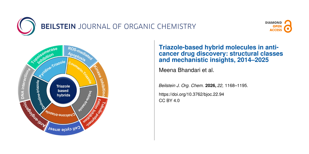
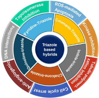
Triazole hybrids are appealing prospective molecules for drug discovery because they provide a multi-target approach to cancer treatment. Anticancer strategies are primarily aimed at disrupting the molecular systems that drive uncontrolled cell proliferation and survival. Among emerging therapeutic agents triazole-based hybrids have gained significant attention due to their ability to interfere with critical cellular processes. These hybrids can act on different targets that collectively disrupt tumor development and progression. They can inhibit topoisomerases, interfere with tublin polymerization, act as kinase inhibitors, trigger apoptosis through oxidative stress and cell cycle arrest, suppress angiogenesis, and interfere with DNA interactions [39-41].
In this review paper, research articles published in English language for the last twelve years were collected by using key words, triazoles, triazole hybrids and anticancer triazole compounds etc. A comprehensive analysis was carried out for the data obtained for the last 12 years (between 2014 and 2025). Only few classes of novel anticancer triazole hybrids were selected which elaborated the mechanism of action of synthesized compounds. This study used a qualitative strategy, a simple research framework with descriptive goals, and a systematic bibliographic approach to investigate the subject.
Review
Triazole-based molecular hybrids
Advances in triazole-based molecular hybrids have revealed promising strategies for drug design and development. The hybrid molecules are created by combining two pharmacophores that are biologically active, with or without a suitable linker. In these hybrids the triazole moiety plays a crucial role in enhancing anticancer activities, improving selectivity, and overcoming resistance. It can act as bioactive core, pharmacophoric linker, or solubility and metabolic modulator [42,43]. Common pharmacophores used in hybridization include chalcone, quinoline, coumarin, indole, and pyridine moieties. Generally, triazole-containing hybrids fall into two major types (Figure 6); Type I: Different cytotoxic pharmacophores interact with the 1,2,3-triazole moiety; Type II: Two chemotherapeutic pharmacophores are connected via triazole linker [44].
![[1860-5397-22-94-6]](/bjoc/content/figures/1860-5397-22-94-6.svg?scale=2.0&max-width=1024&background=FFFFFF)
Figure 6: Types of triazole containing hybrids.
Figure 6: Types of triazole containing hybrids.
These design strategies, including structural modification, incorporation of diverse pharmacophores, and exploration of different linkers, lead to the formation of numerous triazole hybrids with better efficacy across various pathological conditions, paving the way for further advancement in targeted drug therapies.
Coumarin–triazole hybrids
Although coumarin and 1,2,3-triazole derivatives individually demonstrated significant anticancer behavior, lead researchers have extensively explored hybrid compounds combining these pharmacophores for an enhanced and broader spectrum of action [45,46]. In recent years, coumarin–triazole hybrid molecules have emerged as promising candidates in drug discovery, owing to synergistic effects derived from both coumarin and triazole moieties. They exhibit diverse biological activities including anticancer, antioxidant, and antimicrobial [47,48]. These hybrid molecules show potential in combating drug resistance by acting on multiple targets, offering a superior alternative to combination therapy [49]. The synergistic effects of the combined pharmacophores contribute to their high therapeutic potential, making coumarin–triazole hybrids valuable lead molecules, recommended for studying in vivo behavior and potential drug advancement. For instance, chalcone–coumarin hybrids connected through a 1,2,3-triazole linker (20, 21, Figure 7) have been investigated for their cytotoxic activity against various human cancer cell lines, including cholangiocarcinoma (HuCCA-1), human lung cancer (A549), hepatocellular carcinoma (HepG2), and malignant T-lymphoblastic (MOLT-3) cells [50]. Most of the compounds, with the exception of compound 20c, demonstrated selective cytotoxic behavior for the MOLT-3 cell line, indicating good therapeutic potential. A structure–activity relationship (SAR) analysis showed that compounds 20b, 20c, and 21b containing a triazole ring in 3- or 4-position of ring A and 2,3-dimethoxy groups on ring B, exhibited a strong activity against HepG2 cells, having a half-maximal inhibitory concentration (IC50) of 15.70 µM, 8.18 µM, and 4.26 µM, respectively. In contrast, compounds 21d with trimethoxy substitution on ring B generally lost cytotoxicity towards HuCCA-1, HepG2 and A549 cells. However, compound 21c reserved activity against HuCCA-1 with an IC50 of 6.13 µM. Compound 22 (Figure 7) 4-(1,2,3-triazol-1-yl)coumarin bearing a (4-fluorophenoxy)methyl substituent at the triazole C-4 exhibited potent antitumor activity against MCF-7, SW480, and A549 cell lines with IC50 values of 5.89 µM, 1.99 µM, and 0.52 µM, respectively [51]. Mechanistic investigations indicated that compound 22 inhibits cancer cell proliferation by promoting apoptosis and inducing G2/M cell-cycle arrest. SAR analysis revealed that a –CH2–O– linker at the C-4 position of the 1,2,3-triazole substantially increases cytotoxicity, whereas the presence of an analogous hydrogen acceptor substituent at the coumarin C-7 position favorably influences anticancer potency. The novel 2-(triazol-1-yl)dihydrofurocoumarin derivative 23 with 4-hydroxy-3-methoxybenzamidomethyl substituent at C-4 of the triazole ring was found active against leukemia cell lines including CEM-13 (acute lymphoblastic leukemia), MT-4 (membrane-type 4 T-cells), and U-937 (myeloid leukemia) and showed selectivity with cytotoxic dose (CTD50) values of 9.00 µM (CEM-13), 10.00 µM (MT-4), 8.00 µM (U-937) [52]. Also, this compound exhibited a strong binding affinity of −9.2 kcal/mol with PDE4B (phosphodiesterase-4B), which is better to the reference drug, rolipram (−8.3 kcal/mol), indicating its potential as an anticancer lead. The curcuminoid–coumarin hybrids tethered through a 1,2,3-triazole linker (24a,b and 25, Figure 7) showed promising cytotoxicity towards THP-1, COLO-205, and HCT-116 cancer cell lines with IC50 values ranging from 0.82 to 9.95 µM but displayed no significant effect on PC-3 cells. Importantly, the two-carbon spacer flanked by coumarin and triazole units was found essential for the activity [53]. The isatin–coumarin hybrids 26a–m (Figure 7) which exhibited potent activity, especially compound 26a, showed IC50 values of 0.73 µM (THP-1), 3.45 µM (COLO-205), and 3.04 µM (HCT-116), along with strong tubulin polymerization inhibition (IC50 = 1.06 µM) [54]. Also, fluorine-substituted isatin derivative 26b showed notable activity against THP-1 (IC50 = 1.99 µM). SAR analysis indicated that extending the linker or modifying the isatin ring reduces the cytotoxic potency. The coumarin–triazole hybrid 27 with 7-hydroxy-4-methylcoumarin connected to C-4 of the triazole ring (Figure 7) demonstrated strong cytotoxicity towards MCF-7 and HeLa cells lines, with IC50 values of 3.12 µM, and 2.77 µM, respectively, comparable to doxorubicin. Another derivative 28 (Figure 7), containing a 4-hydroxycoumarin moiety, also showed antiproliferative action for MCF-7 and Hela cells but both molecules have been found to be largely inactive against HEK-293 cells [55].
![[1860-5397-22-94-7]](/bjoc/content/figures/1860-5397-22-94-7.svg?scale=2.0&max-width=1024&background=FFFFFF)
Figure 7: Structures of diverse coumarin–1,2,3-triazole hybrids 20–29.
Figure 7: Structures of diverse coumarin–1,2,3-triazole hybrids 20–29.
The coumarin-linked β-lactam–triazole hybrids 29a,b (Figure 7) displayed modest action for MCF-7 having an IC50 concentration of 53.55 µM and 58.62 µM, respectively, and exhibited strong binding affinity to the estrogen receptor-α (ER-α). The presence of a nitro and chloro moiety at the C-3 position of the phenyl ring attached to N-1 of the β-lactam ring increased both potency and selectivity of these compounds for MCF-7 cancer cell lines [56]. The 4-anilinomethyl-1,2,3-triazole moiety tethered to the 6-position of coumarin 30a–f (Figure 8) showed strong inhibitory activity (Ki < 100 nM) towards carbonic anhydrase isoforms IX and XIII which are involved in tumorigenesis [57]. The novel triazole–coumarin–glycosyl hybrids 31a–c (Figure 8) have been evaluated for cytotoxic action towards different cancer cells. These compounds demonstrated promising growth inhibition with IC50 values ranging from 14.6–33.4 µM (Paca-2), 4.1–65.1 µM (MeI-501), 12.5–51.3 µM (PC-3), 9.9–14.5 µM (A-375) and 20.4–70.2 µM (Caco-2). Notably, compounds 31a and 31c are more effective against Paca-2 cells than doxorubicin (IC50 = 19.4 µM) [58]. Six new coumarin–triazole hybrids 32–34 (Figure 8) were studied for their action towards MCF-7 (breast cancer) and HeLa (cervical cancer) cells, among these candidates, 34b, bearing a phenyl group, showed the strongest cytotoxicity. To enhance its delivery and efficacy, compound 34b was encapsulated in poly(lactic-co-glycolic acid) (PGLA) nanoparticles. Importantly, the 34b-loaded PGLA NPs showed significant higher anticancer activity than the free compound 34b. The IC50 value dropped from 5.74 to 0.42 mg/mL against MCF-7 and from 1.32 to 0.77 mg/mL against Hela cells. These results pronounce the probability of use of coumarin–triazole derivatives, with nanoparticles-based delivery system, for enhanced cancer therapy [59].
![[1860-5397-22-94-8]](/bjoc/content/figures/1860-5397-22-94-8.svg?scale=2.0&max-width=1024&background=FFFFFF)
Figure 8: Structures of diverse coumarin–1,2,3-triazole hybrids 30–34.
Figure 8: Structures of diverse coumarin–1,2,3-triazole hybrids 30–34.
Beyond the 1,2,3-triazole-based system, 1,2,4-triazole hybrids have also garnered significant attention owing to their versatile pharmacological profiles and potent anticancer potential. Researchers have explored different synthetic methods and introduced structural modifications to enhance their therapeutic potential [60]. Here, several coumarin-1,2,4-triazole hybrids are discussed that have exhibited noteworthy anticancer activity. A series of N-acetyl carbohydrazide-linked 1,2,4-triazole–coumarin hybrids (35a–r, Figure 9) were evaluated for their cytotoxic activity against four human cancer cell lines: BT20 (breast carcinoma), A549 (lung carcinoma), SK-Mel-128 (melanoma), and DU-145 (prostate carcinoma) [61]. Their performance was compared with the standard chemotherapeutic agent cisplatin. These derivatives displayed anticancer action ranging from moderate to high efficacy across the tested cell lines. Specifically, compounds 35f, 35h–i, 35k, 35p, and 35r showed superior anticancer action towards the BT20 cell line, with cytotoxic concentration (CC50) values fluctuating between 3.6 and 13.9 µg/mL and selectivity indices between 2.4 and 5.2. Compound 35f demonstrated enhanced activity against the DU-145 cell line (CC50 = 3.7 µg/mL, SI = 9.9), while compounds 35h and 35i were more active than cisplatin against A549 cells (CC50 = 7.5 and 8.1 µg/mL, SI = 4.2 and 3.3). However, none of the tested molecules surpassed cisplatin in efficacy against the SK-Mel-128 melanoma cell line. Novel coumarin derivatives connected to 1,2,4-triazole (36a–c, Figure 9) or 1,2,4-triazolo[3,4-b][1,3,4] thiadiazole groups (37a–c, Figure 9) via a methylene linker have been assessed as anticancer agents in human colon cancer cells (HCT116). Compounds 36c and 37c exhibit promising anticancer action having IC50 values of 4.363 µM and 2.656 µM, respectively. Molecular docking of active compounds proposes the interface with tyrosine kinase (CDK2) as a potential mechanism of action [62]. The methylene-bridged coumarin–1,2,4-triazole hybrids 38a,b and 39a,b (Figure 9) were assessed for their cytotoxic effects against several cancer cell lines, including HepG2 (liver), MCF-7 (breast), HeLa (cervical), and SW620 (colorectal) [63]. Among these, compound 39b, containing a carboxamide group at C-3 of the triazole ring and a methoxy substituent at C-7 of the coumarin ring, displayed moderate cytotoxicity toward HeLa cells, with an IC50 value of 35.5 ± 13.5 µM.
![[1860-5397-22-94-9]](/bjoc/content/figures/1860-5397-22-94-9.svg?scale=2.0&max-width=1024&background=FFFFFF)
Figure 9: Structures of diverse coumarin–1,2,4-triazole hybrids 35–41.
Figure 9: Structures of diverse coumarin–1,2,4-triazole hybrids 35–41.
The other compounds exhibited no significant effect on HeLa cells, displaying IC50 values greater than 100 µM, and none showed measurable effects on the remaining three cancer cell lines. Importantly, compound 39b was non-toxic toward normal fibroblast cells, highlighting its selective action against cancer cells. A novel series of coumarin–triazole hybrids containing a C7-methoxy linker (40a–m and 41a–e, Figure 9) were evaluated for anticancer activity against six human cancer cell lines: ASG, MGC-803, HCT-116, A549, HepG2, and HeLa. Among the synthesized candidates, 40d exhibited the most potent activity against ASG and MGC-803 cells, with IC50 values of 2.63 ± 0.17 µM and 3.05 ± 0.29 µM, respectively. Compound 40c showed strong inhibition toward HCT-116 cells (IC50 = 3.66 ± 0.24 µM). In addition, compound 40j demonstrated cytotoxic effects against A549 (IC50 = 17.32 ± 0.63 µM) and HeLa (IC50 = 7.20 ± 0.54 µM) cells, while compound 40h was found to be active against HepG2 cells (IC50 = 8.33 ± 0.27 µM) and compound 40m showed selective toxicity towards Human gastric cancer (AGS) cells (IC50 = 9.00 ± 0.64 µM). The mechanistic evaluation showed that compound 40d suppressed ASG cell proliferation by triggering apoptotic cell death and inducing cell cycle arrest at the G2/M phase. The replacement of the coumarin phenyl moiety with 4-methoxyphenyl (41a–e) was found to decrease the antiproliferative potency. Furthermore, compounds substituted with para-chloro and para-methyl groups demonstrated potent and wide-ranging anticancer efficacy across multiple cell lines [64]. These studies of coumarin-triazole hybrids represent their anticancer potential by combining the bioactive properties of both scaffolds.
Indole–triazole hybrids
The indole ring is a highly prevalent, biologically active heterocyclic structure present in numerous natural compounds. It exhibits anticancer, antioxidant, anti-inflammatory, and antibacterial activity [65,66]. Due to diverse bioactivities, researchers have shown significant interest in designing and synthesizing indole-based hybrid molecules, such as indole–triazole, indole–coumarin, indole–isatin, and indole–pyrimidine to enhance therapeutic efficacy, target specificity, and reduce adverse effect [67]. Recent research has explored the potential of indole–triazole hybrids as anticancer agents. Indole and triazole units have been connected using several synthetic approaches, including click chemistry and various coupling reactions, to generate a diverse array of structurally distinct molecules for evaluating biological activity [68]. The flexible nature of indole–triazole conjugates allows for strategic modifications that can significantly influence their ADME profile as well as their interactions with biological targets. This adaptability makes them valuable scaffolds in medicinal chemistry, where fine tuning of their molecular architecture can lead to enhanced bioavailability, target specificity, and reduced toxicity.
The cytotoxic study of etodolac-based derivatives containing a 1,2,3-triazole linkage (42a–l, Figure 10) against human lung cancer cells A549 showed that compounds 42e, 42f, 42h, 42j, and 42l exhibited significant anticancer activity, surpassing the efficacy of the standard chemotherapeutic agent doxorubicin. Compound 42l exhibited the greatest cytotoxic potency, showed an IC50 value of 3.29 ± 0.7 µM, closely followed by compound 42f, which showed an IC50 of 3.65 ± 0.4 µM. The anticancer behavior of compound 42l can be ascribed to inhibition of human topoisomerase II through specific molecular interactions [69]. 1,2,3-Triazole tangled indole analogues 43a–l (Figure 10) have been tested for anticancer action with two human cancer cell lines MCF-7 and HepG2. The derivatives bearing 4-hydroxy, 4-methoxy, 2-methyl, and 3-acetyl substituents displayed superior cytotoxic action, with IC50 values lower than that of the standard chemotherapeutic agent, doxorubicin. In silico molecular docking analysis explained that the active molecules show robust binding affinities and key molecular connections through the active sites of aurora kinase-1 and DNA topoisomerase IIα, surpassing those of doxorubicin [70]. The molecular hybrids 44 containing 1,2,4-triazole and indole scaffolds were evaluated for their cytotoxic potential against the MCF-7 cell line. Out of synthesized candidates, 44d and 44e exhibited cytotoxicity with IC50 values of 17.67 ± 0.34 µM and 17.01 ± 0.53 µM, respectively. Compound 44e demonstrated aromatase inhibition (IC50 = 0.026 µM), comparable to letrozole (IC50 = 0.024 µM) [71]. The study on anticancer activity of 1,2,4-triazole-tethered indolyl–chalcone scaffolds 45 (Figure 10) against NCI 60 cells demonstrated broad-spectrum antiproliferative effects of compounds 45a and 45d, having GI50 values from 3.69 to 20.40 µM and 0.29 to >100 µM, respectively [72]. Further examined was the inhibitory activity of selected hybrids against the epidermal growth factor receptor (EGFR) and c-MET (mesenchymal-epithelial transition factor) kinases. Compound 45b displayed strong c-MET inhibition, with an IC50 value of 4.70 nM, comparable to the reference inhibitor foretinib (IC50 = 2.5 nM). Similarly, compound 45c exhibited EGFR inhibition equivalent to that of erlotinib (IC50 = 0.052 µM) along with potent c-MET inhibitory activity (IC50 = 4.90 nM). These results highlight the promise of this scaffold as a potential dual kinase-targeted therapeutic agent. The indole–chalcone hybrid derivative functionalized with an N-substituted 1,2,3-triazole unit (46, Figure 10) exhibited strong anticancer activity against SiHa and SW620 cells having IC50 values of 67.99 and 48.96 µg/mL, respectively, while exhibiting minimal toxicity in normal human embryonic kidney HEK293 cells at comparable concentration. Mechanistic studies showed that compound 46 interacts with DNA through a non-covalent intercalative mechanism and contributes to its antiproliferative effects [73].
![[1860-5397-22-94-10]](/bjoc/content/figures/1860-5397-22-94-10.svg?scale=2.0&max-width=1024&background=FFFFFF)
Figure 10: Structures of indole–triazole hybrids.
Figure 10: Structures of indole–triazole hybrids.
Lingaswamy et al. (2025) evaluated the anticancer potential of indole-piperazine-1,2,3-triazole derivatives 47a–m (Figure 10) and found compounds 47d, 47e, and 47f demonstrated superior cytotoxicity towards MCF-7, HCT-116 and HepG2 cancer cells, surpassing the efficacy of standard EGFR inhibitor erlotinib. Also, these compounds showed negligible toxicity towards normal breast epithelial cells (MCF-10A), demonstrating discriminatory anticancer action. Pharmacokinetics analysis showed that 47e fully met major drug-likeness criteria, highlighting its promise as a selective EGFR-targeting anticancer agent [74]. The N-phenylacetamide derivatives incorporating an indole-1,2,4-triazole scaffold 48a–f (Figure 10) were evaluated for cytotoxicity using doxorubicin and ellipticine as reference drugs. Compounds 48a, 48c, and 48f exhibited remarkable anti-HepG2 activity, with cell-capability values of 11.72 ± 0.53, 18.92 ± 1.48, and 12.93 ± 0.55 µg/mL, respectively, demonstrating good to exceptional chemotherapeutic potential. Among them, the 3,4-dichloro-substituted derivative 48b displayed pronounced cytotoxicity against HepG2 liver cancer cells, indicating its strong efficacy as a cellular-damage agent [75]. Indole-2-one-1,2,3-triazoles 49 and 50 (Figure 10) exhibited promising anti-angiogenesis activity as VEGFR-2 inhibitor and low toxicity towards human umbilical vein endothelial cells (HUVECs). Specifically, compound 49d showed better kinase suppression (IC50 = 26.38 nM) than sunitinib (IC50 = 83.20 nM). Also, it effectively suppressed HT-29 and MKN-45 cancer cell growth. Molecular docking and molecular dynamics confirmed stable binding of 49d to the VEGFR-2 active site [76].
The indoly-1,2,4-triazole hybrids 51–55 (Figure 11) showed potent VEGFR-2 kinase inhibition and exhibited good antirenal cancer action. These derivatives exhibited superior VEGFR-2 inhibitory activity relative to the reference drug, sunitinib and revealed strong anticancer behaviour against CAKI-1 and A498 renal cancer cells [77]. Elsawi et al. (2024) developed 1,2,4-triazole-indolin-2-one derivatives 56 and 57 (Figure 11) as potential anti-hepatocellular and anti-pancreatic cancer agents, exhibiting VEGFR-2 inhibitory activity. These compounds demonstrated notable anticancer effects, with IC50 values ranging from 0.17 to 4.29 µM against PANC-1 cells and 0.58 to 4.49 µM against HepG2 cells. Several derivatives, specifically 56d, 56e, 56g, 56k, and 57c showed potent VEGFR-2 inhibition, with IC50 values comparable to or exceeding that of the reference drug, sorafenib [78]. Indole-functionalized 1,2,4-triazole derivatives 58 and 59 (Figure 11) exhibited potent cytotoxicity against MCF-7 and HepG2 cells with IC50 values outperforming the standard drug erlotinib. Compound 59b induced apoptosis caused cell cycle arrest at the G2/M and S phase in MCF-7 and HepG2 cells, respectively and demonstrated dual inhibition of PARP-1 and EGFR with IC50 values of 62.4 nM and 1.24 nM, surpassing erlotinib and olaparib. Furthermore, 59b inhibited cancer cell proliferation by 66.7%, exceeding 65.7% efficacy [79]. These studies on indole–triazole hybrids have shown remarkable potential of these hybrids as anticancer candidates. Continued SAR studies and biological evaluation may pave the way for their advancement into potent therapeutic agents.
![[1860-5397-22-94-11]](/bjoc/content/figures/1860-5397-22-94-11.svg?scale=2.0&max-width=1024&background=FFFFFF)
Figure 11: Structures of indoly–1,2,4-triazole hybrids and indolin-2-one-1,2,4-triazoles.
Figure 11: Structures of indoly–1,2,4-triazole hybrids and indolin-2-one-1,2,4-triazoles.
Quinoline–triazole hybrids
In medicinal chemistry, quinoline is also recognized as highly valuable scaffold due to its presence in numerous bioactive natural products and its broad pharmacological profile, including anticancer, antimalarial, antibacterial, and anti-inflammatory effects [80-84]. Quinoline derivatives display anticancer activity through different mechanisms for instance apoptosis induction, cell cycle arrest, and angiogenesis inhibition [85,86]. However, some quinoline-containing drugs like chloroquine and fluoroquinolones are linked to cardiac side effects such as QT interval prolongation [87]. To overcome these limitations and enhance therapeutic potential, researchers are exploring quinoline-based hybrid molecules. The hybrid molecules containing quinoline and triazole rings have demonstrated significant potential across a wide range of biological activities [88].
Mohassab et al. (2021) evaluated the anticancer potential of quinoline-chalone-1,2,4-triazole hybrids 60–62 (Figure 12) across a diverse panel of cancer cell lines, including several types of cancers such as leukemia, ovarian, renal, prostate, melanoma, lung, CNS, colon, and breast. Several compounds, particularly 60a, 60b, 62a, 62b, and 62d exhibited significant antiproliferative effects and inhibited both EGFR and BRAFV600E kinases. The most promising compound 62b exhibited pronounced anticancer activity by promoting apoptosis and triggered G2/M phase cell cycle arrest. Also, the molecular docking analysis disclosed the significant binding connections with EGFR and BRAFV600E, with binding energies surpassing those of standard inhibitors like erlotinib and vemurafenib, respectively [89]. The quinolone–triazole hybrids 63 and 64 (Figure 12) exhibited moderate activity against MCF-7 breast cancer cells, while few of them displayed similar activity analogous to that of cisplatin. The specific substituents significantly influencing biological activity such as compounds with 2-chloro (63a) and 4-chloro (63b) substituents exhibit activity having IC50 values of 7.46 µM and 6.45 µM, respectively [90]. The new quinolone–1,2,3-triazole compounds 65a–l (Figure 12) were assessed for anticancer activity towards HT-1080 and A-549 cells [91]. Here, many compounds exhibited weak to moderate cytotoxicity; however, derivatives, 65c, 65d, and 65j bearing 2-chlorophenyl, 4-chlorophenyl and 3-acetylphenyl substituents, respectively demonstrated significant activity with IC50 values comparable to that of reference drug, doxorubicin (IC50 = 10.32 ± 1.32µM). El Malah et al. (2024) reported the anticancer potential of 1,2,3-triazolo-benzoquinoline-3-carbonitrile derivatives 66a–f (Figure 12) against three human cancer cells HCT-116 (colon), HepG2 (liver), MCF-7 (breast) and a healthy cell line (BJ-1) and found compound 66b demonstrated selective cytotoxicity against colon cancer (HCT-116), showing higher toxicity to cancer cells over normal ones. Against HepG2 liver cancer cells, compound 66c exhibited potency surpassing the reference standard, doxorubicin [92].
![[1860-5397-22-94-12]](/bjoc/content/figures/1860-5397-22-94-12.svg?scale=2.0&max-width=1024&background=FFFFFF)
Figure 12: Structures of quinoline/quinolone–triazole hybrids 60–68.
Figure 12: Structures of quinoline/quinolone–triazole hybrids 60–68.
The 1,2,3-triazole compound incorporating a quinolone–benzimidazole framework 67 (Figure 12) exhibited notable anticancer activity towards a panel of NCI-60 human cancer cells, with favorable GI50, TGI, and LC50 values. This compound displayed a strong antiproliferative effect towards the BT-474 breast cancer cell-line, with an IC50 value of 0.59 ± 0.01 µM. The anticancer effect is induced primarily through ROS-mediated apoptosis in cancer cells [93]. The novel quinoline-triazole-linked peptidomimetic scaffolds 68 (Figure 12) showed inhibitory properties for CDK2, a protein implicated in various malignancies. An in vitro cytotoxicity assay against MCF-7 demonstrated that 68 repressed cell propagation with an IC50 value of 8 µM, highlighting its anticancer potential [94]. The two distinct quinolone–triazole hybrids 69 and 70 (Figure 13) were assessed for antiproliferative activity towards MCF-7 breast cancer cells. Among these synthesized hybrids, several derivatives such as 69h, 69i, 69l and 69n have exhibited promising antiproliferative action with IC50 values of 0.39–3.65 µg/mL, higher than the reference drug doxorubicin (IC50 = 3.69 µg/mL). Molecular docking analysis demonstrated powerful binding to the CDK2 enzyme with affinities ranging between −9.7 to −10 kcal/mol, emphasizing on the anticancer efficacy [95]. The novel quinolone conjugated with a triazolo[4,3-b]pyridazine moiety 71 (Figure 13) demonstrated antiproliferative activity across multiple cancer cell lines with GI50 values from 1.48–25.4 μM. The hybrid acts as an PIM-1/PIM-3 kinase inhibitor, exhibited IC50 values of 7 nM/70 nM, respectively [96]. The study on the anticancer potential of 7-methoxyquinoline–1,2,3-triazole hybrids 72 (Figure 13) across multiple cancer cell lines such as breast (BT-20, MDA-MB231, MCF-7) and colon (HT-29) cancers revealed that compounds 72a and 72b showed cytotoxicity, with IC50 values of 4.49 ± 0.68 µM and 19.05 ± 1.58 µM in BT20 and HT29 cells, respectively. In addition, interaction of MDA-MB-231 cells with 72c resulted in pronounced nuclear morphological alteration, indicating their potential to act via apoptotic pathways [97]. These studies on quinolone–triazole hybrids have demonstrated their significant potential as anticancer agents. Their ability to act through multiple mechanisms and structural adaptability makes them a valuable scaffold for developing novel and effective anticancer therapeutics.
![[1860-5397-22-94-13]](/bjoc/content/figures/1860-5397-22-94-13.svg?scale=2.0&max-width=1024&background=FFFFFF)
Figure 13: Structures of quinoline/quinolone–triazole hybrids 69–72.
Figure 13: Structures of quinoline/quinolone–triazole hybrids 69–72.
Chalcone–triazole hybrids
These hybrid molecules integrate two pharmacological active components: chalcones, which contain an α,β-unsaturated carbonyl group, known to show interaction with various cellular targets involved in cancer progression, and 1,2,3-triazoles, which are recognized for their strong hydrogen bonding ability and metabolic stability. Together, these functionalities work synergistically to produce compounds with enhanced anticancer potential [98-100].
Utilizing click chemistry under eco-friendly conditions, the chalcone–triazole hybrid 73 (Figure 14) was produced and assessed for its cytotoxicity against MCF-7, MIA PaCa-2, A549, and HepG2 cells [101]. The hybrid demonstrated the maximum effective cytotoxic activity, having IC50 values oscillating between 4–11 µM towards all tested cell lines. The mechanistic studies exposed that compound 73 brings apoptosis and causes G2/S phase cell cycle detention in MIA PaCa2 cells and, induces a mitochondrial potential loss in MIA PaCa-2 cells. Mallikarjun et al. (2023) evaluated the anticancer potential of novel coumarin-triazole-chalcone 74 against various human cancer cells, including HEK293 (human embryonic kidney cells), HeLa (cervix carcinoma), PANC1 (pancreatic cancer), HT1080 (fibrosarcoma), and A549 (lung cancer). Among synthesized hybrids, para-nitrile chalcone 74a exhibited the most potent cytotoxicity with IC50 values ranging between 3.1 to 7.02 µg/mL against tested cell lines. In silico studies demonstrated that compound 74a binds effectively to cancer-relevant molecular targets PI3K and AKT, showing specificity for AKT with binding interactions comparable to known AKT inhibitors [102].
![[1860-5397-22-94-14]](/bjoc/content/figures/1860-5397-22-94-14.svg?scale=2.0&max-width=1024&background=FFFFFF)
Figure 14: Structures of chalcone–triazole hybrids 73–79.
Figure 14: Structures of chalcone–triazole hybrids 73–79.
The cytotoxic activity of 1,2,4-triazole–chalcone derivatives 75a and 75b (Figure 14) was assessed against several human cancer cell lines, including MCF-7, HeLa, A549, and SW620. Among these, compound 75a demonstrated notable selectivity toward SW620 cells, exhibiting an IC50 value of 21.55 µM, while compound 75b showed significant cytotoxicity against A549 cells, with an IC50 value of 25.58 µM [103]. A series of chalcone derivatives 76 containing a triazolo[4,3-a]quinoxaline scaffold (Figure 14) was investigated for their potential as chemotherapeutic agents, capable of simultaneously inhibiting tubulin polymerization and EGFR kinase activity. Among these, compound 76g demonstrated the highest potency, attributed to its favorable interactions with the colchicine-binding site of tubulin. It exhibited IC50 values of 1.65, 3.61, and 8.58 µM against MCF-7, HCT-116, and HepG2 cancer cell lines, respectively [104]. The anticancer potential of 1,2,3-triazole–chalcone derivatives 77 (Figure 14) was evaluated against A549 cancer cell line, exhibiting IC50 values ranging from 8.67 to 11.62 µM. SAR studies indicated that the presence of a bromo substituent is critical for cytotoxic activity, while modifications on the heterocyclic ring led to a marked reduction in potency. Among the series, compounds 77a, and 77c demonstrated the highest antiproliferative effect, with IC50 values of 8.67 and 9.74 μM, respectively, approaching the efficacy of the reference chemotherapeutic agent, doxorubicin (IC50: 3.24 μM) [105]. The methoxy-bridged 1,2,3-triazole–chalcone derivatives 78a–c (Figure 14) exhibit enhanced cytotoxic activity against A540 lung cancer cells (IC50 = 35.81–50.82 μM) compared to the standard chemotherapeutic agent doxorubicin (IC50: 69.33 μM), and demonstrated their potential as lead compound for lung cancer therapy [106]. The triazole–tethered chalcone–matrine hybrids 79a–c (Figure 14) with electron-withdrawing groups exhibited enhanced antiproliferative activity (IC50 = 5.01–12.72 μM) against A549 cells. The hybrid 79a was showing the most potent cytotoxicity (IC50 = 5.01–7.31 μM) across evaluated cell lines, while showing minimal cytotoxicity against normal NIH/3T3 fibroblast cells (IC50 = 39.21 μM), indicating a favorable therapeutic index. Mechanistically, 79a induces dose-dependent apoptosis in A549 cells [107].
The 1,2,3-triazole–chalcone derivatives 80a–k (Figure 15) were assessed for cytotoxic activity against a diverse panel of 60 human cancer cell lines using the NCI-60 screening protocol. Among the synthesized candidates, 80d, featuring methoxy groups at 3 and 4 position of the phenyl ring in the chalcone moiety, exhibited the most promising anticancer activity. At a concentration of 10 μM, 80d achieved greater than 94% growth suppression in RPMI-8226 and SR leukemia cell lines, and demonstrated over 80% inhibition against M14 melanoma, K-562 leukemia, and MCF-7 breast cancer cells. Also, derivative 80d displayed an IC50 value below 1 μM in six cancer cell lines, indicating high potency and selectivity. Compound 80d induces G2/M phase cell cycle arrest and triggers apoptosis through the mitochondrial pathway [108]. Djemoui et al. (2020) elucidated the anticancer potential of the triazole–benzimidazole–chalcone hybrids 81 and 82 (Figure 15) against T47-D, MDA-MB-231 and PC-3 cancer cell lines. The presence of chlorine group on the phenyl ring of the chalcone led to a marked enhancement in cytotoxic activity. Additionally, the incorporation of a benzyl group on the triazole moiety was found to further potentiate the compound’s antiproliferative effects [109]. The chalcone hybrid tethered to a 1,2,3-triazole ring 83 (Figure 15) specifically 83d, 83g, and 83i exhibited strong action towards MCF-7, MDA-MB-231 cancer cells, and HeLa cancer cells with lower IC50 value as compared to the standard drug, cisplatin. Also, these potent compounds exhibited low toxicity for normal breast epithelial cells (MCF-10A), indicating their selective behaviour [110]. The chalcone–thymol–triazole hybrids 84a–h (Figure 15) were synthesized and subjected to in vitro antiproliferative evaluation towards four cancer cell lines: HT-1080, A-549, MCF-7 and MDA-MB-231. These hybrids showed cytotoxic effects, having IC50 concentrations ranging from 4.61 to 50 μM. Furthermore, hybrids bearing phenyl and cyano substituents on the triazole moiety particularly showed strong inhibition of HT-1080 cell proliferation, with IC50 values of 4.61 μM and 9.08 μM, respectively [111]. A recent study on chalcone-based derivatives 85a–l containing two 1,2,3-triazole units (Figure 15) showed anticancer potential having IC50 values ranging from 44.7 to 86.2 μM, which is in contrast with the standard drug doxorubicin. The observed anticancer activity may be attributed to ERK2 kinase inhibition, which disrupts cell growth signaling and promotes cancer cell death [112].
![[1860-5397-22-94-15]](/bjoc/content/figures/1860-5397-22-94-15.svg?scale=2.0&max-width=1024&background=FFFFFF)
Figure 15: Structures of chalcone–triazole hybrids 80–85.
Figure 15: Structures of chalcone–triazole hybrids 80–85.
Pyridine–triazole hybrids
Pyridine scaffolds are widely recognized in medicinal chemistry for their crucial role in numerous clinically approved drugs, owing to their heteroaromatic nature and ability to intermingle to various biological targets such as kinases, enzymes, and nucleic acid [113,114]. Incorporation of a triazole moiety into these frameworks further enhances their pharmacological profile by improving metabolic stability, hydrogen bonding capacity, and target affinity. The novel 1,2,3-triazole–pyridine–benzo[d]imidazole hybrids 86 (Figure 16) were studied for their anticancer activity as prospective anticancer medications for the breast (MCF-7), colon (HCT116), human cell line (BJ1), and hepatocellular carcinoma (HepG2). Among synthesized candidates, 86g showed superior cytotoxicity against HepG-2 (IC50 = 3.3 μM), hybrids 86c, 86g, and 86h showed cytotoxicity towards MCF-7 with IC50 in between 3.0 to 3.5 μM. Against HCT-116, all synthesized hybrids showed better cytotoxic activity (IC50 = 5.2 μM to 6.2 μM) than doxorubicin (IC50 = 6.5 μM) [115]. A series of imidazo[1,2-a]pyridines-1,2,3-triazole conjugates 87 (Figure 16) were explored for cytotoxic effects across multiple cancer cell lines, including MCF-7, A549, HepG2, and T98G. The methoxy substituted derivatives exhibited the most potent antiproliferative activity and showed negligible cytotoxicity to normal human fibroblast cells (MRC-5), indicating a favorable therapeutic index. The mechanistic studies revealed a significant reduction in PARP protein levels in MCF-7 cells, suggested that apoptosis is mediated through PARP-dependent pathways [116]. The 1,2,3-triazoles linked via a methylene group to the imidazo[1,2-a]pyridine scaffold 88 (Figure 16) were tested towards two types of cancer cells (Hela and MCF-7) to determine their antitumor capabilities. Several of the synthesized compounds exhibited significant cytotoxicity, with IC50 values varying between 2.35 to 120.46 μM against the panel of tested cancer cell lines. Among them, compound 88d demonstrated superior antiproliferative efficacy compared to cisplatin, exhibiting IC50 values of 10.89 μM against Hela and 2.35 μM against MCF-7 cell lines [117]. The oxazolo[4,5-b]pyridine conjugated 1,2,3-triazole hybrids 89a–i (Figure 16) assessed for anticancer effects against PC3, DU-145, A549, and MCF-7 cancer cell lines. Although, the majority of the synthesized candidates exhibited good to moderate anticancer effects in comparison with the reference drug, etoposide, compounds 89a–e, and 89i demonstrated the strongest anticancer effects across all tested compounds. Notably, hybrid molecule 89a showed superior anticancer activity against PC3, DU-145, A549, and MCF-7 cells with IC50 values of 0.01 ± 0.0074 μM, 0.043 ± 0.009 μM, 0.15 ± 0.038 μM, and 0.045 ± 0.0021 μM, respectively. These oxazolo[4,5-b]pyridine-based triazoles exhibited inhibitory activity against human dihydroorotate dehydrogenase (hDHODH), suggesting a possible mechanism underlying their anticancer effects [118].
![[1860-5397-22-94-16]](/bjoc/content/figures/1860-5397-22-94-16.svg?scale=2.0&max-width=1024&background=FFFFFF)
Figure 16: Structures of 1,2,3-triazole–pyridine derivatives 86–89.
Figure 16: Structures of 1,2,3-triazole–pyridine derivatives 86–89.
The estimation of the antihepatic cancer potential of 1,2,3-triazolyl–pyridine derivatives 90–96 (Figure 17) revealed notable cytotoxicity against human hepatoblastoma (HepG2) cells [119]. Most of the candidates exhibited promising anticancer activity, while hybrid molecule 93 (IC50 = 0.64 ± 0.15 μg/mL) and 94 (IC50 = 1.08 ± 0.95 μg/mL) demonstrated superior efficacy in comparison to the reference medicine, doxorubicin (IC50 = 3.56 ± 0.46 μg/mL). Further, these hybrids showed negligible cytotoxicity towards the normal BALAB/3T3 cell line, indicating a favorable selectivity profile and potential therapeutic window for hepatocellular carcinoma treatment. The anticancer potential of novel pyridine–imidazole–triazole hybrids 97a–l (Figure 18) was evaluated against HT-1080 (fibrosarcoma) and Caco-2 (colorectal adenocarcinoma) cell lines. Among synthesized derivatives, compounds 97b, 97c, and 97f displayed the strongest activity towards HT-1080, while compounds 97b and 97c exhibited significant cytotoxicity towards Caco-2 cells [120].
![[1860-5397-22-94-17]](/bjoc/content/figures/1860-5397-22-94-17.svg?scale=2.0&max-width=1024&background=FFFFFF)
Figure 17: Structures of 1,2,3-triazolyl–3-cyanopyridine hybrids 90–96.
Figure 17: Structures of 1,2,3-triazolyl–3-cyanopyridine hybrids 90–96.
![[1860-5397-22-94-18]](/bjoc/content/figures/1860-5397-22-94-18.svg?scale=2.0&max-width=1024&background=FFFFFF)
Figure 18: Structures of pyridine/pyrimidine–triazole hybrids 97–102.
Figure 18: Structures of pyridine/pyrimidine–triazole hybrids 97–102.
Thiophene-1,2,3-triazole–pyridine hybrid 98 (Figure 18) exhibited potent cytotoxicity towards A549 (IC50 = 0.68 μM), PC-3 (IC50 = 1.03 μM), and MDAMB-231 (IC50 = 0.88 μM). Molecular docking analysis revealed strong binding affinity of the compound to the ATP-binding site of human topoisomerase IIα [121]. The novel pyridine-1,2,4-triazoles 99a–j tethered via an amide linker (Figure 18) were evaluated for their cytotoxic potential towards gastrointestinal cancer cell lines (EPG, Caco-2, LoVo, LoVo/Dx, HT-29), alongside normal colonic epithelial cells (CCD841 CoTr) for comparative assessment. Among the synthesized compounds, derivative 99c featuring a phenyl substituent at the N-4 position and a 4-methylphenylpiperazine group at the N-2 of the 1,2,4-triazole ring demonstrated the most promising anticancer profile. It exhibited potent cytotoxicity against EPG and Caco-2 cancer cell lines while showing reduced toxicity towards normal epithelial cells, indicating a high degree of selectivity. Additionally, compound 99c effectively inhibited P-glycoprotein in P-gp-expressing cell lines (HT-29, LoVo, and LoVo/Dx). Mechanistic studies further revealed that treatment with compound 99c induced apoptosis in HT-29 cells via upregulation of caspase-3 and p53 protein levels [122]. The study on anticancer properties of a series of pyridine-1,2,3-triazole-N-glycoside derivatives 100 (Figure 18) found that compounds containing biphenyl or thienyl substituents on the phenyl ring (100b and 100c) exhibited the most potent cytotoxic activity against A549, MCF-7, and PC3 cell lines. Molecular docking studies further revealed strong binding interactions of these derivatives within the EGFR active site, corroborating their proposed mode of action [123]. The novel pyridine–1,2,4-triazolyl–pyrimidine hybrids 101a–j (Figure 18) were evaluated for cytotoxic potential against MCF-7, A549, Colo-205, and A2780 cancer cell lines. Most of the synthesized candidates demonstrated moderate to significant cytotoxicity relative to the reference drug, etoposide. However, compound 101a showed particularly promising activity across the tested cell lines and showed IC50 values of 0.1 ± 0.075 μM, 0.26 ± 0.037 μM, 0.48 ± 0.048 μM, and 0.13 ± 0.023 μM on MCF-7, A549, Colo-205, and A2780 cell lines, respectively [124]. A series of novel thieno[2,3-d][1,2,4]triazolo[1,5-a]pyrimidine derivatives 102a–e (Figure 18) was evaluated for cytotoxic activity against PC-3 (prostate), HCT-116 (colon), and MCF-7 (breast) cancer cell lines. Within the evaluated series, compounds 102b and 102e showed noticeable growth-inhibitory effects on the MCF-7 breast cancer cell line, with IC50 values of 19.4 ± 0.22 and 14.5 ± 0.30 μM, respectively, outperforming the standard chemotherapeutic agent, doxorubicin (IC50 = 40.0 ± 3.9 μM) [125].
Miscellaneous
In addition to the above well-defined classes, several other triazole-based compounds with diverse scaffolds have also shown notable anticancer activity. These structurally varied hybrids often combine triazole with pharmacophores like benzimidazole, pyrimidine, or steroidal backbones. For instance, bistriazolyl derivative 103 (Figure 19) linked to a 2-mercaptobenzimidazole scaffold demonstrated significant cytotoxic activity against HepG2 and A549 cancer cells with IC50 values of 10.9 μM and 8.9 μM, respectively. Compound 103 exhibited aurora-A kinase inhibition that resulted in cell-cycle arrest and apoptosis induction [126]. Two different benzimidazole–triazole conjugates 104 and 105 (Figure 19) were reported as potent inhibitors targeting EGFR, VEGFR-2 and topoisomerase II. Among the synthesized hybrids, 104a and 105g emerged as lead candidates, showing significant cytotoxicity against HCT-116, MCF-7, HepG-2, and HeLa cancer cell lines. Compound 104a demonstrated strong inhibition of EGFR with an IC50 value of 0.086 μM, comparable to gefitinib, along with moderate VEGFR-2 inhibition (IC50 = 0.109 μM) as compared to sorafenib, and enhanced topo II suppression (IC50 = 2.52 μM) relative to the standard anticancer agent doxorubicin (IC50 = 3.62 μM). Whereas compound 105g showed a less potent profile across the three targets [127]. In a study by Chandrakar et al. (2024) benzimidazole-1,2,4-triazole derivatives 106a–k (Figure 19) were tested for anticancer potential against various human cancer cell lines. Among the screened compounds, 106b and 106g demonstrated remarkable cytotoxicity on the HCA-7 colorectal cancer cell line, exhibiting IC50 values of 8.8 ± 0.9 μM and 9.2 ± 1.5 μM, respectively. Significant antiproliferative effects were also observed for 106d and 106i against MCF-7 cancer cells, with IC50 values of 8.3 ± 2.1 μM and 10.6 ± 1.2 μM, respectively. Also, 106d and 106g showed potent inhibitory activity against HT-29 colorectal cancer cells with IC50 values of 7.1 ± 0.9 and 7.7 ± 1.2 μM, respectively. Molecular docking revealed that 106d interacts with the binding site of tyrosine phosphate (PDB ID: 1WCH), a critical enzyme involved in colorectal cancer signaling pathways [128]. The 1,2,4-triazole coupled acetamide derivative 107 (Figure 19), bearing two ortho-methyl substituents on the N-phenyl ring displayed antiproliferative activity on the HepG2 cell line with an IC50 value of 16.782 µg/mL. Molecular docking studies further revealed strong binding affinities of compound 107 towards c-kit tyrosine kinase and protein kinase B with values of −170.066 kcal/mol and −176.749 kcal/mol, respectively [129]. The antitumor potential of newly synthesized oxazol–1,2,4-triazole hybrids 108 (Figure 19) was evaluated against PC-93 and HBT-55 cell lines representing prostate and lung cancers, respectively. The study revealed that several compounds exhibited notable cytotoxic effects on the PC-93 cell lines, exhibited IC50 values varying from 13.12 to 24.80 μM, depending on the nature of the substituent. Furthermore, these derivatives demonstrated significant anticancer activity against the HBT-55 cell line, showing comparable or superior efficacy to the reference drug, doxorubicin (IC50 = 19.41 μM) [130].
![[1860-5397-22-94-19]](/bjoc/content/figures/1860-5397-22-94-19.svg?scale=2.0&max-width=1024&background=FFFFFF)
Figure 19: Structures of bistriazolyl-2-mercaptobenzimidazole 103, benzimidazole-1,2,3-triazole conjugates 104, 105, benzimidazole–1,2,4-triazole derivative 106, 1,2,4-triazole–acetamide hybrid 107, and oxazol–1,2,4-triazole derivative 108.
Figure 19: Structures of bistriazolyl-2-mercaptobenzimidazole 103, benzimidazole-1,2,3-triazole conjugates 104...
A triazole derivative of betulone (109, Figure 20), exhibited exceptional cytotoxicity against the glioblastoma SNB-19 cell line, with an IC50 value of 0.17 μM, and demonstrated approximately five-fold higher efficacy than the standard drug cisplatin. The same derivative exhibited cytotoxic activity against amelanotic melanoma C-32 and exhibited an IC50 value of 0.81 μM [131]. This compound, therefore, represents a potential lead molecule for further development as an effective anticancer agent. The botulin–triazole derivatives 110 and 111 effectively suppressed the proliferation of SKOV-3 ovarian cancer cells by approximately 75% and 70%, respectively, at a concentration of 50 μg/mL. Despite exhibiting lower overall toxicity compared to betulin, their effective dosage indicated a strong influence in cell growth suppression. Compound 112 demonstrated pronounced cytotoxicity against T47D breast cancer cells, with an IC50 value of 1.3 μM, approximately 19 times more potent than cisplatin. Furthermore, the bistriazole derivative 113 exhibited exceptional cytotoxic activity against SNB-19, MCF-7, and T47D cell lines, with IC50 values of 0.08 μM, 0.09 μM, and 0.05 μM, respectively, underscoring its strong anticancer potential [132]. The N-acetyltriazole derivative betulin 114 showed significant cytotoxic activity against HCT 116, HEp-2, MS, and RD TE32 cancer cell lines, with IC50 values ranging from 2.3 to 7.5 µM [133]. Novel diacetylbetulin-1,2,4-triazole 115 exhibited anticancer activity against A375, MCF-7, and HT-29 cancer cell lines, with IC50 values ranging from 22.41 to 46.92 μM. Compound 115 causes apoptosis in tested cell lines, as confirmed by nuclear morphological alterations [134]. Triazole-tethered betulinic acid 116 demonstrated superior anticancer potential compared to betulinic acid, exhibiting IC50 values in the range of 5–7 µM against HL-60, MIA PaCa-2, PC-43, and A549 cell lines. In human leukemia HL-60 cells, it was found to induce apoptosis through both intrinsic and extrinsic pathways. This demonstrated the significance of triazole-tethered betulinic acids as promising candidates for anticancer drug development [135].
![[1860-5397-22-94-20]](/bjoc/content/figures/1860-5397-22-94-20.svg?scale=2.0&max-width=1024&background=FFFFFF)
Figure 20: Structures of botulin-based triazole hybrids 109–116.
Figure 20: Structures of botulin-based triazole hybrids 109–116.
Conclusion and Future Scope
Triazole-based hybrids have proven to be a multipurpose and promising class of heterocyclic compounds in anticancer drug development. Their unique ability to integrate diverse pharmacophores into a single molecular framework enables multi-target interactions and improved biological efficacy. This article emphasized the different classes of triazole hybrids such as coumarin–triazole, indole–triazole, chalcone–triazole, pyridine–triazoles etc., exhibiting cytotoxicity against a wide range of cancer cell lines. These hybrids often exhibit their anticancer effects via mechanisms such as kinase inhibition, topoisomerase inhibition, cell cycle arrest, and induction of apoptosis etc.
Though triazole-based hybrids exhibit encouraging in vitro cytotoxic potential; nevertheless, thorough in vivo studies remain relatively few. Prospective research ought to comprise:
- In vivo validation and translational studies through appropriate use of orthotopic and xenograft animal models, pharmacokinetic and pharmacodynamic profiling, risk evaluation and toxicological analysis and studying their bioavailability, metabolic pathways, and tissue dispersion.
- Use of proteolysis-targeting chimeras (PROTACs), a novel therapeutic approach for selectively degrading oncological proteins as triazole linkers provide excellent stability and biorthogonality. Nevertheless, traditional PROTAC synthesis is frequently time-consuming, including several synthetic stages, pre-assembled linkers, lengthy reaction durations, and low total yields. However, the one-pot synthetic approach is extremely effective, step-economic, easy to use, and environmentally benign, making it a useful platform for quickening PROTAC-based drug development [136].
- Incorporation of artificial intelligence and machine learning techniques for accelerating lead identification and optimization, predicting biological activity and toxicity, structure-based virtual screening & molecular docking and optimization of potency, selectivity, ADMET properties, and synthetic feasibility.
- Combining triazole hybrids with nanocarriers to lower off-target toxicity, increase the therapeutic index, targeted drug release, enhanced permeability and retention and improve their solubility in water.
- Adoption of a multi target drug design approach for synthesis of novel triazole hybrids that can modulate several biological pathways involving cellular proliferation, angiogenesis, apoptosis, metastasis, and drug resistance etc. These multi-target-directed triazole-based agonists would be safer, more efficient, and therapeutically transferable.
Despite the encouraging progress, challenges remain in translating these leads into clinical candidates. Overall, the structural diversity and therapeutic potential of triazole hybrids make them valuable candidates for further development as promising anticancer agents.
Vitae
![[Graphic 1]](/bjoc/content/inline/1860-5397-22-94-i1.png?max-width=637&scale=0.945456)
Prof. (Dr.) Meena Bhandari: Professor (Dr.) Meena Bhandari serves as the Dean, School of Basic and Applied Sciences, K.R. Mangalam University, and brings 27 years of distinguished teaching and research experience. She earned her Ph.D in Chemistry from IIT Delhi with specialization in calixarenes, along with an M.Phil. from Panjab University. Her research focuses on heterocyclic chemistry and green chemistry. She has successfully supervised four Ph.D scholars and is a recipient of the CSIR Fellowship and the AICTE Career Award for Young Teachers. Dr. Bhandari has published and presented 45 research papers in reputed SCIE, Scopus, and Web of Science indexed journals.
![[Graphic 2]](/bjoc/content/inline/1860-5397-22-94-i2.png?max-width=637&scale=0.945456)
Ms. Akshi Goyal: Akshi Goyal is a Research Scholar in the Department of Chemistry, School of Basic and Applied Sciences at K.R. Mangalam University, Gurugram, Haryana. She completed her B.Sc. (2007) from Kurukshetra University, followed by M.Sc. in Chemistry (2009) from Kurukshetra University, where she earned a Gold Medal for academic excellence. Since 2019, she has been serving as an Assistant Professor of Chemistry at Government College for Girls, Manesar, Gurugram. Her academic and research interests focus on heterocyclic chemistry.
![[Graphic 3]](/bjoc/content/inline/1860-5397-22-94-i3.png?max-width=637&scale=0.945456)
Dr. Deepak Yadav: Dr. Deepak Yadav is working as an Assistant Professor of Chemistry at Rajdhani College, University of Delhi. He obtained his Bachelor (2013) and Master (2015) from the University of Delhi. In 2015, he was awarded a research fellowship by CSIR-India. He earned his Ph.D degree in Chemistry in the year 2021 from the Central University of Haryana, Mahendergarh. He was a visiting fellow in the years 2019 and 2020 at Freie University of Berlin, Germany. His research work focuses on Chemistry of unsaturated sulfones, benzannulation reactions, and heterocyclic compounds. Dr. Yadav has published extensively in reputed journals and has also authored multiple book chapters in edited books.
Data Availability Statement
Data sharing is not applicable as no new data was generated or analyzed in this study.
References
-
Bray, F.; Laversanne, M.; Sung, H.; Ferlay, J.; Siegel, R. L.; Soerjomataram, I.; Jemal, A. Ca-Cancer J. Clin. 2024, 74, 229–263. doi:10.3322/caac.21834
Return to citation in text: [1] [2] -
López-Lázaro, M. Crit. Rev. Oncol. Hematol. 2018, 123, 95–113. doi:10.1016/j.critrevonc.2018.01.010
Return to citation in text: [1] -
Hatta, M. N. A.; Mohamad Hanif, E. A.; Chin, S.-F.; Neoh, H.-m. Biology (Basel, Switz.) 2021, 10, 533. doi:10.3390/biology10060533
Return to citation in text: [1] [2] -
Wu, Z.; Xia, F.; Lin, R. J. Hematol. Oncol. 2024, 17, 119. doi:10.1186/s13045-024-01640-8
Return to citation in text: [1] [2] -
Siegel, R. L.; Miller, K. D.; Fuchs, H. E.; Jemal, A. Ca-Cancer J. Clin. 2022, 72, 7–33. doi:10.3322/caac.21708
Return to citation in text: [1] -
Yang, J.-K.; Kwon, H.; Kim, S. J. Mater. Chem. B 2024, 12, 2650–2669. doi:10.1039/d3tb02842a
Return to citation in text: [1] -
Zugazagoitia, J.; Guedes, C.; Ponce, S.; Ferrer, I.; Molina-Pinelo, S.; Paz-Ares, L. Clin. Ther. 2016, 38, 1551–1566. doi:10.1016/j.clinthera.2016.03.026
Return to citation in text: [1] -
Bienia, A.; Wiecheć-Cudak, O.; Murzyn, A. A.; Krzykawska-Serda, M. Pharmaceutics 2021, 13, 1147. doi:10.3390/pharmaceutics13081147
Return to citation in text: [1] -
Ferrante, K.; Winograd, B.; Canetta, R. Cancer Chemother. Pharmacol. 1999, 43, S61–S68. doi:10.1007/s002800051100
Return to citation in text: [1] -
Behranvand, N.; Nasri, F.; Zolfaghari Emameh, R.; Khani, P.; Hosseini, A.; Garssen, J.; Falak, R. Cancer Immunol. Immunother. 2022, 71, 507–526. doi:10.1007/s00262-021-03013-3
Return to citation in text: [1] -
Zhong, L.; Li, Y.; Xiong, L.; Wang, W.; Wu, M.; Yuan, T.; Yang, W.; Tian, C.; Miao, Z.; Wang, T.; Yang, S. Signal Transduction Targeted Ther. 2021, 6, 201. doi:10.1038/s41392-021-00572-w
Return to citation in text: [1] -
Choi, H. Y.; Chang, J.-E. Int. J. Mol. Sci. 2023, 24, 13618. doi:10.3390/ijms241713618
Return to citation in text: [1] -
Olivier, T.; Haslam, A.; Prasad, V. JAMA Network Open 2021, 4, e2138793. doi:10.1001/jamanetworkopen.2021.38793
Return to citation in text: [1] -
Ostios-Garcia, L.; Pérez, D. M.; Castelo, B.; Herradón, N. H.; Zamora, P.; Feliu, J.; Espinosa, E. Cancer Metastasis Rev. 2024, 43, 1561–1571. doi:10.1007/s10555-024-10188-5
Return to citation in text: [1] -
Basak, D.; Arrighi, S.; Darwiche, Y.; Deb, S. Life 2021, 12, 48. doi:10.3390/life12010048
Return to citation in text: [1] -
Saloustros, E.; Mavroudis, D.; Georgoulias, V. Expert Opin. Pharmacother. 2008, 9, 2603–2616. doi:10.1517/14656566.9.15.2603
Return to citation in text: [1] -
Dhyani, P.; Quispe, C.; Sharma, E.; Bahukhandi, A.; Sati, P.; Attri, D. C.; Szopa, A.; Sharifi-Rad, J.; Docea, A. O.; Mardare, I.; Calina, D.; Cho, W. C. Cancer Cell Int. 2022, 22, 206. doi:10.1186/s12935-022-02624-9
Return to citation in text: [1] -
Sriram, D.; Yogeeswari, P.; Thirumurugan, R.; Ratan Bal, T. Nat. Prod. Res. 2005, 19, 393–412. doi:10.1080/14786410412331299005
Return to citation in text: [1] -
Wang, X.; Zhang, H.; Chen, X. Cancer Drug Resist. 2019, 2, 141–160. doi:10.20517/cdr.2019.10
Return to citation in text: [1] -
Chopra, D.; Rehan, H. S.; Sharma, V.; Mishra, R. Indian J. Med. Paediatr. Oncol. 2016, 37, 42–46. doi:10.4103/0971-5851.177015
Return to citation in text: [1] -
Dar, B. A.; Bhat, A. R.; Ahmed, S.; Sheikh, F. A.; Jamalis, J.; Kawsar, S. M. A.; Sheikh, S. A.; Patil, R. C.; Abu-Rayyan, A. InSilico Res. Biomed. 2025, 1, 100094. doi:10.1016/j.insi.2025.100094
Return to citation in text: [1] -
Jaiswal, S.; Devi, M.; Sharma, N.; Rathi, K.; Dwivedi, J.; Sharma, S. Org. Prep. Proced. Int. 2022, 54, 387–422. doi:10.1080/00304948.2022.2069456
Return to citation in text: [1] -
Matin, M. M.; Matin, P.; Rahman, M. R.; Ben Hadda, T.; Almalki, F. A.; Mahmud, S.; Ghoneim, M. M.; Alruwaily, M.; Alshehri, S. Front. Mol. Biosci. 2022, 9, 864286. doi:10.3389/fmolb.2022.864286
Return to citation in text: [1] -
Goyal, A.; Bhandari, M. Heterocycl. Commun. 2024, 30, 20220174. doi:10.1515/hc-2022-0174
Return to citation in text: [1] -
Bonandi, E.; Christodoulou, M. S.; Fumagalli, G.; Perdicchia, D.; Rastelli, G.; Passarella, D. Drug Discovery Today 2017, 22, 1572–1581. doi:10.1016/j.drudis.2017.05.014
Return to citation in text: [1] -
Horne, W. S.; Yadav, M. K.; Stout, C. D.; Ghadiri, M. R. J. Am. Chem. Soc. 2004, 126, 15366–15367. doi:10.1021/ja0450408
Return to citation in text: [1] -
Abdul Rahman, S. M.; Bhatti, J. S.; Thareja, S.; Monga, V. Eur. J. Med. Chem. 2023, 259, 115699. doi:10.1016/j.ejmech.2023.115699
Return to citation in text: [1] -
Belay, Y.; Muller, A.; Mokoena, F. S.; Adeyinka, A. S.; Motadi, L. R.; Oyebamiji, A. K. Sci. Rep. 2024, 14, 6951. doi:10.1038/s41598-024-57689-5
Return to citation in text: [1] -
Dumitriu, G.-M.; Belei, D.; Dascalu, A.-E.; Thuru, X.; Bouafia, H.; Floquet, E.; Ghinet, A.; Bîcu, E. Results Chem. 2026, 26, 103356. doi:10.1016/j.rechem.2026.103356
Return to citation in text: [1] -
Bhatnagar, A. S. Breast Cancer Res. Treat. 2007, 105 (Suppl. 1), 7–17. doi:10.1007/s10549-007-9696-3
Return to citation in text: [1] -
Sanford, M.; Plosker, G. L. Drugs 2008, 68, 1319–1340. doi:10.2165/00003495-200868090-00007
Return to citation in text: [1] -
Goss, P. E. Breast Cancer Res. Treat. 1998, 49 (Suppl. 1), S59–S65. doi:10.1023/a:1006052923468
Return to citation in text: [1] -
Romagnoli, R.; Baraldi, P. G.; Cruz-Lopez, O.; Lopez Cara, C.; Carrion, M. D.; Brancale, A.; Hamel, E.; Chen, L.; Bortolozzi, R.; Basso, G.; Viola, G. J. Med. Chem. 2010, 53, 4248–4258. doi:10.1021/jm100245q
Return to citation in text: [1] -
Elsawi, A. E.; Elbadawi, M. M.; Nocentini, A.; Almahli, H.; Giovannuzzi, S.; Shaldam, M.; Salem, R.; Ibrahim, T. M.; Abdel-Aziz, H. A.; Supuran, C. T.; Eldehna, W. M. J. Med. Chem. 2023, 66, 10558–10578. doi:10.1021/acs.jmedchem.3c00721
Return to citation in text: [1] -
Alam, M. M.; Malebari, A. M.; Syed, N.; Neamatallah, T.; Almalki, A. S. A.; Elhenawy, A. A.; Obaid, R. J.; Alsharif, M. A. Bioorg. Med. Chem. 2021, 38, 116136. doi:10.1016/j.bmc.2021.116136
Return to citation in text: [1] -
Odlo, K.; Hentzen, J.; dit Chabert, J. F.; Ducki, S.; Gani, O. A. B. S. M.; Sylte, I.; Skrede, M.; Flørenes, V. A.; Hansen, T. V. Bioorg. Med. Chem. 2008, 16, 4829–4838. doi:10.1016/j.bmc.2008.03.049
Return to citation in text: [1] -
Szumilak, M.; Wiktorowska-Owczarek, A.; Stanczak, A. Molecules 2021, 26, 2601. doi:10.3390/molecules26092601
Return to citation in text: [1] -
Kerru, N.; Singh, P.; Koorbanally, N.; Raj, R.; Kumar, V. Eur. J. Med. Chem. 2017, 142, 179–212. doi:10.1016/j.ejmech.2017.07.033
Return to citation in text: [1] -
Khasawneh, H. E. N.; Hassan, H. M.; Alharbi, A. A.; Almagharbeh, W. T.; Al-Aouadi, R. F. A.; Kaur, K.; Maranan, R.; Farhan, H. M.; Hashem, H.; Hassan, M. M.; Bräse, S.; Al-Emam, A. RSC Adv. 2025, 15, 40998–41047. doi:10.1039/d5ra03819g
Return to citation in text: [1] -
Alam, M. M. Arch. Pharm. (Weinheim, Ger.) 2022, 355, e2100158. doi:10.1002/ardp.202100158
Return to citation in text: [1] -
Xu, Z.; Zhao, S.-J.; Liu, Y. Eur. J. Med. Chem. 2019, 183, 111700. doi:10.1016/j.ejmech.2019.111700
Return to citation in text: [1] -
Guan, Q.; Gao, Z.; Chen, Y.; Guo, C.; Chen, Y.; Sun, H. Eur. J. Med. Chem. 2024, 275, 116578. doi:10.1016/j.ejmech.2024.116578
Return to citation in text: [1] -
Kaur, R.; Ranjan Dwivedi, A.; Kumar, B.; Kumar, V. Anti-Cancer Agents Med. Chem. 2016, 16, 465–489. doi:10.2174/1871520615666150819121106
Return to citation in text: [1] -
Zhao, S.; Liu, J.; Lv, Z.; Zhang, G.; Xu, Z. Eur. J. Med. Chem. 2023, 251, 115254. doi:10.1016/j.ejmech.2023.115254
Return to citation in text: [1] -
Kubrak, T. P.; Makuch-Kocka, A.; Aebisher, D. Pharmaceutics 2025, 17, 595. doi:10.3390/pharmaceutics17050595
Return to citation in text: [1] -
Bhagat, D. S.; Bumbrah, G. S.; Chawla, P. A.; Gurnule, W. B.; Shejul, S. K. Anticancer Agents Med. Chem. 2022, 22, 2852–2875. doi:10.2174/1871520622666220217161346
Return to citation in text: [1] -
Chandrakanth, M.; Thomas, N. M.; G, A. C.; K, F.; Banothu, J. J. Mol. Struct. 2024, 1299, 137197. doi:10.1016/j.molstruc.2023.137197
Return to citation in text: [1] -
Patil, S. A.; Nesaragi, A. R.; Rodríguez-Berrios, R. R.; Hampton, S. M.; Bugarin, A.; Patil, S. A. Antibiotics (Basel, Switz.) 2023, 12, 160. doi:10.3390/antibiotics12010160
Return to citation in text: [1] -
Upadhyay, H. C. Curr. Top. Med. Chem. 2021, 21, 737–752. doi:10.2174/1568026621666210303145759
Return to citation in text: [1] -
Pingaew, R.; Saekee, A.; Mandi, P.; Nantasenamat, C.; Prachayasittikul, S.; Ruchirawat, S.; Prachayasittikul, V. Eur. J. Med. Chem. 2014, 85, 65–76. doi:10.1016/j.ejmech.2014.07.087
Return to citation in text: [1] -
Zhang, W.; Li, Z.; Zhou, M.; Wu, F.; Hou, X.; Luo, H.; Liu, H.; Han, X.; Yan, G.; Ding, Z.; Li, R. Bioorg. Med. Chem. Lett. 2014, 24, 799–807. doi:10.1016/j.bmcl.2013.12.095
Return to citation in text: [1] -
Lipeeva, A. V.; Pokrovsky, M. A.; Baev, D. S.; Shakirov, M. M.; Bagryanskaya, I. Y.; Tolstikova, T. G.; Pokrovsky, A. G.; Shults, E. E. Eur. J. Med. Chem. 2015, 100, 119–128. doi:10.1016/j.ejmech.2015.05.016
Return to citation in text: [1] -
Singh, H.; Kumar, M.; Nepali, K.; Gupta, M. K.; Saxena, A. K.; Sharma, S.; Bedi, P. M. S. Eur. J. Med. Chem. 2016, 116, 102–115. doi:10.1016/j.ejmech.2016.03.050
Return to citation in text: [1] -
Singh, H.; Singh, J. V.; Gupta, M. K.; Saxena, A. K.; Sharma, S.; Nepali, K.; Bedi, P. M. S. Bioorg. Med. Chem. Lett. 2017, 27, 3974–3979. doi:10.1016/j.bmcl.2017.07.069
Return to citation in text: [1] -
Narsimha, S.; Nukala, S. K.; Savitha Jyostna, T.; Ravinder, M.; Srinivasa Rao, M.; Vasudeva Reddy, N. J. Heterocycl. Chem. 2020, 57, 1655–1665. doi:10.1002/jhet.3890
Return to citation in text: [1] -
Dhawan, S.; Awolade, P.; Kisten, P.; Cele, N.; Pillay, A.-S.; Saha, S.; Kaur, M.; Jonnalagadda, S. B.; Singh, P. Chem. Biodiversity 2020, 17, e1900462. doi:10.1002/cbdv.201900462
Return to citation in text: [1] -
Thacker, P. S.; Tiwari, P. L.; Angeli, A.; Srikanth, D.; Swain, B.; Arifuddin, M.; Supuran, C. T. Metabolites 2021, 11, 225. doi:10.3390/metabo11040225
Return to citation in text: [1] -
El-Sayed, W. A.; Alminderej, F. M.; Mounier, M. M.; Nossier, E. S.; Saleh, S. M.; Kassem, A. F. Molecules 2022, 27, 2047. doi:10.3390/molecules27072047
Return to citation in text: [1] -
Arvas, B.; Ucar, B.; Acar, T.; Varli, H. S.; Arvas, M. B.; Aydogan, F.; Yolacan, C. Nanotechnology 2024, 35, 305602. doi:10.1088/1361-6528/ad403e
Return to citation in text: [1] -
Aggarwal, R.; Sumran, G. Eur. J. Med. Chem. 2020, 205, 112652. doi:10.1016/j.ejmech.2020.112652
Return to citation in text: [1] -
Kahveci, B.; Yılmaz, F.; Menteşe, E.; Ülker, S. Arch. Pharm. (Weinheim, Ger.) 2017, 350, 1600369. doi:10.1002/ardp.201600369
Return to citation in text: [1] -
Al-Wahaibi, L. H.; Abu-Melha, H. M.; Ibrahim, D. A. J. Chem. 2018, 5201374. doi:10.1155/2018/5201374
Return to citation in text: [1] -
Benci, K.; Mandić, L.; Suhina, T.; Sedić, M.; Klobučar, M.; Kraljević Pavelić, S.; Pavelić, K.; Wittine, K.; Mintas, M. Molecules 2012, 17, 11010–11025. doi:10.3390/molecules170911010
Return to citation in text: [1] -
Liu, C.-F.; Shen, Q.-K.; Li, J.-J.; Tian, Y.-S.; Quan, Z. J. Enzyme Inhib. Med. Chem. 2017, 32, 1111–1119. doi:10.1080/14756366.2017.1344982
Return to citation in text: [1] -
Babalola, B. A.; Malik, M.; Olowokere, O.; Adebesin, A.; Sharma, L. Eur. J. Med. Chem. Rep. 2025, 13, 100252. doi:10.1016/j.ejmcr.2025.100252
Return to citation in text: [1] -
Mo, X.; Rao, D. P.; Kaur, K.; Hassan, R.; Abdel-Samea, A. S.; Farhan, S. M.; Bräse, S.; Hashem, H. Molecules 2024, 29, 4770. doi:10.3390/molecules29194770
Return to citation in text: [1] -
Mahmoud, E.; Hayallah, A. M.; Kovacic, S.; Abdelhamid, D.; Abdel-Aziz, M. Pharmacol. Rep. 2022, 74, 570–582. doi:10.1007/s43440-022-00370-3
Return to citation in text: [1] -
Rohilla, S.; Goyal, G.; Berwal, P.; Mathur, N. Curr. Top. Med. Chem. 2024, 24, 1557–1588. doi:10.2174/0115680266307132240509065351
Return to citation in text: [1] -
Kummari, B.; Polkam, N.; Ramesh, P.; Anantaraju, H.; Yogeeswari, P.; Anireddy, J. S.; Guggilapu, S. D.; Babu, B. N. RSC Adv. 2017, 7, 23680–23686. doi:10.1039/c6ra28525b
Return to citation in text: [1] -
Veeranna, D.; Ramdas, L.; Ravi, G.; Bujji, S.; Thumma, V.; Ramchander, J. ChemistrySelect 2022, 7, e202201758. doi:10.1002/slct.202201758
Return to citation in text: [1] -
Elrashedy, A.; Ibrahim, N. E.; Abo-Salem, H.; Elaasser, M. M.; El-Sawy, E. R. Bioorg. Chem. 2025, 163, 108677. doi:10.1016/j.bioorg.2025.108677
Return to citation in text: [1] -
Mahmoud, E.; Abdelhamid, D.; Youssif, B. G. M.; Gomaa, H. A. M.; Hayallah, A. M.; Abdel‐Aziz, M. Arch. Pharm. (Weinheim, Ger.) 2024, 357, 2300562. doi:10.1002/ardp.202300562
Return to citation in text: [1] -
Aneja, B.; Arif, R.; Perwez, A.; Napoleon, J. V.; Hasan, P.; Rizvi, M. M. A.; Azam, A.; Rahisuddin; Abid, M. ChemistrySelect 2018, 3, 2638–2645. doi:10.1002/slct.201702913
Return to citation in text: [1] -
Lingaswamy, V.; Nukala, S. K.; Srinivas, B.; Bokkala, K.; Manchal, R. ChemistrySelect 2025, 10, e202405930. doi:10.1002/slct.202405930
Return to citation in text: [1] -
Zahoor, A. F.; Saeed, S.; Rasul, A.; Noreen, R.; Irfan, A.; Ahmad, S.; Faisal, S.; Al-Hussain, S. A.; Saeed, M. A.; Muhammed, M. T.; Muhammad, Z. A.; Zaki, M. E. A. Biomedicines 2023, 11, 3078. doi:10.3390/biomedicines11113078
Return to citation in text: [1] -
Wang, D.-p.; Liu, K.-l.; Li, X.-y.; Lu, G.-q.; Xue, W.-h.; Qian, X.-h.; Mohamed O, K.; Meng, F.-h. Eur. J. Med. Chem. 2021, 211, 113083. doi:10.1016/j.ejmech.2020.113083
Return to citation in text: [1] -
Al-Hussain, S. A.; Farghaly, T. A.; Zaki, M. E. A.; Abdulwahab, H. G.; Al-Qurashi, N. T.; Muhammad, Z. A. Bioorg. Chem. 2020, 105, 104330. doi:10.1016/j.bioorg.2020.104330
Return to citation in text: [1] -
Elsawi, A. E.; Shahin, M. I.; Elbendary, H. A.; Al-Warhi, T.; Hassan, F. E.; Eldehna, W. M. Pharmaceuticals 2024, 17, 81. doi:10.3390/ph17010081
Return to citation in text: [1] -
Youssef, M. F.; Nafie, M. S.; Salama, E. E.; Boraei, A. T. A.; Gad, E. M. ACS Omega 2022, 7, 45665–45677. doi:10.1021/acsomega.2c06531
Return to citation in text: [1] -
Kumari, L.; Salahuddin; Mazumder, A.; Pandey, D.; Yar, M. S.; Kumar, R.; Mazumder, R.; Sarafroz, M.; Ahsan, M. J.; Kumar, V.; Gupta, S. Mini-Rev. Org. Chem. 2019, 16, 653–688. doi:10.2174/1570193x16666190213105146
Return to citation in text: [1] -
Weyesa, A.; Mulugeta, E. RSC Adv. 2020, 10, 20784–20793. doi:10.1039/d0ra03763j
Return to citation in text: [1] -
Orozco, D.; Kouznetsov, V. V.; Bermúdez, A.; Vargas Méndez, L. Y.; Mendoza Salgado, A. R.; Meléndez Gómez, C. M. RSC Adv. 2020, 10, 4876–4898. doi:10.1039/c9ra09905k
Return to citation in text: [1] -
Dorababu, A. Arch. Pharm. (Weinheim, Ger.) 2021, 354, 2000232. doi:10.1002/ardp.202000232
Return to citation in text: [1] -
Panda, P.; Chakroborty, S. ChemistrySelect 2020, 5, 10187–10199. doi:10.1002/slct.202002790
Return to citation in text: [1] -
Jain, S.; Chandra, V.; Kumar Jain, P.; Pathak, K.; Pathak, D.; Vaidya, A. Arabian J. Chem. 2019, 12, 4920–4946. doi:10.1016/j.arabjc.2016.10.009
Return to citation in text: [1] -
Gao, F.; Zhang, X.; Wang, T.; Xiao, J. Eur. J. Med. Chem. 2019, 165, 59–79. doi:10.1016/j.ejmech.2019.01.017
Return to citation in text: [1] -
Haeusler, I. L.; Chan, X. H. S.; Guérin, P. J.; White, N. J. BMC Med. 2018, 16, 200. doi:10.1186/s12916-018-1188-2
Return to citation in text: [1] -
Xu, J.-H.; Fan, Y.-L.; Zhou, J. J. Heterocycl. Chem. 2018, 55, 1854–1862. doi:10.1002/jhet.3234
Return to citation in text: [1] -
Mohassab, A. M.; Hassan, H. A.; Abdelhamid, D.; Gouda, A. M.; Youssif, B. G. M.; Tateishi, H.; Fujita, M.; Otsuka, M.; Abdel-Aziz, M. Bioorg. Chem. 2021, 106, 104510. doi:10.1016/j.bioorg.2020.104510
Return to citation in text: [1] -
Chaitanya, V. K.; Jalapathi, P.; Chandar, M. R.; Vishnu, T.; Veerabhadraiah, M.; Raghavender, M. J. Iran. Chem. Soc. 2023, 20, 995–1006. doi:10.1007/s13738-022-02737-y
Return to citation in text: [1] -
Sunkara, S.; Baka, D. P.; Edigi, P. K.; Kishore, R. Asian J. Chem. 2024, 36, 2675–2682. doi:10.14233/ajchem.2024.32707
Return to citation in text: [1] -
El Malah, T.; Farag, H.; Awad, H. M.; Soliman, H. A. New J. Chem. 2024, 48, 1567–1577. doi:10.1039/d3nj05003c
Return to citation in text: [1] -
Gaikwad, N. B.; Bansode, S.; Biradar, S.; Ban, M.; Srinivas, N.; Godugu, C.; Yaddanapudi, V. M. Arch. Pharm. (Weinheim, Ger.) 2021, 354, 2100074. doi:10.1002/ardp.202100074
Return to citation in text: [1] -
Ramya, K.; Chakko, S.; Shinu, V. S.; Varghese, D.; Pullukkara, R. J.; Arun, S. Results Chem. 2025, 15, 102172. doi:10.1016/j.rechem.2025.102172
Return to citation in text: [1] -
Bakale, R. D.; Bhagat, A. N.; Mhetre, U. V.; Londhe, S. V.; Rathod, S. S.; Choudhari, P. B.; Haval, K. P. J. Mol. Struct. 2025, 1321, 140072. doi:10.1016/j.molstruc.2024.140072
Return to citation in text: [1] -
Martínez-González, S.; Rodríguez-Arístegui, S.; Gómez de la Oliva, C. A.; Hernández, A. I.; González Cantalapiedra, E.; Varela, C.; García, A. B.; Rabal, O.; Oyarzabal, J.; Bischoff, J. R.; Klett, J.; Albarrán, M. I.; Cebriá, A.; Ajenjo, N.; García-Serelde, B.; Gómez-Casero, E.; Cuadrado-Urbano, M.; Cebrián, D.; Blanco-Aparicio, C.; Pastor, J. Eur. J. Med. Chem. 2019, 168, 87–109. doi:10.1016/j.ejmech.2019.02.022
Return to citation in text: [1] -
Allıto, L.; Comert Onder, F.; Demirel, R.; Onder, A.; Özden, Ö.; Erdoğan, M. Future Med. Chem. 2025, 17, 1559–1573. doi:10.1080/17568919.2025.2533003
Return to citation in text: [1] -
Priya, S.; Islam, M. M.; Kasana, S.; Kurmi, B. D.; Gupta, G. D.; Patel, P. Future Med. Chem. 2025, 17, 449–465. doi:10.1080/17568919.2025.2458450
Return to citation in text: [1] -
Bhukal, A.; Kumar, V.; Kumar, L.; Lal, K. Results Chem. 2023, 6, 101173. doi:10.1016/j.rechem.2023.101173
Return to citation in text: [1] -
Gao, F.; Huang, G.; Xiao, J. Med. Res. Rev. 2020, 40, 2049–2084. doi:10.1002/med.21698
Return to citation in text: [1] -
Yadav, P.; Lal, K.; Kumar, A.; Guru, S. K.; Jaglan, S.; Bhushan, S. Eur. J. Med. Chem. 2017, 126, 944–953. doi:10.1016/j.ejmech.2016.11.030
Return to citation in text: [1] -
Mallikarjun, E.; Suneesha, D.; Kumar, A. N.; Singh, A.; Dutta, H.; Kumar, J. K.; Srinivas, K. V. N. S.; Meena, A.; Venkatesh, B.; Jain, N.; Sravanthi; Radhika, T. Indian J. Chem. 2023, 62, 1162–1170. doi:10.56042/ijc.v62i11.2610
Return to citation in text: [1] -
Li, J.; Fan, P.; Liu, Y.; Zhao, Y.; Shi, C.; Zhou, S.; Qiu, H. Heterocycl. Commun. 2023, 29, 20220165. doi:10.1515/hc-2022-0165
Return to citation in text: [1] -
Alswah, M.; Bayoumi, A. H.; Elgamal, K.; Elmorsy, A.; Ihmaid, S.; Ahmed, H. E. A. Molecules 2018, 23, 48. doi:10.3390/molecules23010048
Return to citation in text: [1] -
Vanaparthi, S.; Bantu, R.; Jain, N.; Janardhan, S.; Nagarapu, L. Bioorg. Med. Chem. Lett. 2020, 30, 127304. doi:10.1016/j.bmcl.2020.127304
Return to citation in text: [1] -
Chinthala, Y.; Thakur, S.; Tirunagari, S.; Chinde, S.; Domatti, A. K.; Arigari, N. K.; K.V.N.S., S.; Alam, S.; Jonnala, K. K.; Khan, F.; Tiwari, A.; Grover, P. Eur. J. Med. Chem. 2015, 93, 564–573. doi:10.1016/j.ejmech.2015.02.027
Return to citation in text: [1] -
Zhao, L.; Mao, L.; Hong, G.; Yang, X.; Liu, T. Bioorg. Med. Chem. Lett. 2015, 25, 2540–2544. doi:10.1016/j.bmcl.2015.04.051
Return to citation in text: [1] -
Ashour, H. F.; Abou-zeid, L. A.; El-Sayed, M. A.-A.; Selim, K. B. Eur. J. Med. Chem. 2020, 189, 112062. doi:10.1016/j.ejmech.2020.112062
Return to citation in text: [1] -
Djemoui, A.; Naouri, A.; Ouahrani, M. R.; Djemoui, D.; Lahcene, S.; Lahrech, M. B.; Boukenna, L.; Albuquerque, H. M. T.; Saher, L.; Rocha, D. H. A.; Monteiro, F. L.; Helguero, L. A.; Bachari, K.; Talhi, O.; Silva, A. M. S. J. Mol. Struct. 2020, 1204, 127487. doi:10.1016/j.molstruc.2019.127487
Return to citation in text: [1] -
Gurrapu, N.; Praveen Kumar, E.; Kolluri, P. K.; Putta, S.; Sivan, S. K.; Subhashini, N. J. P. J. Mol. Struct. 2020, 1217, 128356. doi:10.1016/j.molstruc.2020.128356
Return to citation in text: [1] -
Laamari, Y.; Fawzi, M.; Oubella, A.; Alotaibi, S. H.; Alotaibi, F. M.; Rohand, T.; Van Meervelt, L.; Morjani, H.; Ait Itto, M. Y.; Auhmani, A. J. Mol. Struct. 2025, 1319, 139648. doi:10.1016/j.molstruc.2024.139648
Return to citation in text: [1] -
Banoji, V.; Kumar Angajala, K.; Vianala, S.; Manne, S.; rao Ravulapelly, K.; Vannada, J. Results Chem. 2024, 7, 101294. doi:10.1016/j.rechem.2023.101294
Return to citation in text: [1] -
De, S.; Kumar S K, A.; Shah, S. K.; Kazi, S.; Sarkar, N.; Banerjee, S.; Dey, S. RSC Adv. 2022, 12, 15385–15406. doi:10.1039/d2ra01571d
Return to citation in text: [1] -
Aishwarya, N. V. S. S.; Matada, G. S. P.; Pal, R.; I, A.; Hosamani, K. R.; B, K.; BV, M.; Ghara, A. J. Mol. Struct. 2025, 1321, 139655. doi:10.1016/j.molstruc.2024.139655
Return to citation in text: [1] -
Al-Qahtani, S. D.; Al-Senani, G. M. J. Mol. Struct. 2024, 1318, 139376. doi:10.1016/j.molstruc.2024.139376
Return to citation in text: [1] -
Albayrak Halac, F.; Essiz, S.; Servili, B.; Altundas, R.; Onur Sucu, B.; Kulu, I. ChemistrySelect 2024, 9, e202304911. doi:10.1002/slct.202304911
Return to citation in text: [1] -
Endoori, S.; Gulipalli, K. C.; Bodige, S.; Ravula, P.; Seelam, N. J. Heterocycl. Chem. 2021, 58, 1311–1320. doi:10.1002/jhet.4259
Return to citation in text: [1] -
Sireesha, R.; Tej, M. B.; Poojith, N.; Sreenivasulu, R.; Musuluri, M.; Subbarao, M. Polycyclic Aromat. Compd. 2023, 43, 915–932. doi:10.1080/10406638.2021.2021256
Return to citation in text: [1] -
Rashdan, H. R. M.; Shehadi, I. A.; Abdelmonsef, A. H. ACS Omega 2021, 6, 1445–1455. doi:10.1021/acsomega.0c05116
Return to citation in text: [1] -
Lakavath, R.; Dharmasothu, V.; Guguloth, R.; Bujji, S.; Ramchader, J. Res. Square 2022, rs.3.rs-1334374/v1. doi:10.21203/rs.3.rs-1334374/v1
Return to citation in text: [1] -
Murugavel, S.; Ravikumar, C.; Jaabil, G.; Alagusundaram, P. Comput. Biol. Chem. 2019, 79, 73–82. doi:10.1016/j.compbiolchem.2019.01.013
Return to citation in text: [1] -
Świątek, P.; Glomb, T.; Wiatrak, B.; Nowotarska, P.; Gębarowski, T.; Wojtkowiak, K.; Jezierska, A.; Strzelecka, M. Int. J. Mol. Sci. 2025, 26, 6572. doi:10.3390/ijms26146572
Return to citation in text: [1] -
Al-Sahaly, N. S. A.; Alghannay, S.; Aouadi, K.; El-Bayaa, M. N.; Alminderej, F. M.; Gomha, S. M.; Elganzory, H. H.; El-Sayed, W. A. Egypt. J. Chem. 2024, 67, 1221–1233. doi:10.21608/ejchem.2024.326738.10597
Return to citation in text: [1] -
Siva Reddy, B.; Purna Chandra Rao, G.; Ramya Devi, E.; Prasad, K. R. S.; Nalla, S. Results Chem. 2024, 8, 101598. doi:10.1016/j.rechem.2024.101598
Return to citation in text: [1] -
Elsenbawy, E. S. M.; Alshehri, Z. S.; Babteen, N. A.; Abdel-Rahman, A. A.-H.; El-Manawaty, M. A.; Nossier, E. S.; Arafa, R. K.; Hassan, N. A. Molecules 2024, 29, 1067. doi:10.3390/molecules29051067
Return to citation in text: [1] -
Soliman, M. A.; Ahmed, H. E. A.; Eltamany, E. H.; Boraei, A. T. A.; Aljuhani, A.; Salama, S. A.; Alghamdi, R.; Aljohani, A. K. B.; Almaghrabi, M.; Aouad, M. R. Future Med. Chem. 2025, 17, 93–107. doi:10.1080/17568919.2024.2437980
Return to citation in text: [1] -
Othman, D. I. A.; Hamdi, A.; Tawfik, S. S.; Elgazar, A. A.; Mostafa, A. S. J. Enzyme Inhib. Med. Chem. 2023, 38, 2166037. doi:10.1080/14756366.2023.2166037
Return to citation in text: [1] -
Chandrakar, K.; Sureddy, N. K.; Chedupaka, R.; Papisetti, V.; Mahapatra, S. P.; Penta, S. ChemistrySelect 2024, 9, e202304958. doi:10.1002/slct.202304958
Return to citation in text: [1] -
Akhter, N.; Batool, S.; Khan, S. G.; Rasool, N.; Anjum, F.; Rasul, A.; Adem, Ş.; Mahmood, S.; Rehman, A. u.; Nisa, M. u.; Razzaq, Z.; Christensen, J. B.; Abourehab, M. A. S.; Shah, S. A. A.; Imran, S. Pharmaceuticals 2023, 16, 211. doi:10.3390/ph16020211
Return to citation in text: [1] -
Maddali, N. K.; Ivaturi, V. K. V.; Murthy Yellajyosula, L. N.; Malkhed, V.; Brahman, P. K.; Pindiprolu, S. K. S. S.; Kondaparthi, V.; Nethinti, S. R. ChemistrySelect 2021, 6, 6788–6796. doi:10.1002/slct.202101387
Return to citation in text: [1] -
Bębenek, E.; Kadela-Tomanek, M.; Chrobak, E.; Latocha, M.; Boryczka, S. Med. Chem. Res. 2018, 27, 2051–2061. doi:10.1007/s00044-018-2213-x
Return to citation in text: [1] -
Bębenek, E.; Jastrzębska, M.; Kadela-Tomanek, M.; Chrobak, E.; Orzechowska, B.; Zwolińska, K.; Latocha, M.; Mertas, A.; Czuba, Z.; Boryczka, S. Molecules 2017, 22, 1876. doi:10.3390/molecules22111876
Return to citation in text: [1] -
Grishko, V. V.; Tolmacheva, I. A.; Nebogatikov, V. O.; Galaiko, N. V.; Nazarov, A. V.; Dmitriev, M. V.; Ivshina, I. B. Eur. J. Med. Chem. 2017, 125, 629–639. doi:10.1016/j.ejmech.2016.09.065
Return to citation in text: [1] -
Prodea, A.; Milan, A.; Mioc, M.; Mioc, A.; Oprean, C.; Racoviceanu, R.; Negrea-Ghiulai, R.; Mardale, G.; Avram, Ș.; Balan-Porcărașu, M.; Rotunjanu, S.; Trandafirescu, C.; Şoica, I.; Șoica, C. Processes 2024, 12, 24. doi:10.3390/pr12010024
Return to citation in text: [1] -
Khan, I.; Guru, S. K.; Rath, S. K.; Chinthakindi, P. K.; Singh, B.; Koul, S.; Bhushan, S.; Sangwan, P. L. Eur. J. Med. Chem. 2016, 108, 104–116. doi:10.1016/j.ejmech.2015.11.018
Return to citation in text: [1] -
Liu, J.; Deng, Y.; Yin, J.; Ji, J.; Guan, C.; Chen, X.; Wu, X.; Zhu, T.; Liu, S. Cell Rep. Phys. Sci. 2024, 5, 102139. doi:10.1016/j.xcrp.2024.102139
Return to citation in text: [1]
| 106. | Chinthala, Y.; Thakur, S.; Tirunagari, S.; Chinde, S.; Domatti, A. K.; Arigari, N. K.; K.V.N.S., S.; Alam, S.; Jonnala, K. K.; Khan, F.; Tiwari, A.; Grover, P. Eur. J. Med. Chem. 2015, 93, 564–573. doi:10.1016/j.ejmech.2015.02.027 |
| 107. | Zhao, L.; Mao, L.; Hong, G.; Yang, X.; Liu, T. Bioorg. Med. Chem. Lett. 2015, 25, 2540–2544. doi:10.1016/j.bmcl.2015.04.051 |
| 104. | Alswah, M.; Bayoumi, A. H.; Elgamal, K.; Elmorsy, A.; Ihmaid, S.; Ahmed, H. E. A. Molecules 2018, 23, 48. doi:10.3390/molecules23010048 |
| 105. | Vanaparthi, S.; Bantu, R.; Jain, N.; Janardhan, S.; Nagarapu, L. Bioorg. Med. Chem. Lett. 2020, 30, 127304. doi:10.1016/j.bmcl.2020.127304 |
| 103. | Li, J.; Fan, P.; Liu, Y.; Zhao, Y.; Shi, C.; Zhou, S.; Qiu, H. Heterocycl. Commun. 2023, 29, 20220165. doi:10.1515/hc-2022-0165 |
| 112. | Banoji, V.; Kumar Angajala, K.; Vianala, S.; Manne, S.; rao Ravulapelly, K.; Vannada, J. Results Chem. 2024, 7, 101294. doi:10.1016/j.rechem.2023.101294 |
| 110. | Gurrapu, N.; Praveen Kumar, E.; Kolluri, P. K.; Putta, S.; Sivan, S. K.; Subhashini, N. J. P. J. Mol. Struct. 2020, 1217, 128356. doi:10.1016/j.molstruc.2020.128356 |
| 111. | Laamari, Y.; Fawzi, M.; Oubella, A.; Alotaibi, S. H.; Alotaibi, F. M.; Rohand, T.; Van Meervelt, L.; Morjani, H.; Ait Itto, M. Y.; Auhmani, A. J. Mol. Struct. 2025, 1319, 139648. doi:10.1016/j.molstruc.2024.139648 |
| 108. | Ashour, H. F.; Abou-zeid, L. A.; El-Sayed, M. A.-A.; Selim, K. B. Eur. J. Med. Chem. 2020, 189, 112062. doi:10.1016/j.ejmech.2020.112062 |
| 109. | Djemoui, A.; Naouri, A.; Ouahrani, M. R.; Djemoui, D.; Lahcene, S.; Lahrech, M. B.; Boukenna, L.; Albuquerque, H. M. T.; Saher, L.; Rocha, D. H. A.; Monteiro, F. L.; Helguero, L. A.; Bachari, K.; Talhi, O.; Silva, A. M. S. J. Mol. Struct. 2020, 1204, 127487. doi:10.1016/j.molstruc.2019.127487 |
| 118. | Sireesha, R.; Tej, M. B.; Poojith, N.; Sreenivasulu, R.; Musuluri, M.; Subbarao, M. Polycyclic Aromat. Compd. 2023, 43, 915–932. doi:10.1080/10406638.2021.2021256 |
| 119. | Rashdan, H. R. M.; Shehadi, I. A.; Abdelmonsef, A. H. ACS Omega 2021, 6, 1445–1455. doi:10.1021/acsomega.0c05116 |
| 116. | Albayrak Halac, F.; Essiz, S.; Servili, B.; Altundas, R.; Onur Sucu, B.; Kulu, I. ChemistrySelect 2024, 9, e202304911. doi:10.1002/slct.202304911 |
| 117. | Endoori, S.; Gulipalli, K. C.; Bodige, S.; Ravula, P.; Seelam, N. J. Heterocycl. Chem. 2021, 58, 1311–1320. doi:10.1002/jhet.4259 |
| 113. | De, S.; Kumar S K, A.; Shah, S. K.; Kazi, S.; Sarkar, N.; Banerjee, S.; Dey, S. RSC Adv. 2022, 12, 15385–15406. doi:10.1039/d2ra01571d |
| 114. | Aishwarya, N. V. S. S.; Matada, G. S. P.; Pal, R.; I, A.; Hosamani, K. R.; B, K.; BV, M.; Ghara, A. J. Mol. Struct. 2025, 1321, 139655. doi:10.1016/j.molstruc.2024.139655 |
| 136. | Liu, J.; Deng, Y.; Yin, J.; Ji, J.; Guan, C.; Chen, X.; Wu, X.; Zhu, T.; Liu, S. Cell Rep. Phys. Sci. 2024, 5, 102139. doi:10.1016/j.xcrp.2024.102139 |
| 115. | Al-Qahtani, S. D.; Al-Senani, G. M. J. Mol. Struct. 2024, 1318, 139376. doi:10.1016/j.molstruc.2024.139376 |
| 135. | Khan, I.; Guru, S. K.; Rath, S. K.; Chinthakindi, P. K.; Singh, B.; Koul, S.; Bhushan, S.; Sangwan, P. L. Eur. J. Med. Chem. 2016, 108, 104–116. doi:10.1016/j.ejmech.2015.11.018 |
| 134. | Prodea, A.; Milan, A.; Mioc, M.; Mioc, A.; Oprean, C.; Racoviceanu, R.; Negrea-Ghiulai, R.; Mardale, G.; Avram, Ș.; Balan-Porcărașu, M.; Rotunjanu, S.; Trandafirescu, C.; Şoica, I.; Șoica, C. Processes 2024, 12, 24. doi:10.3390/pr12010024 |
| 122. | Świątek, P.; Glomb, T.; Wiatrak, B.; Nowotarska, P.; Gębarowski, T.; Wojtkowiak, K.; Jezierska, A.; Strzelecka, M. Int. J. Mol. Sci. 2025, 26, 6572. doi:10.3390/ijms26146572 |
| 123. | Al-Sahaly, N. S. A.; Alghannay, S.; Aouadi, K.; El-Bayaa, M. N.; Alminderej, F. M.; Gomha, S. M.; Elganzory, H. H.; El-Sayed, W. A. Egypt. J. Chem. 2024, 67, 1221–1233. doi:10.21608/ejchem.2024.326738.10597 |
| 120. | Lakavath, R.; Dharmasothu, V.; Guguloth, R.; Bujji, S.; Ramchader, J. Res. Square 2022, rs.3.rs-1334374/v1. doi:10.21203/rs.3.rs-1334374/v1 |
| 121. | Murugavel, S.; Ravikumar, C.; Jaabil, G.; Alagusundaram, P. Comput. Biol. Chem. 2019, 79, 73–82. doi:10.1016/j.compbiolchem.2019.01.013 |
| 77. | Al-Hussain, S. A.; Farghaly, T. A.; Zaki, M. E. A.; Abdulwahab, H. G.; Al-Qurashi, N. T.; Muhammad, Z. A. Bioorg. Chem. 2020, 105, 104330. doi:10.1016/j.bioorg.2020.104330 |
| 78. | Elsawi, A. E.; Shahin, M. I.; Elbendary, H. A.; Al-Warhi, T.; Hassan, F. E.; Eldehna, W. M. Pharmaceuticals 2024, 17, 81. doi:10.3390/ph17010081 |
| 76. | Wang, D.-p.; Liu, K.-l.; Li, X.-y.; Lu, G.-q.; Xue, W.-h.; Qian, X.-h.; Mohamed O, K.; Meng, F.-h. Eur. J. Med. Chem. 2021, 211, 113083. doi:10.1016/j.ejmech.2020.113083 |
| 1. | Bray, F.; Laversanne, M.; Sung, H.; Ferlay, J.; Siegel, R. L.; Soerjomataram, I.; Jemal, A. Ca-Cancer J. Clin. 2024, 74, 229–263. doi:10.3322/caac.21834 |
| 9. | Ferrante, K.; Winograd, B.; Canetta, R. Cancer Chemother. Pharmacol. 1999, 43, S61–S68. doi:10.1007/s002800051100 |
| 10. | Behranvand, N.; Nasri, F.; Zolfaghari Emameh, R.; Khani, P.; Hosseini, A.; Garssen, J.; Falak, R. Cancer Immunol. Immunother. 2022, 71, 507–526. doi:10.1007/s00262-021-03013-3 |
| 90. | Chaitanya, V. K.; Jalapathi, P.; Chandar, M. R.; Vishnu, T.; Veerabhadraiah, M.; Raghavender, M. J. Iran. Chem. Soc. 2023, 20, 995–1006. doi:10.1007/s13738-022-02737-y |
| 6. | Yang, J.-K.; Kwon, H.; Kim, S. J. Mater. Chem. B 2024, 12, 2650–2669. doi:10.1039/d3tb02842a |
| 7. | Zugazagoitia, J.; Guedes, C.; Ponce, S.; Ferrer, I.; Molina-Pinelo, S.; Paz-Ares, L. Clin. Ther. 2016, 38, 1551–1566. doi:10.1016/j.clinthera.2016.03.026 |
| 8. | Bienia, A.; Wiecheć-Cudak, O.; Murzyn, A. A.; Krzykawska-Serda, M. Pharmaceutics 2021, 13, 1147. doi:10.3390/pharmaceutics13081147 |
| 4. | Wu, Z.; Xia, F.; Lin, R. J. Hematol. Oncol. 2024, 17, 119. doi:10.1186/s13045-024-01640-8 |
| 5. | Siegel, R. L.; Miller, K. D.; Fuchs, H. E.; Jemal, A. Ca-Cancer J. Clin. 2022, 72, 7–33. doi:10.3322/caac.21708 |
| 88. | Xu, J.-H.; Fan, Y.-L.; Zhou, J. J. Heterocycl. Chem. 2018, 55, 1854–1862. doi:10.1002/jhet.3234 |
| 2. | López-Lázaro, M. Crit. Rev. Oncol. Hematol. 2018, 123, 95–113. doi:10.1016/j.critrevonc.2018.01.010 |
| 3. | Hatta, M. N. A.; Mohamad Hanif, E. A.; Chin, S.-F.; Neoh, H.-m. Biology (Basel, Switz.) 2021, 10, 533. doi:10.3390/biology10060533 |
| 89. | Mohassab, A. M.; Hassan, H. A.; Abdelhamid, D.; Gouda, A. M.; Youssif, B. G. M.; Tateishi, H.; Fujita, M.; Otsuka, M.; Abdel-Aziz, M. Bioorg. Chem. 2021, 106, 104510. doi:10.1016/j.bioorg.2020.104510 |
| 21. | Dar, B. A.; Bhat, A. R.; Ahmed, S.; Sheikh, F. A.; Jamalis, J.; Kawsar, S. M. A.; Sheikh, S. A.; Patil, R. C.; Abu-Rayyan, A. InSilico Res. Biomed. 2025, 1, 100094. doi:10.1016/j.insi.2025.100094 |
| 85. | Jain, S.; Chandra, V.; Kumar Jain, P.; Pathak, K.; Pathak, D.; Vaidya, A. Arabian J. Chem. 2019, 12, 4920–4946. doi:10.1016/j.arabjc.2016.10.009 |
| 86. | Gao, F.; Zhang, X.; Wang, T.; Xiao, J. Eur. J. Med. Chem. 2019, 165, 59–79. doi:10.1016/j.ejmech.2019.01.017 |
| 19. | Wang, X.; Zhang, H.; Chen, X. Cancer Drug Resist. 2019, 2, 141–160. doi:10.20517/cdr.2019.10 |
| 20. | Chopra, D.; Rehan, H. S.; Sharma, V.; Mishra, R. Indian J. Med. Paediatr. Oncol. 2016, 37, 42–46. doi:10.4103/0971-5851.177015 |
| 87. | Haeusler, I. L.; Chan, X. H. S.; Guérin, P. J.; White, N. J. BMC Med. 2018, 16, 200. doi:10.1186/s12916-018-1188-2 |
| 16. | Saloustros, E.; Mavroudis, D.; Georgoulias, V. Expert Opin. Pharmacother. 2008, 9, 2603–2616. doi:10.1517/14656566.9.15.2603 |
| 17. | Dhyani, P.; Quispe, C.; Sharma, E.; Bahukhandi, A.; Sati, P.; Attri, D. C.; Szopa, A.; Sharifi-Rad, J.; Docea, A. O.; Mardare, I.; Calina, D.; Cho, W. C. Cancer Cell Int. 2022, 22, 206. doi:10.1186/s12935-022-02624-9 |
| 18. | Sriram, D.; Yogeeswari, P.; Thirumurugan, R.; Ratan Bal, T. Nat. Prod. Res. 2005, 19, 393–412. doi:10.1080/14786410412331299005 |
| 79. | Youssef, M. F.; Nafie, M. S.; Salama, E. E.; Boraei, A. T. A.; Gad, E. M. ACS Omega 2022, 7, 45665–45677. doi:10.1021/acsomega.2c06531 |
| 11. | Zhong, L.; Li, Y.; Xiong, L.; Wang, W.; Wu, M.; Yuan, T.; Yang, W.; Tian, C.; Miao, Z.; Wang, T.; Yang, S. Signal Transduction Targeted Ther. 2021, 6, 201. doi:10.1038/s41392-021-00572-w |
| 12. | Choi, H. Y.; Chang, J.-E. Int. J. Mol. Sci. 2023, 24, 13618. doi:10.3390/ijms241713618 |
| 13. | Olivier, T.; Haslam, A.; Prasad, V. JAMA Network Open 2021, 4, e2138793. doi:10.1001/jamanetworkopen.2021.38793 |
| 14. | Ostios-Garcia, L.; Pérez, D. M.; Castelo, B.; Herradón, N. H.; Zamora, P.; Feliu, J.; Espinosa, E. Cancer Metastasis Rev. 2024, 43, 1561–1571. doi:10.1007/s10555-024-10188-5 |
| 15. | Basak, D.; Arrighi, S.; Darwiche, Y.; Deb, S. Life 2021, 12, 48. doi:10.3390/life12010048 |
| 80. | Kumari, L.; Salahuddin; Mazumder, A.; Pandey, D.; Yar, M. S.; Kumar, R.; Mazumder, R.; Sarafroz, M.; Ahsan, M. J.; Kumar, V.; Gupta, S. Mini-Rev. Org. Chem. 2019, 16, 653–688. doi:10.2174/1570193x16666190213105146 |
| 81. | Weyesa, A.; Mulugeta, E. RSC Adv. 2020, 10, 20784–20793. doi:10.1039/d0ra03763j |
| 82. | Orozco, D.; Kouznetsov, V. V.; Bermúdez, A.; Vargas Méndez, L. Y.; Mendoza Salgado, A. R.; Meléndez Gómez, C. M. RSC Adv. 2020, 10, 4876–4898. doi:10.1039/c9ra09905k |
| 83. | Dorababu, A. Arch. Pharm. (Weinheim, Ger.) 2021, 354, 2000232. doi:10.1002/ardp.202000232 |
| 84. | Panda, P.; Chakroborty, S. ChemistrySelect 2020, 5, 10187–10199. doi:10.1002/slct.202002790 |
| 93. | Gaikwad, N. B.; Bansode, S.; Biradar, S.; Ban, M.; Srinivas, N.; Godugu, C.; Yaddanapudi, V. M. Arch. Pharm. (Weinheim, Ger.) 2021, 354, 2100074. doi:10.1002/ardp.202100074 |
| 94. | Ramya, K.; Chakko, S.; Shinu, V. S.; Varghese, D.; Pullukkara, R. J.; Arun, S. Results Chem. 2025, 15, 102172. doi:10.1016/j.rechem.2025.102172 |
| 91. | Sunkara, S.; Baka, D. P.; Edigi, P. K.; Kishore, R. Asian J. Chem. 2024, 36, 2675–2682. doi:10.14233/ajchem.2024.32707 |
| 92. | El Malah, T.; Farag, H.; Awad, H. M.; Soliman, H. A. New J. Chem. 2024, 48, 1567–1577. doi:10.1039/d3nj05003c |
| 101. | Yadav, P.; Lal, K.; Kumar, A.; Guru, S. K.; Jaglan, S.; Bhushan, S. Eur. J. Med. Chem. 2017, 126, 944–953. doi:10.1016/j.ejmech.2016.11.030 |
| 102. | Mallikarjun, E.; Suneesha, D.; Kumar, A. N.; Singh, A.; Dutta, H.; Kumar, J. K.; Srinivas, K. V. N. S.; Meena, A.; Venkatesh, B.; Jain, N.; Sravanthi; Radhika, T. Indian J. Chem. 2023, 62, 1162–1170. doi:10.56042/ijc.v62i11.2610 |
| 97. | Allıto, L.; Comert Onder, F.; Demirel, R.; Onder, A.; Özden, Ö.; Erdoğan, M. Future Med. Chem. 2025, 17, 1559–1573. doi:10.1080/17568919.2025.2533003 |
| 98. | Priya, S.; Islam, M. M.; Kasana, S.; Kurmi, B. D.; Gupta, G. D.; Patel, P. Future Med. Chem. 2025, 17, 449–465. doi:10.1080/17568919.2025.2458450 |
| 99. | Bhukal, A.; Kumar, V.; Kumar, L.; Lal, K. Results Chem. 2023, 6, 101173. doi:10.1016/j.rechem.2023.101173 |
| 100. | Gao, F.; Huang, G.; Xiao, J. Med. Res. Rev. 2020, 40, 2049–2084. doi:10.1002/med.21698 |
| 95. | Bakale, R. D.; Bhagat, A. N.; Mhetre, U. V.; Londhe, S. V.; Rathod, S. S.; Choudhari, P. B.; Haval, K. P. J. Mol. Struct. 2025, 1321, 140072. doi:10.1016/j.molstruc.2024.140072 |
| 96. | Martínez-González, S.; Rodríguez-Arístegui, S.; Gómez de la Oliva, C. A.; Hernández, A. I.; González Cantalapiedra, E.; Varela, C.; García, A. B.; Rabal, O.; Oyarzabal, J.; Bischoff, J. R.; Klett, J.; Albarrán, M. I.; Cebriá, A.; Ajenjo, N.; García-Serelde, B.; Gómez-Casero, E.; Cuadrado-Urbano, M.; Cebrián, D.; Blanco-Aparicio, C.; Pastor, J. Eur. J. Med. Chem. 2019, 168, 87–109. doi:10.1016/j.ejmech.2019.02.022 |
| 54. | Singh, H.; Singh, J. V.; Gupta, M. K.; Saxena, A. K.; Sharma, S.; Nepali, K.; Bedi, P. M. S. Bioorg. Med. Chem. Lett. 2017, 27, 3974–3979. doi:10.1016/j.bmcl.2017.07.069 |
| 55. | Narsimha, S.; Nukala, S. K.; Savitha Jyostna, T.; Ravinder, M.; Srinivasa Rao, M.; Vasudeva Reddy, N. J. Heterocycl. Chem. 2020, 57, 1655–1665. doi:10.1002/jhet.3890 |
| 56. | Dhawan, S.; Awolade, P.; Kisten, P.; Cele, N.; Pillay, A.-S.; Saha, S.; Kaur, M.; Jonnalagadda, S. B.; Singh, P. Chem. Biodiversity 2020, 17, e1900462. doi:10.1002/cbdv.201900462 |
| 62. | Al-Wahaibi, L. H.; Abu-Melha, H. M.; Ibrahim, D. A. J. Chem. 2018, 5201374. doi:10.1155/2018/5201374 |
| 63. | Benci, K.; Mandić, L.; Suhina, T.; Sedić, M.; Klobučar, M.; Kraljević Pavelić, S.; Pavelić, K.; Wittine, K.; Mintas, M. Molecules 2012, 17, 11010–11025. doi:10.3390/molecules170911010 |
| 61. | Kahveci, B.; Yılmaz, F.; Menteşe, E.; Ülker, S. Arch. Pharm. (Weinheim, Ger.) 2017, 350, 1600369. doi:10.1002/ardp.201600369 |
| 1. | Bray, F.; Laversanne, M.; Sung, H.; Ferlay, J.; Siegel, R. L.; Soerjomataram, I.; Jemal, A. Ca-Cancer J. Clin. 2024, 74, 229–263. doi:10.3322/caac.21834 |
| 3. | Hatta, M. N. A.; Mohamad Hanif, E. A.; Chin, S.-F.; Neoh, H.-m. Biology (Basel, Switz.) 2021, 10, 533. doi:10.3390/biology10060533 |
| 4. | Wu, Z.; Xia, F.; Lin, R. J. Hematol. Oncol. 2024, 17, 119. doi:10.1186/s13045-024-01640-8 |
| 59. | Arvas, B.; Ucar, B.; Acar, T.; Varli, H. S.; Arvas, M. B.; Aydogan, F.; Yolacan, C. Nanotechnology 2024, 35, 305602. doi:10.1088/1361-6528/ad403e |
| 60. | Aggarwal, R.; Sumran, G. Eur. J. Med. Chem. 2020, 205, 112652. doi:10.1016/j.ejmech.2020.112652 |
| 57. | Thacker, P. S.; Tiwari, P. L.; Angeli, A.; Srikanth, D.; Swain, B.; Arifuddin, M.; Supuran, C. T. Metabolites 2021, 11, 225. doi:10.3390/metabo11040225 |
| 58. | El-Sayed, W. A.; Alminderej, F. M.; Mounier, M. M.; Nossier, E. S.; Saleh, S. M.; Kassem, A. F. Molecules 2022, 27, 2047. doi:10.3390/molecules27072047 |
| 64. | Liu, C.-F.; Shen, Q.-K.; Li, J.-J.; Tian, Y.-S.; Quan, Z. J. Enzyme Inhib. Med. Chem. 2017, 32, 1111–1119. doi:10.1080/14756366.2017.1344982 |
| 65. | Babalola, B. A.; Malik, M.; Olowokere, O.; Adebesin, A.; Sharma, L. Eur. J. Med. Chem. Rep. 2025, 13, 100252. doi:10.1016/j.ejmcr.2025.100252 |
| 66. | Mo, X.; Rao, D. P.; Kaur, K.; Hassan, R.; Abdel-Samea, A. S.; Farhan, S. M.; Bräse, S.; Hashem, H. Molecules 2024, 29, 4770. doi:10.3390/molecules29194770 |
| 67. | Mahmoud, E.; Hayallah, A. M.; Kovacic, S.; Abdelhamid, D.; Abdel-Aziz, M. Pharmacol. Rep. 2022, 74, 570–582. doi:10.1007/s43440-022-00370-3 |
| 74. | Lingaswamy, V.; Nukala, S. K.; Srinivas, B.; Bokkala, K.; Manchal, R. ChemistrySelect 2025, 10, e202405930. doi:10.1002/slct.202405930 |
| 75. | Zahoor, A. F.; Saeed, S.; Rasul, A.; Noreen, R.; Irfan, A.; Ahmad, S.; Faisal, S.; Al-Hussain, S. A.; Saeed, M. A.; Muhammed, M. T.; Muhammad, Z. A.; Zaki, M. E. A. Biomedicines 2023, 11, 3078. doi:10.3390/biomedicines11113078 |
| 72. | Mahmoud, E.; Abdelhamid, D.; Youssif, B. G. M.; Gomaa, H. A. M.; Hayallah, A. M.; Abdel‐Aziz, M. Arch. Pharm. (Weinheim, Ger.) 2024, 357, 2300562. doi:10.1002/ardp.202300562 |
| 73. | Aneja, B.; Arif, R.; Perwez, A.; Napoleon, J. V.; Hasan, P.; Rizvi, M. M. A.; Azam, A.; Rahisuddin; Abid, M. ChemistrySelect 2018, 3, 2638–2645. doi:10.1002/slct.201702913 |
| 70. | Veeranna, D.; Ramdas, L.; Ravi, G.; Bujji, S.; Thumma, V.; Ramchander, J. ChemistrySelect 2022, 7, e202201758. doi:10.1002/slct.202201758 |
| 71. | Elrashedy, A.; Ibrahim, N. E.; Abo-Salem, H.; Elaasser, M. M.; El-Sawy, E. R. Bioorg. Chem. 2025, 163, 108677. doi:10.1016/j.bioorg.2025.108677 |
| 68. | Rohilla, S.; Goyal, G.; Berwal, P.; Mathur, N. Curr. Top. Med. Chem. 2024, 24, 1557–1588. doi:10.2174/0115680266307132240509065351 |
| 69. | Kummari, B.; Polkam, N.; Ramesh, P.; Anantaraju, H.; Yogeeswari, P.; Anireddy, J. S.; Guggilapu, S. D.; Babu, B. N. RSC Adv. 2017, 7, 23680–23686. doi:10.1039/c6ra28525b |
| 129. | Akhter, N.; Batool, S.; Khan, S. G.; Rasool, N.; Anjum, F.; Rasul, A.; Adem, Ş.; Mahmood, S.; Rehman, A. u.; Nisa, M. u.; Razzaq, Z.; Christensen, J. B.; Abourehab, M. A. S.; Shah, S. A. A.; Imran, S. Pharmaceuticals 2023, 16, 211. doi:10.3390/ph16020211 |
| 130. | Maddali, N. K.; Ivaturi, V. K. V.; Murthy Yellajyosula, L. N.; Malkhed, V.; Brahman, P. K.; Pindiprolu, S. K. S. S.; Kondaparthi, V.; Nethinti, S. R. ChemistrySelect 2021, 6, 6788–6796. doi:10.1002/slct.202101387 |
| 127. | Othman, D. I. A.; Hamdi, A.; Tawfik, S. S.; Elgazar, A. A.; Mostafa, A. S. J. Enzyme Inhib. Med. Chem. 2023, 38, 2166037. doi:10.1080/14756366.2023.2166037 |
| 128. | Chandrakar, K.; Sureddy, N. K.; Chedupaka, R.; Papisetti, V.; Mahapatra, S. P.; Penta, S. ChemistrySelect 2024, 9, e202304958. doi:10.1002/slct.202304958 |
| 125. | Elsenbawy, E. S. M.; Alshehri, Z. S.; Babteen, N. A.; Abdel-Rahman, A. A.-H.; El-Manawaty, M. A.; Nossier, E. S.; Arafa, R. K.; Hassan, N. A. Molecules 2024, 29, 1067. doi:10.3390/molecules29051067 |
| 126. | Soliman, M. A.; Ahmed, H. E. A.; Eltamany, E. H.; Boraei, A. T. A.; Aljuhani, A.; Salama, S. A.; Alghamdi, R.; Aljohani, A. K. B.; Almaghrabi, M.; Aouad, M. R. Future Med. Chem. 2025, 17, 93–107. doi:10.1080/17568919.2024.2437980 |
| 124. | Siva Reddy, B.; Purna Chandra Rao, G.; Ramya Devi, E.; Prasad, K. R. S.; Nalla, S. Results Chem. 2024, 8, 101598. doi:10.1016/j.rechem.2024.101598 |
| 33. | Romagnoli, R.; Baraldi, P. G.; Cruz-Lopez, O.; Lopez Cara, C.; Carrion, M. D.; Brancale, A.; Hamel, E.; Chen, L.; Bortolozzi, R.; Basso, G.; Viola, G. J. Med. Chem. 2010, 53, 4248–4258. doi:10.1021/jm100245q |
| 34. | Elsawi, A. E.; Elbadawi, M. M.; Nocentini, A.; Almahli, H.; Giovannuzzi, S.; Shaldam, M.; Salem, R.; Ibrahim, T. M.; Abdel-Aziz, H. A.; Supuran, C. T.; Eldehna, W. M. J. Med. Chem. 2023, 66, 10558–10578. doi:10.1021/acs.jmedchem.3c00721 |
| 35. | Alam, M. M.; Malebari, A. M.; Syed, N.; Neamatallah, T.; Almalki, A. S. A.; Elhenawy, A. A.; Obaid, R. J.; Alsharif, M. A. Bioorg. Med. Chem. 2021, 38, 116136. doi:10.1016/j.bmc.2021.116136 |
| 36. | Odlo, K.; Hentzen, J.; dit Chabert, J. F.; Ducki, S.; Gani, O. A. B. S. M.; Sylte, I.; Skrede, M.; Flørenes, V. A.; Hansen, T. V. Bioorg. Med. Chem. 2008, 16, 4829–4838. doi:10.1016/j.bmc.2008.03.049 |
| 37. | Szumilak, M.; Wiktorowska-Owczarek, A.; Stanczak, A. Molecules 2021, 26, 2601. doi:10.3390/molecules26092601 |
| 28. | Belay, Y.; Muller, A.; Mokoena, F. S.; Adeyinka, A. S.; Motadi, L. R.; Oyebamiji, A. K. Sci. Rep. 2024, 14, 6951. doi:10.1038/s41598-024-57689-5 |
| 29. | Dumitriu, G.-M.; Belei, D.; Dascalu, A.-E.; Thuru, X.; Bouafia, H.; Floquet, E.; Ghinet, A.; Bîcu, E. Results Chem. 2026, 26, 103356. doi:10.1016/j.rechem.2026.103356 |
| 30. | Bhatnagar, A. S. Breast Cancer Res. Treat. 2007, 105 (Suppl. 1), 7–17. doi:10.1007/s10549-007-9696-3 |
| 31. | Sanford, M.; Plosker, G. L. Drugs 2008, 68, 1319–1340. doi:10.2165/00003495-200868090-00007 |
| 32. | Goss, P. E. Breast Cancer Res. Treat. 1998, 49 (Suppl. 1), S59–S65. doi:10.1023/a:1006052923468 |
| 25. | Bonandi, E.; Christodoulou, M. S.; Fumagalli, G.; Perdicchia, D.; Rastelli, G.; Passarella, D. Drug Discovery Today 2017, 22, 1572–1581. doi:10.1016/j.drudis.2017.05.014 |
| 133. | Grishko, V. V.; Tolmacheva, I. A.; Nebogatikov, V. O.; Galaiko, N. V.; Nazarov, A. V.; Dmitriev, M. V.; Ivshina, I. B. Eur. J. Med. Chem. 2017, 125, 629–639. doi:10.1016/j.ejmech.2016.09.065 |
| 26. | Horne, W. S.; Yadav, M. K.; Stout, C. D.; Ghadiri, M. R. J. Am. Chem. Soc. 2004, 126, 15366–15367. doi:10.1021/ja0450408 |
| 27. | Abdul Rahman, S. M.; Bhatti, J. S.; Thareja, S.; Monga, V. Eur. J. Med. Chem. 2023, 259, 115699. doi:10.1016/j.ejmech.2023.115699 |
| 131. | Bębenek, E.; Kadela-Tomanek, M.; Chrobak, E.; Latocha, M.; Boryczka, S. Med. Chem. Res. 2018, 27, 2051–2061. doi:10.1007/s00044-018-2213-x |
| 22. | Jaiswal, S.; Devi, M.; Sharma, N.; Rathi, K.; Dwivedi, J.; Sharma, S. Org. Prep. Proced. Int. 2022, 54, 387–422. doi:10.1080/00304948.2022.2069456 |
| 23. | Matin, M. M.; Matin, P.; Rahman, M. R.; Ben Hadda, T.; Almalki, F. A.; Mahmud, S.; Ghoneim, M. M.; Alruwaily, M.; Alshehri, S. Front. Mol. Biosci. 2022, 9, 864286. doi:10.3389/fmolb.2022.864286 |
| 24. | Goyal, A.; Bhandari, M. Heterocycl. Commun. 2024, 30, 20220174. doi:10.1515/hc-2022-0174 |
| 132. | Bębenek, E.; Jastrzębska, M.; Kadela-Tomanek, M.; Chrobak, E.; Orzechowska, B.; Zwolińska, K.; Latocha, M.; Mertas, A.; Czuba, Z.; Boryczka, S. Molecules 2017, 22, 1876. doi:10.3390/molecules22111876 |
| 42. | Guan, Q.; Gao, Z.; Chen, Y.; Guo, C.; Chen, Y.; Sun, H. Eur. J. Med. Chem. 2024, 275, 116578. doi:10.1016/j.ejmech.2024.116578 |
| 43. | Kaur, R.; Ranjan Dwivedi, A.; Kumar, B.; Kumar, V. Anti-Cancer Agents Med. Chem. 2016, 16, 465–489. doi:10.2174/1871520615666150819121106 |
| 38. | Kerru, N.; Singh, P.; Koorbanally, N.; Raj, R.; Kumar, V. Eur. J. Med. Chem. 2017, 142, 179–212. doi:10.1016/j.ejmech.2017.07.033 |
| 39. | Khasawneh, H. E. N.; Hassan, H. M.; Alharbi, A. A.; Almagharbeh, W. T.; Al-Aouadi, R. F. A.; Kaur, K.; Maranan, R.; Farhan, H. M.; Hashem, H.; Hassan, M. M.; Bräse, S.; Al-Emam, A. RSC Adv. 2025, 15, 40998–41047. doi:10.1039/d5ra03819g |
| 40. | Alam, M. M. Arch. Pharm. (Weinheim, Ger.) 2022, 355, e2100158. doi:10.1002/ardp.202100158 |
| 41. | Xu, Z.; Zhao, S.-J.; Liu, Y. Eur. J. Med. Chem. 2019, 183, 111700. doi:10.1016/j.ejmech.2019.111700 |
| 52. | Lipeeva, A. V.; Pokrovsky, M. A.; Baev, D. S.; Shakirov, M. M.; Bagryanskaya, I. Y.; Tolstikova, T. G.; Pokrovsky, A. G.; Shults, E. E. Eur. J. Med. Chem. 2015, 100, 119–128. doi:10.1016/j.ejmech.2015.05.016 |
| 53. | Singh, H.; Kumar, M.; Nepali, K.; Gupta, M. K.; Saxena, A. K.; Sharma, S.; Bedi, P. M. S. Eur. J. Med. Chem. 2016, 116, 102–115. doi:10.1016/j.ejmech.2016.03.050 |
| 50. | Pingaew, R.; Saekee, A.; Mandi, P.; Nantasenamat, C.; Prachayasittikul, S.; Ruchirawat, S.; Prachayasittikul, V. Eur. J. Med. Chem. 2014, 85, 65–76. doi:10.1016/j.ejmech.2014.07.087 |
| 51. | Zhang, W.; Li, Z.; Zhou, M.; Wu, F.; Hou, X.; Luo, H.; Liu, H.; Han, X.; Yan, G.; Ding, Z.; Li, R. Bioorg. Med. Chem. Lett. 2014, 24, 799–807. doi:10.1016/j.bmcl.2013.12.095 |
| 47. | Chandrakanth, M.; Thomas, N. M.; G, A. C.; K, F.; Banothu, J. J. Mol. Struct. 2024, 1299, 137197. doi:10.1016/j.molstruc.2023.137197 |
| 48. | Patil, S. A.; Nesaragi, A. R.; Rodríguez-Berrios, R. R.; Hampton, S. M.; Bugarin, A.; Patil, S. A. Antibiotics (Basel, Switz.) 2023, 12, 160. doi:10.3390/antibiotics12010160 |
| 49. | Upadhyay, H. C. Curr. Top. Med. Chem. 2021, 21, 737–752. doi:10.2174/1568026621666210303145759 |
| 44. | Zhao, S.; Liu, J.; Lv, Z.; Zhang, G.; Xu, Z. Eur. J. Med. Chem. 2023, 251, 115254. doi:10.1016/j.ejmech.2023.115254 |
| 45. | Kubrak, T. P.; Makuch-Kocka, A.; Aebisher, D. Pharmaceutics 2025, 17, 595. doi:10.3390/pharmaceutics17050595 |
| 46. | Bhagat, D. S.; Bumbrah, G. S.; Chawla, P. A.; Gurnule, W. B.; Shejul, S. K. Anticancer Agents Med. Chem. 2022, 22, 2852–2875. doi:10.2174/1871520622666220217161346 |
© 2026 Bhandari et al.; licensee Beilstein-Institut.
This is an open access article licensed under the terms of the Beilstein-Institut Open Access License Agreement (https://www.beilstein-journals.org/bjoc/terms), which is identical to the Creative Commons Attribution 4.0 International License (https://creativecommons.org/licenses/by/4.0). The reuse of material under this license requires that the author(s), source and license are credited. Third-party material in this article could be subject to other licenses (typically indicated in the credit line), and in this case, users are required to obtain permission from the license holder to reuse the material.