Supporting Information
| Supporting Information File 1: Additional experimental data. | ||
| Format: PDF | Size: 757.1 KB | Download |
Cite the Following Article
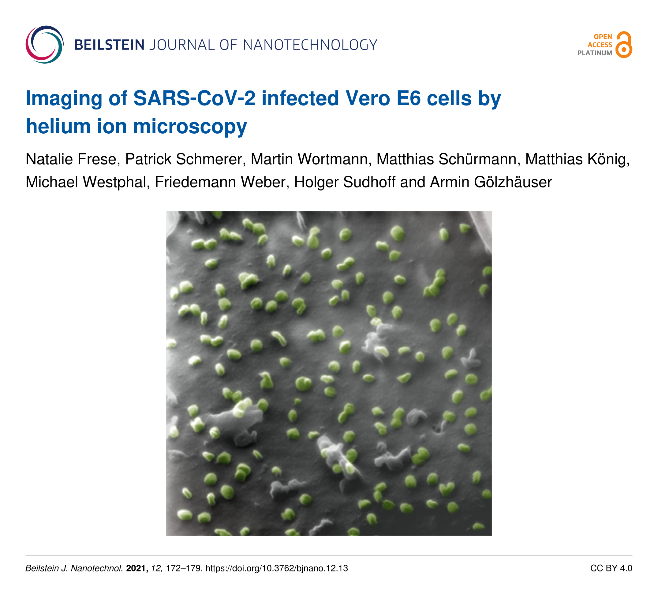
Imaging of SARS-CoV-2 infected Vero E6 cells by helium ion microscopy
Natalie Frese, Patrick Schmerer, Martin Wortmann, Matthias Schürmann, Matthias König, Michael Westphal, Friedemann Weber, Holger Sudhoff and Armin Gölzhäuser
Beilstein J. Nanotechnol. 2021, 12, 172–179.
https://doi.org/10.3762/bjnano.12.13
How to Cite
Frese, N.; Schmerer, P.; Wortmann, M.; Schürmann, M.; König, M.; Westphal, M.; Weber, F.; Sudhoff, H.; Gölzhäuser, A. Beilstein J. Nanotechnol. 2021, 12, 172–179. doi:10.3762/bjnano.12.13
Download Citation
Citation data can be downloaded as file using the "Download" button or used for copy/paste from the text window
below.
Citation data in RIS format can be imported by all major citation management software, including EndNote,
ProCite, RefWorks, and Zotero.
Presentation Graphic
| Picture with graphical abstract, title and authors for social media postings and presentations. | ||
| Format: PNG | Size: 12.1 MB | Download |
Citations to This Article
Up to 20 of the most recent references are displayed here.
Scholarly Works
- Caldas, L. A.; Carneiro, F. A.; Monteiro, F. L. L.; Salles, T. S.; Gonçalves, N. R.; Higa, L. M.; Moreira, M. F.; Azevedo, R. C.; Tanuri, A.; de Souza, W. A comparative analysis of virus-cell interactions for two early Brazilian SARS-CoV-2 variants. Virology 2026, 617, 110822. doi:10.1016/j.virol.2026.110822
- Arriaga-Hernández, J.; Cuevas-Otahola, B.; Oliveros-Oliveros, J. J.; Morín-Castillo, M. M. Phase analysis simulating the Takeda method to obtain a 3D profile of SARS-CoV-2 cells. Pattern Analysis and Applications 2024, 27. doi:10.1007/s10044-024-01225-8
- Quaranta, P.; Scabia, G.; Storti, B.; Dattilo, A.; Quintino, L.; Perrera, P.; Di Primio, C.; Costa, M.; Pistello, M.; Bizzarri, R.; Maffei, M. SARS-CoV-2 Infection Alters the Phenotype and Gene Expression of Adipocytes. International journal of molecular sciences 2024, 25, 2086. doi:10.3390/ijms25042086
- Benchimol, M. Scanning Ion Microscopy and Its Application in Microbiology. Journal of Clinical Immunology & Microbiology 2023, 1–9. doi:10.46889/jcim.2023.4309
- Höflich, K.; Hobler, G.; Allen, F. I.; Wirtz, T.; Rius, G.; McElwee-White, L.; Krasheninnikov, A. V.; Schmidt, M.; Utke, I.; Klingner, N.; Osenberg, M.; Córdoba, R.; Djurabekova, F.; Manke, I.; Moll, P.; Manoccio, M.; De Teresa, J. M.; Bischoff, L.; Michler, J.; De Castro, O.; Delobbe, A.; Dunne, P.; Dobrovolskiy, O. V.; Frese, N.; Gölzhäuser, A.; Mazarov, P.; Koelle, D.; Möller, W.; Pérez-Murano, F.; Philipp, P.; Vollnhals, F.; Hlawacek, G. Roadmap for focused ion beam technologies. Applied Physics Reviews 2023, 10. doi:10.1063/5.0162597
- Wortmann, M.; Westphal, M.; Kaltschmidt, B.; Klöcker, M.; Layland, A. S.; Brockhagen, B.; Hütten, A.; Frese, N.; Ehrmann, A. Nanofibers are a matter of perspective: effects of methodology and subjectivity on diameter measurements. Nanoscale advances 2023, 5, 5900–5906. doi:10.1039/d3na00528c
- Cuevas Otahola, B.; Arriaga-Hernández, J.; Morín Castillo, M.; Oliveros Oliveros, J. 3D solid of SARS-CoV-2 viral particles applying Legendre polynomials from tomography Fourier analysis. Journal of the Optical Society of America. A, Optics, image science, and vision 2023, 40, 1994. doi:10.1364/josaa.498859
- Wortmann, M.; Keil, W.; Diestelhorst, E.; Westphal, M.; Haverkamp, R.; Brockhagen, B.; Biedinger, J.; Bondzio, L.; Weinberger, C.; Baier, D.; Tiemann, M.; Hütten, A.; Hellweg, T.; Reiss, G.; Schmidt, C.; Sattler, K.; Frese, N. Hard carbon microspheres with bimodal size distribution and hierarchical porosity via hydrothermal carbonization of trehalose. RSC advances 2023, 13, 14181–14189. doi:10.1039/d3ra01301d
- Mousley, M.; Tabean, S.; Bouton, O.; Hoang, Q. H.; Wirtz, T.; Eswara, S. Scanning Transmission Ion Microscopy Time-of-Flight Spectroscopy Using 20 keV Helium Ions. Microscopy and microanalysis : the official journal of Microscopy Society of America, Microbeam Analysis Society, Microscopical Society of Canada 2023, 29, 563–573. doi:10.1093/micmic/ozac049
- Sinnberg, T.; Lichtensteiger, C.; Hill-Mündel, K.; Leischner, C.; Niessner, H.; Busch, C.; Renner, O.; Wyss, N.; Flatz, L.; Lauer, U. M.; Hoelzle, L. E.; Nohr, D.; Burkard, M.; Marongiu, L.; Venturelli, S. Vitamin C Deficiency in Blood Samples of COVID-19 Patients. Antioxidants (Basel, Switzerland) 2022, 11, 1580. doi:10.3390/antiox11081580
- Gölzhäuser, A. Using the Helium Ion Microscope for Imaging and Modification of Nanostructures, 2D Materials, and SARS-CoV-2 Infected Cells. Microscopy and Microanalysis 2022, 28, 28. doi:10.1017/s1431927622001027
- Deroubaix, A.; Kramvis, A. Imaging Techniques: Essential Tools for the Study of SARS-CoV-2 Infection. Frontiers in cellular and infection microbiology 2022, 12, 794264. doi:10.3389/fcimb.2022.794264
- Schmidt, M. Hochaufgelöste Mikrobenporträts mit hoher Tiefenschärfe. Biospektrum : Zeitschrift der Gesellschaft fur Biologishe Chemie (GBCH) und der Vereinigung fur Allgemeine und Angewandte Mikrobiologie (VAAM) 2022, 28, 377–380. doi:10.1007/s12268-022-1772-z
- Merolli, A.; Kasaei, L.; Ramasamy, S.; Kolloli, A.; Kumar, R.; Subbian, S.; Feldman, L. C. An intra-cytoplasmic route for SARS-CoV-2 transmission unveiled by Helium-ion microscopy. Scientific reports 2022, 12, 3794. doi:10.1038/s41598-022-07867-0
- Wortmann, M.; Frese, N. Industrial‐scale vacuum casting with silicone molds: A review. Applied Research 2022, 1. doi:10.1002/appl.202100012
- Wortmann, M.; Keil, W.; Brockhagen, B.; Biedinger, J.; Westphal, M.; Weinberger, C.; Diestelhorst, E.; Hachmann, W.; Zhao, Y.; Tiemann, M.; Reiss, G.; Hüsgen, B.; Schmidt, C.; Sattler, K.; Frese, N. Pyrolysis of Sucrose-Derived Hydrochar. Journal of Analytical and Applied Pyrolysis 2022, 161, 105404. doi:10.1016/j.jaap.2021.105404
- Schmidt, M.; Bandara, C. D.; Tamisier, M.; Maasilta, I.; Byrne, J. M. Imaging and Ion-Beam Milling of Biological Specimens with the Helium-Ion Microscope. Microscopy and Microanalysis 2021, 27, 768–769. doi:10.1017/s1431927621003068