


Search results
Search for "endocytosis" in Full Text gives 105 result(s) in Beilstein Journal of Nanotechnology.
Cardiomyocyte uptake mechanism of a hydroxyapatite nanoparticle mediated gene delivery system
Beilstein J. Nanotechnol. 2020, 11, 1685–1692, doi:10.3762/bjnano.11.150
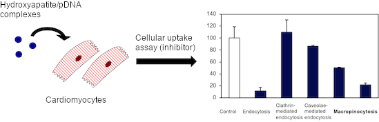
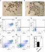
- vector for transfecting cardiomyocyte-derived HL-1 cells. HAp exhibited particles on the nanoscale and with a low cytotoxicity in HL-1 cells. The transfection assay performed with several endocytosis inhibitors suggested that the HAp/pDNA complexes were internalized by HL-1 cells through macropinocytosis
- . Furthermore, this HL-1 cell uptake was generated in response to HAp stimulation. Thus, HAp is a positive regulator of macropinocytosis in HL-1 cells and a good system for gene delivery in cardiomyocytes. Keywords: cardiomyocyte; endocytosis; gene delivery system; hydroxyapatite nanoparticles
- degradation by facilitating the release of the gene into the cytoplasm. Endocytosis has been well known as the main mechanism for cellular uptake of nanoparticles into mammalian cells [13][14]. The endocytosis process encompasses four main routes [15]: phagocytosis, clathrin-mediated endocytosis, caveolae


Applications of superparamagnetic iron oxide nanoparticles in drug and therapeutic delivery, and biotechnological advancements
Beilstein J. Nanotechnol. 2020, 11, 1092–1109, doi:10.3762/bjnano.11.94
- therapeutic efficacy, and safety studies. Keywords: drug delivery; drug targeting; endocytosis; medical; nanoparticles; superparamagnetic iron oxide nanoparticles (SPIONs); toxicity; Introduction Nanoencapsulation technologies have been researched over the past several decades and have been widely
- reticuloendothelial system [85][96]. Hennig et al. [97] obtained lauric acid/BSA-coated and dextran-coated SPIONs with neutral surface charge, which remained colloidally stable without forming any aggregates. This feature is very important for cell endocytosis, as aggregates precipitate between cells in vitro and
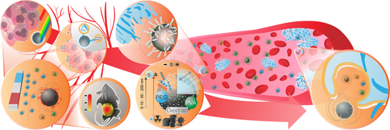
- readily detected. Figure 3 gives a schematic overview of how the physical and chemical characteristics of SPIONs influence biological response and biomedical applications. Endocytosis, exocytosis, retention and clearance There are a number of possible cell entry mechanisms for nanoparticles, such as


Key for crossing the BBB with nanoparticles: the rational design
Beilstein J. Nanotechnol. 2020, 11, 866–883, doi:10.3762/bjnano.11.72
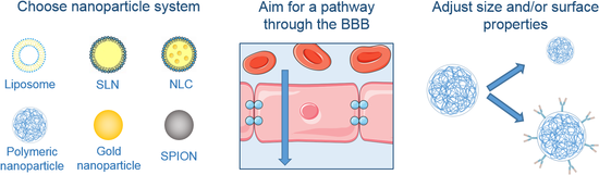
- [39]. However, this pathway remains one of the most promising for drug delivery through the BBB. Transcytosis includes three steps: endocytosis, intracellular vesicular trafficking and exocytosis [40]. Indeed, molecules bind to their receptors on the luminal side of the endothelial cells and
- endocytosis is initiated. The receptor–ligand complex is invaginated, which leads to the formation of intracellular transport vesicles. The vesicles are then sorted and the ones sorted for exocytosis cross the cell to release the ligand to its basolateral side. The receptor is then recycled [41]. Some of the
- endocytic vesicles have been identified and described in the brain endothelial cells: clathrin-coated pits, caveolae and macropinocytosis vesicles. Clathrin-coated pits are involved in most of the internalization processes mediated by receptors such as TfR or insulin receptors [39][40]. After endocytosis


Luminescent gold nanoclusters for bioimaging applications
Beilstein J. Nanotechnol. 2020, 11, 533–546, doi:10.3762/bjnano.11.42
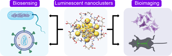
- endocytosis. The superparamagnetic nature of the PML-MF allowed for the magnetic targeting of the nanocarriers. Further, the ability of BSA to encapsulate drug molecules was explored to load doxorubicin (DPML-MF) in the nanocarriers. The release kinetics of doxorubicin studied at pH 7.4 and 4.4 were found to
- endocytosis of NCs. The generation of ROS upon X-ray irradiation in the presence of [Au8(C21H27O2)8] significantly suppressed the tumorigenicity in vivo after one radiotherapy treatment in mouse models. Because of their well-defined surface functionalities and small size, the dispersion behavior of NCs is
- the rational design of nanocarriers encapsulating poorly soluble drugs for targeted delivery. Cell line-based studies have provided enough evidence that the internalization of AuNCs occurs through endocytosis. However, there are several challenges to utilize AuNCs in bacterial sensing due to their


Brome mosaic virus-like particles as siRNA nanocarriers for biomedical purposes
Beilstein J. Nanotechnol. 2020, 11, 372–382, doi:10.3762/bjnano.11.28
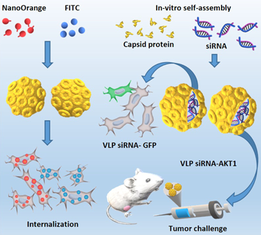
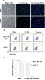
- . These results differ from those reported for cell internalization of cowpea mosaic virus (CPMV) in the same cell lines, which do not show high levels of vimentin on the cell surface [20][34]. Vimentin is a receptor that facilitates caveolar endocytosis [35][36], and it has been reported to promote the
- cellular internalization of CPMV [32][35][36][37]. Therefore, the cellular internalization of the CPMV virus occurs via endocytosis, which includes multiple routes: clathrin-dependent, caveolar and micropinocytosis [36][37][38]. Our results showed that the presence of low levels of vimentin on the cell


Interactions at the cell membrane and pathways of internalization of nano-sized materials for nanomedicine
Beilstein J. Nanotechnol. 2020, 11, 338–353, doi:10.3762/bjnano.11.25
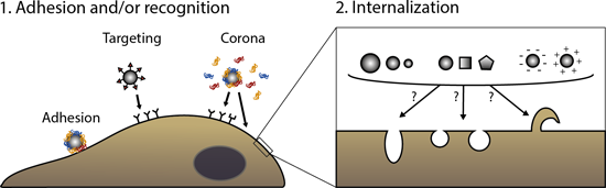
- receptors, and the subsequent mechanisms cells use to internalize them. The factors affecting these initial events are discussed. Then, we briefly describe the different pathways of endocytosis in cells and illustrate with some examples the challenges in understanding how nanomaterial properties, such as
- . Keywords: cell receptors; drug targeting; endocytosis; nanoparticle corona; nanoparticle uptake; Introduction Nano-sized materials are widely studied in nanomedicine for their potential use as drug carriers, in imaging, and for diagnostic purposes [1][2][3]. Because of their size, they can interact with
- of cellular internalization, i.e., endocytosis. Then, examples of works trying to understand how nanomaterial properties affect the mechanisms of uptake by cells are presented to illustrate the complexity of outcomes observed and the difficulties in drawing conclusions. 2.1 Pathways of endocytosis in


Rational design of block copolymer self-assemblies in photodynamic therapy
Beilstein J. Nanotechnol. 2020, 11, 180–212, doi:10.3762/bjnano.11.15
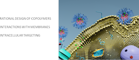
- carcinoma, ovarian, kidney, and pancreatic cancer). Chang et al. have explored the potential of PLA-PEG-AntiEGFR self-assemblies loaded with chlorin-e6 as a photosensitizer and found that this construct led to the increased internalization of the micelles through receptor-mediated endocytosis, which in turn
- efficiency using elongated vectors compared to spherical ones [137][138][139]. Simulations [139][140][141] showed that from a thermodynamic standpoint it is always more favorable to encapsulate spherocylindrical particles instead of spheres with the same radius and that endocytosis of spherocylinders occurs




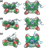


Internalization mechanisms of cell-penetrating peptides
Beilstein J. Nanotechnol. 2020, 11, 101–123, doi:10.3762/bjnano.11.10
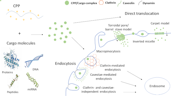
- the cell interior. Nevertheless, the mechanism they use to enter cells still remains an unsolved piece of the puzzle. Endocytosis and direct penetration have been suggested as the two major mechanisms used for internalization, however, it is not all black and white in the nanoworld. Studies have shown
- influence the cellular uptake mechanism. Keywords: cell-penetrating peptides; direct translocation; drug delivery; endocytosis; internalization; Introduction The cell membrane is a semipermeable barrier, serving as a protective layer for the cells. It is an essential organelle for cell survival and
- , just a few out of many, guide the internalization paths of CPPs to two major routes: endocytosis (active or energy-dependent uptake) and membrane translocation (direct/passive or energy-independent uptake). Overall, the type of uptake which will be selected mainly depends on the physicochemical


Bombesin receptor-targeted liposomes for enhanced delivery to lung cancer cells
Beilstein J. Nanotechnol. 2019, 10, 2553–2562, doi:10.3762/bjnano.10.246
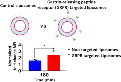
- -bound through GRPR binding cf. non-targeted liposomes, which exchange rapidly between the free and cell surface-bound state, would indirectly increase the relative intracellular accumulation. Access to the intracellular compartment would be achieved by constitutive plasma membrane endocytosis (e.g
- relative cellular accumulation (data not shown). To overcome this, the density of targeting lipid was increased to 3 mol %. To examine for active internalisation and intracellular accumulation of liposomes at the endocytosis permissive temperature of 37 °C, we subtracted the median fluorescence intensity

Small protein sequences can induce cellular uptake of complex nanohybrids
Beilstein J. Nanotechnol. 2019, 10, 2477–2482, doi:10.3762/bjnano.10.238
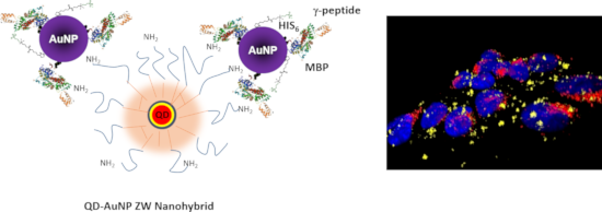
- the processes involved in the intracellular uptake and ensuing distribution of various nanomaterials [9]. Several studies have consistently found that NPs are very often taken up by endocytosis, and once inside the cells they remain trapped within endosomal compartments [10][12]. A few other studies
- reported that a sizable fraction of the delivered nanoparticles can end up in the cytoplasm, by either circumventing endocytosis through the use of virus-derived peptide sequences, or non-disruptively penetrating the cellular membranes [13]. Escape from endosomal vesicles of once endocytosed nanoparticles
- may not be driven by endocytosis [22]. Nonetheless, the distribution of the QD fluorescence is still different from that expected for a pure cytosolic delivery, where a more homogeneous distribution of the signal would be expected [33]. Whether these findings are due to the cellular response on the NP

Synthesis and potent cytotoxic activity of a novel diosgenin derivative and its phytosomes against lung cancer cells
Beilstein J. Nanotechnol. 2019, 10, 1933–1942, doi:10.3762/bjnano.10.189
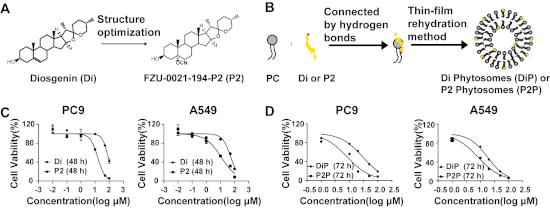
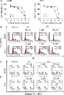
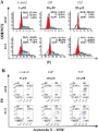
- of apoptosis, suggesting that the actions of P2 on induction of cell apoptosis could be altered after loading in phytosomes. One reason might be the changed drug uptake pathway. The free drugs were taken up by passive transport, while the phytosomes were taken up through endocytosis [39]. Besides



Engineered superparamagnetic iron oxide nanoparticles (SPIONs) for dual-modality imaging of intracranial glioblastoma via EGFRvIII targeting
Beilstein J. Nanotechnol. 2019, 10, 1860–1872, doi:10.3762/bjnano.10.181
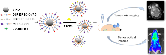
- enhanced accumulation of PNPs in EGFRvIII-positive tumors. Further TEM imaging demonstrated that plenty of the PNP nanoprobes accumulated in U87-EGFRvIII cells, suggesting the increased endocytosis of PNPs in U87-EGFRvIII cells (Figure 8b). Primary safety evaluation of PNPs The cytotoxicity of nanoprobes




Targeting strategies for improving the efficacy of nanomedicine in oncology
Beilstein J. Nanotechnol. 2019, 10, 168–181, doi:10.3762/bjnano.10.16
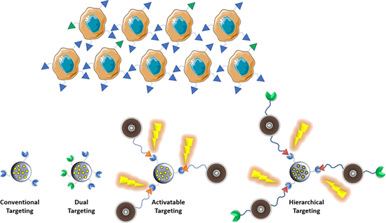
- strategy is the so-called “cellular or secondary targeting”, because it is generally based on a ligand–receptor-mediated endocytosis, triggered by the strong interaction of the targeting group with the membrane receptor of the tumoral cell. It improves the selectivity of the treatment achieving a drastic
- the action of a cell surface-associated protease exposing the RGD, which then binds to neuropilin-1 triggering the particle endocytosis. Another cell-penetrating peptide (or CPP, which is the usual name of the peptides used for targeting purposes in nanomedicine) closely related to RGD is the
- is endocytosis [51]. Usually, the nanocarriers enter into the cells into endosomes, which evolve into lysosomes, which can lead to the degradation of the transported cargo, especially in the case of sensitive agents such as genes or siRNA. Therefore, it is necessary to design mechanisms to induce the


Cytotoxicity of doxorubicin-conjugated poly[N-(2-hydroxypropyl)methacrylamide]-modified γ-Fe2O3 nanoparticles towards human tumor cells
Beilstein J. Nanotechnol. 2018, 9, 2533–2545, doi:10.3762/bjnano.9.236
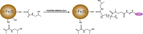
- their enhanced accumulation inside the cells. The particles are able to penetrate cells by the selected type of endocytosis mechanism: phagocytosis, pinocytosis, or receptor mediated endocytosis [25]. In order to check this hypothesis, cellular uptake of γ-Fe2O3@P(HPMA-MMAA)-Dox nanoparticles was


Nanoparticle delivery to metastatic breast cancer cells by nanoengineered mesenchymal stem cells
Beilstein J. Nanotechnol. 2018, 9, 321–332, doi:10.3762/bjnano.9.32
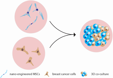
- ) compared with 3D culture (Figure 4B). To the contrary, MDA-MB-231 internalised 6-fold more QDs in 3D culture compared with 2D (Figure 4B). Such discrepancy in uptake efficacy might be associated with different endocytosis pathways. MCF7 cells internalised QDs through phagocytosis and clathrin/caveolae
- -dependent endocytosis, whereas the clathrin/caveolae-dependent pathway dominated in MDA-MB-231 cells in monocultures (Supporting Information File 1, Figure S2). Cell viability in 3D culture Cells in a 3D culture formed floating and dense spheroids. Therefore, we sought to analyse the effect of the 3D
- facilitating the uptake of MSC-excreted QDs by cancer cells. Our data demonstrate that phagocytosis and clathrin/caveolae-dependent endocytosis are the major QD uptake pathways in MCF7 cells, whereas the clathrin/caveolae-dependent pathway dominated in MDA-MB-231 cells in monocultures (see Supporting



Involvement of two uptake mechanisms of gold and iron oxide nanoparticles in a co-exposure scenario using mouse macrophages
Beilstein J. Nanotechnol. 2017, 8, 2396–2409, doi:10.3762/bjnano.8.239
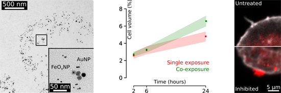
- negative, must be considered for nanotechnology and nanomedicine in particular to develop to its full potential. Keywords: co-exposure; endocytosis; live cell imaging; nanoparticles; quantitative microscopy; Introduction Over the past two decades, improvements in nanomaterial research were followed by a
- , which can be subdivided into macro- and microscale processes [8]. The first, phagocytosis, involves the ingestion of large particles: NPs or agglomerates typically larger than 0.25 μm in diameter. The second, pinocytosis, includes micro- and macropinocytosis, clathrin-, and caveolin-mediated endocytosis
- , and clathrin- and caveolin-independent endocytosis, involving the ingestion of fluid, molecules, and NPs via small vesicles (<0.15 µm in diameter) [9]. Although NPs have been shown to be taken up by the cells mainly by pinocytotis [9], many factors have been shown to influence the interaction with



Uptake and intracellular accumulation of diamond nanoparticles – a metabolic and cytotoxic study
Beilstein J. Nanotechnol. 2017, 8, 1649–1657, doi:10.3762/bjnano.8.165
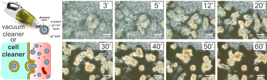
- in cultures exposed and unexposed to photoluminescent nanodiamonds. This positive effect can be attributed to the fact that the mechanism of the ND uptake was clathrin-mediated endocytosis, that is, a physiological cellular mechanism for internalization of various bioactive substances from the


Nano-engineered skin mesenchymal stem cells: potential vehicles for tumour-targeted quantum-dot delivery
Beilstein J. Nanotechnol. 2017, 8, 1218–1230, doi:10.3762/bjnano.8.123
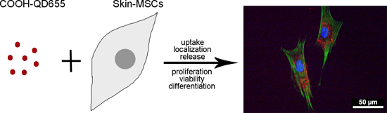
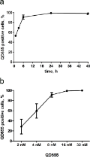
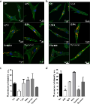
- examined via fluorescence imaging using endocytosis inhibitors for the micropinocytosis, phagocytosis, lipid-raft, clathrin- and caveolin-dependent endocytosis pathways. These data showed that QDs were efficiently accumulated in the cytoplasm of MSCs after incubation for 6 h. The main uptake route of QDs
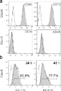
- in skin MSCs was clathrin-mediated endocytosis. QDs were mainly localized in early endosomes after 6 h as well as in late endosomes and lysosomes after 24 h. QDs in concentrations ranging from 0.5 to 64 nM had no effect on cell viability and proliferation. The expression of MSC markers, CD73 and CD90
- suggest that QD-labelled MSCs could be used for targeted drug delivery studies. Keywords: endocytosis; mesenchymal stem cells; quantum dots; stem cell differentiation; Introduction Despite remarkable advances in targeted therapies of various human malignancies, cancer is one of the leading causes of





Uptake of the proteins HTRA1 and HTRA2 by cells mediated by calcium phosphate nanoparticles
Beilstein J. Nanotechnol. 2017, 8, 381–393, doi:10.3762/bjnano.8.40
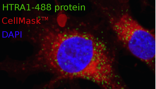
- flow cytometry. All proteins were readily transported into the cells by cationic calcium phosphate nanoparticles. Notably, only HTRA1 was able to penetrate the cell membrane of MG-63 cells in dissolved form. However, the application of endocytosis inhibitors revealed that the uptake pathway was
- different for dissolved HTRA1 and HTRA1-loaded nanoparticles. Keywords: calcium phosphate; endocytosis; nanoparticles; proteins; Introduction Many receptors for drugs or proteins are located inside cells [1][2]. However, because many biomolecules are not able to penetrate the cell membrane on their own, a
- suitable carrier is required [3][4]. Nanoparticles are readily taken up by cells via endocytosis and are easily able to deliver their cargo into cells across the cell membrane [5][6][7]. Calcium phosphate nanoparticles have demonstrated to be very efficient to transport (bio)molecules into cells [8][9


Viability and proliferation of endothelial cells upon exposure to GaN nanoparticles
Beilstein J. Nanotechnol. 2016, 7, 1330–1337, doi:10.3762/bjnano.7.124
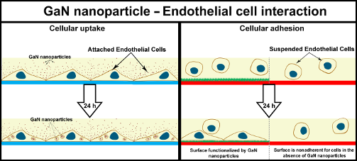
- susceptible for them. In contrast to this situation, the cells take up all the GaN nanoparticles floating in the medium, thus burning energy via the endocytosis process and slowing down the proliferation activity. Conclusion We found that the interaction of porcine endothelial cells with GaN nanoparticles



On the pathway of cellular uptake: new insight into the interaction between the cell membrane and very small nanoparticles
Beilstein J. Nanotechnol. 2016, 7, 1296–1311, doi:10.3762/bjnano.7.121
- : ATP depletion; calcium crystallization; cytotoxicity; endocytosis; HeLa cells; LDH; mesenchymal stem cells; morphology; necrosis; particle size; silica nanoparticles; TEM; Introduction Silicon dioxide nanoparticles (SiNPs) are used in a wide range of commercially available products to improve product
- see a size dependency of NP endocytosis (Figure 4, Figure 10, Figure 11). The endocytic route for the larger of our examined NPs (SiNP-22 and SiNP-12) can be described as following: First the NPs are taken up as individuals or in small groups and are found in the cytosol with tightly wrapping membrane
- additional silica layer is likely to change the viscoelastic properties of the cell membrane yielding an increased maximum bending radius. Nevertheless, the cell manages to perform the pinch-off which is indicative for an active endocytic process. Moreover, when suppressing any active endocytosis by cooling




Improved biocompatibility and efficient labeling of neural stem cells with poly(L-lysine)-coated maghemite nanoparticles
Beilstein J. Nanotechnol. 2016, 7, 926–936, doi:10.3762/bjnano.7.84
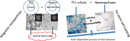
- ]. Endocytosis as a process of internalization of foreign materials can be divided into two major groups, phagocytosis for larger particles and pinocytosis for nanoparticles. Pinocytosis can be further subdivided depending on the size of particles into clathrin-mediated, caveolae, and macropinocytosis [25]. To
- -dependent process macropinocytosis cytochalasine D, inhibitor of microtubule function involved in intracellular vesicle trafficking nocodazole, inhibitor of the clathrin-mediated endocytosis phenylarsine oxide and filipin, which blocks caveolae pathways [25]. Our results suggested that the internalization
- determine the mechanism of nanoparticle uptake, NSCs were pre-treated with inhibitors of endocytosis for 30 min and then incubated with nanoparticles for 48 h in the presence of the inhibitor [25]. The inhibitors were phenylarsine oxide (12 nM), cytochalasin D (60 nM), nocodazole (20 nM) and filipin (0.3 µg




The role of morphology and coupling of gold nanoparticles in optical breakdown during picosecond pulse exposures
Beilstein J. Nanotechnol. 2016, 7, 869–880, doi:10.3762/bjnano.7.79
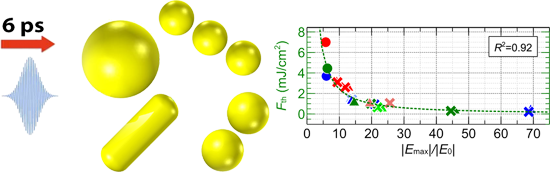
- surface of cell [6][7][8][9], modification of gold nanoparticle surfaces to cause binding to each other [10][11] or endocytosis [12]. Nanoparticle assemblies exhibit optical behavior that differs from single particles. The changes in the optical behavior of nanoparticle assemblies are governed by surface




Tight junction between endothelial cells: the interaction between nanoparticles and blood vessels
Beilstein J. Nanotechnol. 2016, 7, 675–684, doi:10.3762/bjnano.7.60
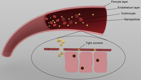
- , while a significant change occurred in myoblasts after 4 h [87]. Recent studies revealed that hydrodynamic conditions influence the endothelial endocytosis of nanocarriers. By using nanocarriers targeted to PECAM-1, the authors found a flow-stimulated endocytosis of nanocarriers through eliciting

pH-Triggered release from surface-modified poly(lactic-co-glycolic acid) nanoparticles
Beilstein J. Nanotechnol. 2015, 6, 2504–2512, doi:10.3762/bjnano.6.260
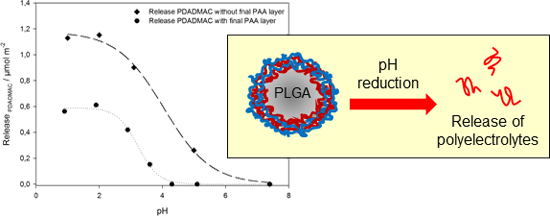
- taken up by endocytosis. During the process of endocytosis, nanoparticular drug carriers most often end up in endolysosomes with a reduced internal pH value. In order to provide improved accessibility of the drug to the whole cell, membrane destruction of the endolysosomal bilayer would be beneficial





