Search results
Search for "X-ray" in Full Text gives 954 result(s) in Beilstein Journal of Nanotechnology. Showing first 200.
N-Heterocyclic carbene-based gold etchants
Beilstein J. Nanotechnol. 2023, 14, 865–871, doi:10.3762/bjnano.14.71

- dissolution of gold. We present scanning electron micrographs and elemental imaging analyses by energy dispersive X-ray spectroscopy to examine the effect of solutions of each species on the gold film. This work highlights the risk of unwanted etching during some routes to NHC-based surface functionalization
- % nitrogen gas. Electron micrographs were recorded using the SE2 detector of a Zeiss SIGMA VP field emission scanning electron microscope (SEM). Energy dispersive X-ray spectroscopy (EDS) spectra were obtained for each sample using an Oxford Instruments X-max 50 mm2 EDS attachment. Peaks were identified


A wearable nanoscale heart sound sensor based on P(VDF-TrFE)/ZnO/GR and its application in cardiac disease detection
Beilstein J. Nanotechnol. 2023, 14, 819–833, doi:10.3762/bjnano.14.67

- . Composition and β-phase content of the piezoelectric composite films were analyzed using X-ray diffraction. The morphology of the composite film fibers was observed through scanning electron microscopy. Finally, the P(VDF-TrFE)/ZnO/graphene composite film was encapsulated in a sandwich-structure heart sound
- underwent characterization through electron microscopy, X-ray diffraction (XRD), and piezoelectric performance testing. The results indicated that the piezoelectric film with a composition ratio of 12% P(VDF-TrFE) + 10% ZnO + 0.1% GR exhibited superior performance regarding various aspects. Consequently, in
- -TrFE)/ZnO flexible piezoelectric composite films without GR. Analysis of X-ray diffraction patterns indicated that the β-phase content of P(VDF-TrFE)/ZnO/GR piezoelectric composite films was higher than that of P(VDF-TrFE)/ZnO and P(VDF-TrFE). P(VDF-TrFE)/ZnO/GR was packaged into a flexible


Nanostructured lipid carriers containing benznidazole: physicochemical, biopharmaceutical and cellular in vitro studies
Beilstein J. Nanotechnol. 2023, 14, 804–818, doi:10.3762/bjnano.14.66

- observed by transmission electron microscopy. Fourier-transform infrared spectroscopy, differential scanning calorimetry, thermogravimetric analysis, and small-angle X-ray scattering analyses of the nanoparticles indicated that BNZ might be dispersed in the nanoparticle matrix in an amorphous state. The
- ]. Structural analysis was performed by selecting different angular regions from the small-angle X-ray scattering (SAXS) and wide-angle X-ray scattering (WAXS) patterns. The WAXS patterns (Figure 5) showed contributions of diffraction peaks from BNZ, myristyl myristate, and NLC. The nanostructured lipid
- medium observed after the initial stage could be attributed to the gradual release of drug molecules from the matrix core, where the drug is mainly located according to X-ray diffraction (XRD) results [33]. Remarkably, although our NLC possess a comparatively lower drug load, the maximal accumulated drug


Silver-based SERS substrates fabricated using a 3D printed microfluidic device
Beilstein J. Nanotechnol. 2023, 14, 793–803, doi:10.3762/bjnano.14.65

- NPs. The SEM image of PS@Ag reveals the presence of nanoscale gaps between the Ag NPs, which act as hot spots with a high electric field intensity when exposed to laser irradiation (Figure S10c). To confirm the distribution of chemical elements on the SERS substrate, energy-dispersive X-ray


Silver nanoparticles loaded on lactose/alginate: in situ synthesis, catalytic degradation, and pH-dependent antibacterial activity
Beilstein J. Nanotechnol. 2023, 14, 781–792, doi:10.3762/bjnano.14.64

- potential measurements, which were measured on a nanoPartica Horiba SZ-100 (Japan). Fourier-transform infrared (FTIR) spectra were obtained using a Bruker Tensor 27 FTIR spectrophotometer (Germany). X-ray diffraction (XRD) patterns were collected using a Bruker D8 Advance X-ray diffractometer. The


Carboxylic acids and light interact to affect nanoceria stability and dissolution in acidic aqueous environments
Beilstein J. Nanotechnol. 2023, 14, 762–780, doi:10.3762/bjnano.14.63

- produced drastically different TEM images, as expected from the DLS results. The number of nanoceria particles exposed to citric acid was reduced between weeks 0 and 1. By week 2, most of the particles completely disappeared with no evidence of cerium determined by energy-dispersive X-ray spectroscopy (EDS

In situ magnesiothermic reduction synthesis of a Ge@C composite for high-performance lithium-ion batterie anodes
Beilstein J. Nanotechnol. 2023, 14, 751–761, doi:10.3762/bjnano.14.62

- and transferred to a ceramic crucible. The mixture was heated at 750 °C in argon gas flow for 3 h. The obtained solid was re-ground and denoted as Ge/C-SS750. Material characterization X-ray diffraction measurements (XRD, Bruker D8 Advance with Cu Kα radiation (λ = 1.5406 Å) at 40 kV and 40 mA) were


Control of morphology and crystallinity of CNTs in flame synthesis with one-dimensional reaction zone
Beilstein J. Nanotechnol. 2023, 14, 741–750, doi:10.3762/bjnano.14.61

- observed, revealing the agglomeration and sintering of particles during high-temperature growth in the flame. The energy dispersive X-ray (EDX) analysis results in Table 1 indicate a high fraction of nickel. A previous study showed that nickel particles of 5 nm and above can be reactive towards CH4, which
- by field-emission scanning electron microscopy (FESEM, Zeiss Crossbeam 340) coupled with energy-dispersive X-ray analysis (EDX) for morphology and elemental analysis. Raman spectroscopy (HORIBA XploRA PLUS, 532 nm) was done to analyze the signature spectra of the grown CNTs. Line-of-sight images of


A graphene quantum dots–glassy carbon electrode-based electrochemical sensor for monitoring malathion
Beilstein J. Nanotechnol. 2023, 14, 701–710, doi:10.3762/bjnano.14.56

- hydrothermal process with glucose as a precursor undergoing carbonization. Different spectroscopic techniques were used to analyze the optical characteristics of GQDs, including UV–visible, photoluminescence, FTIR, and Raman spectroscopy. Atomic force microscopy, transmission electron microscopy, and X-ray
- synthesized GQDs using the Debye–Scherrer formula, D = 0.9λ/(β·cos θ), where D is the average crystallite size of the synthesized GQDs, λ is the X-ray wavelength, θ is the Bragg diffraction angle, and β is the FWHM. The elemental analysis of GQDs from EDX measurements is shown in Figure 4b. The EDX spectrum


The microstrain-accompanied structural phase transition from h-MoO3 to α-MoO3 investigated by in situ X-ray diffraction
Beilstein J. Nanotechnol. 2023, 14, 692–700, doi:10.3762/bjnano.14.55

- Sciences, Beijing, 100190, China 10.3762/bjnano.14.55 Abstract In situ X-ray diffraction indicates that the structural phase transition from h-MoO3 to α-MoO3 is a first-order transition with a phase transition temperature range of 378.5–443.1 °C. The linear coefficients of thermal expansion of h-MoO3 are
- α-MoO3 is still unclear. Here, to reveal the features of the structural phase transition from h-MoO3 to α-MoO3, we performed in situ X-ray diffraction experiments at temperatures ranging from 30 to 450 °C. The Rietveld refinement results indicate water molecules at the (0 0 0.25) site inside the
- α-MoO3 To observe the crystal structure evolution of h-MoO3 induced by temperature, a thoroughly powdered sample was used to perform in situ X-ray diffraction measurements during heating from 30 to 450 °C, as shown in Figure 1a. At 30 °C, all diffraction peaks can be well indexed to the hexagonal


Titania nanoparticles for photocatalytic degradation of ethanol under simulated solar light
Beilstein J. Nanotechnol. 2023, 14, 616–630, doi:10.3762/bjnano.14.51

- pyrolysis from TiCl4 vapor in air in the presence of ethylene as sensitizer at different working pressures (250–850 mbar) with and without further calcination at 450 °C. The obtained powders were analyzed by energy-dispersive X-ray spectroscopy, X-ray photoelectron spectroscopy, X-ray diffractometry, and
- described as: The raw TiO2 powders contain some carbon (from the decomposition of the ethylene sensitizer) and chlorine impurities, whose amount it is greatly diminished by calcination in air at 450 °C for 5 h. To certify this, a composition investigation by energy-dispersive X-ray spectroscopy (EDS) has
- (point defects), there are some deviations. The small oxygen deficiency observed even after calcination is related to remaining chlorine impurities and Ti3+ ions that resisted calcination. Phase composition and crystallites sizes of the TiO2 powders were investigated. The X-ray diffractograms of the


Carbon nanotube-cellulose ink for rapid solvent identification
Beilstein J. Nanotechnol. 2023, 14, 535–543, doi:10.3762/bjnano.14.44

- rapid test, identification, and monitoring of liquid samples in various fields such as fuel adulteration, water quality, solvents, and beverages [6][7][8][9]. Usually, the testing of liquids requires conventional analytical techniques such as absorption/emission spectroscopy (AAS/AES), X-ray


Nanoarchitectonics to entrap living cells in silica-based systems: encapsulations with yolk–shell and sepiolite nanomaterials
Beilstein J. Nanotechnol. 2023, 14, 522–534, doi:10.3762/bjnano.14.43

- without staining. Electron microscopy imaging was conducted using a field-emission scanning electron microscope FEI-NOVA NanoSEM 230 equipped with an Apollo XL silicon drift detector from EDAX-Ametek or using a high-resolution JEOL IT500HR/LA microscope equipped with an energy dispersive X-ray


On the use of Raman spectroscopy to characterize mass-produced graphene nanoplatelets
Beilstein J. Nanotechnol. 2023, 14, 509–521, doi:10.3762/bjnano.14.42

- %), transmission electron microscopy (TEM) (7.2%) or X-ray photoelectron spectroscopy (XPS) (5.6%). It has the advantages of relatively low cost, simple sample preparation, quick measurements, and automated analysis, offering clear benefits for quality control applications. It has been demonstrated in several


Mixed oxides with corundum-type structure obtained from recycling can seals as paint pigments: color stability
Beilstein J. Nanotechnol. 2023, 14, 467–477, doi:10.3762/bjnano.14.37

- structure. The pigments are obtained via the addition of coloring ions to boehmite from recycled metallic aluminium. X-ray diffractometry (XRD) and Raman spectroscopy confirmed the crystallographic phase. Additionally, the oxidation state 3+ responsible for the greenish (chromium) and reddish (iron
- obtaining boehmite, we added chromium and iron ions to obtain colored mixed oxides with a corundum-type structure. The stability of the synthesized pigments in acid and alkaline environments was evaluated by colorimetric measurements. Results and Discussion X-ray diffractometry (XRD) The XRD of the pristine
- characteristic of α-Fe2O3 nanoparticles [21]. The same morphology was observed for concentrations of 5 and 20 wt % of coloring ions (Figure S2, Supporting Information File 1). X-ray photoelectron spectroscopy (XPS) The elemental composition of the samples evaluated by the analysis of XPS spectra is shown in


Evaluation of electrosynthesized reduced graphene oxide–Ni/Fe/Co-based (oxy)hydroxide catalysts towards the oxygen evolution reaction
Beilstein J. Nanotechnol. 2023, 14, 420–433, doi:10.3762/bjnano.14.34
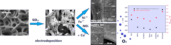
- magnifications of the morphology of NiFe and CoNiFe after GO addition are presented in Figure S1 and S2, respectively, in Supporting Information File 1. Figure 2 presents the energy-dispersive X-ray (EDX) maps with corresponding SEM images of the catalysts. The analysis confirms the presence of the following
- probably inhibited the electrodeposition process of NiFe and CoNiFe on its surface. This may be the reason for the slower stabilization of the synthesis current density observed in the chronoamperograms (Figure 1a). X-ray diffraction, X-ray photoemission spectroscopy and X-ray absorption spectroscopy
- Figure 3a–d shows the X-ray absorption spectra (XAS) of the L3 edge of nickel (a), iron (b), cobalt (c), and carbon (d) in the studied catalysts. The appearance of a shoulder peak at the L3 edge of the nickel (Figure 3a) at 855 eV indicates the presence of oxides in the structure of the catalysts (Ni in


Recent progress in cancer cell membrane-based nanoparticles for biomedical applications
Beilstein J. Nanotechnol. 2023, 14, 262–279, doi:10.3762/bjnano.14.24

- anticancer effects [76]. After QT was delivered to tumor tissue by the active targeting ability of the membrane, the sensitivity to radiotherapy was effectively improved, and a strong anticancer effect was exerted under X-ray irradiation [76]. Gong et al. designed a pH-responsive multifunctional biomimetic


A novel approach to pulsed laser deposition of platinum catalyst on carbon particles for use in polymer electrolyte membrane fuel cells
Beilstein J. Nanotechnol. 2023, 14, 190–204, doi:10.3762/bjnano.14.19

- accelerating voltage of 20 kV and identical work parameters of the EDS detector. The quality of platinum coating on the carbon material was assessed based on images made using a transmission electron microscope (TEM) equipped with an energy-dispersive X-ray spectrometer (EDX). TEM measurements were carried out
- the prepared platinum-coated carbon support, X-ray photoelectron spectroscopy (XPS) was used (Prevac, Poland) with an R3000 VG analyzer (Scienta, Sweden) and an X-ray source with an Al Kα anode (Prevac, Poland) emitting X-rays with a photon energy of 1486.6 eV. The analysis of the XPS spectra


Formation of nanoflowers: Au and Ni silicide cores surrounded by SiOx branches
Beilstein J. Nanotechnol. 2023, 14, 133–140, doi:10.3762/bjnano.14.14

- annealed at 1050 °C was named 15Au5Ni. The morphology was imaged by optical microscopy (OM, Zeiss Axiotech) and high-resolution scanning electron microscopy (HR-SEM, Hitachi S-4800) equipped with energy-dispersive X-ray spectroscopy (EDS, Thermo Scientific). The SEM images were recorded by using mixed
- atomic numbers show brighter contrasts. EDS measurements were performed to obtain the element distribution in the target areas. X-ray diffraction (XRD, Siemens D-5000) analyses were conducted in Bragg–Brentano mode using Cu Kα irradiation at 40 kV. The height distribution of the areas of interest was


Characterisation of a micrometer-scale active plasmonic element by means of complementary computational and experimental methods
Beilstein J. Nanotechnol. 2023, 14, 110–122, doi:10.3762/bjnano.14.12

- reference light is recorded after the aperture and reflected from a cube beam splitter, with the signal photodiode placed on the 2θ arm of a high-accuracy (18 arcsec resolution) Siemens θ–2θ X-ray diffractometer stage with inbuilt goniometer to collect light reflected from the interface. The absolute


Combining physical vapor deposition structuration with dealloying for the creation of a highly efficient SERS platform
Beilstein J. Nanotechnol. 2023, 14, 83–94, doi:10.3762/bjnano.14.10

- SERS properties of the nanoporous structure. Using scanning electron microscopy (SEM) and X-ray photoelectron spectroscopy (XPS) the morphology and surface composition of each nanoporous structure were respectively evaluated and used to describe the SERS properties of the samples. Results and
- to the alloy thin film. Three different silver compositions were selected, namely 18, 30, and 36 atom %, and characterized by SEM/energy-dispersive X-ray spectroscopy (EDX). Figure 1 displays the SEM micrograph of the as-deposited thin films. The thin films exhibit a columnar morphology (see the
- a sample and on two samples made in the same conditions. To evaluate the surface composition and oxidation state, XPS was used. The XPS measurements were carried out on a PHI 5000 VersaProbe using a monochromatic Al Kα X-ray source (1486.6 eV). The high-resolution spectra were recorded with a pass


Liquid phase exfoliation of talc: effect of the medium on flake size and shape
Beilstein J. Nanotechnol. 2023, 14, 68–78, doi:10.3762/bjnano.14.8

- powder was exfoliated in each liquid medium by exposure to mechanical energy provided by an ultrasonic bath (full details in the Experimental section). Talc was manually milled down to a fine powder and characterized by X-ray diffraction (XRD). Figure 1a displays the results. All peaks are assigned to
- obtaining information on thousands of flakes and using appropriate statistical descriptions to analyze the data. Experimental Materials. Talc was obtained through a donation of a sample from Minas Gerais state, Brazil. X-ray diffraction (XRD) was performed to characterize the sample composition. The rock
- measurements were performed on silicon substrates with an oxide layer, Si/SiOx. Substrates were functionalized with (3-aminopropyl)triethoxysilane (APTES) following the procedure reported by Fernandes and co-workers [24]. X-ray diffraction. XRD was performed in a Rigaku Geigerflex 2037 diffractometer with a


Gap-directed chemical lift-off lithographic nanoarchitectonics for arbitrary sub-micrometer patterning
Beilstein J. Nanotechnol. 2023, 14, 34–44, doi:10.3762/bjnano.14.4

- the rectangle function in ZEN 2012 Service Pack 2 software (Carl Zeiss Microscopy, Jena, Germany). XPS spectra after each surface modification step were collected with ULVAC-PHI X-ray photoelectron spectrometer (PHI QuanteraII, Kanagawa, Japan). Selective wet chemical etching processes and metal


Two-step single-reactor synthesis of oleic acid- or undecylenic acid-stabilized magnetic nanoparticles by thermal decomposition
Beilstein J. Nanotechnol. 2023, 14, 11–22, doi:10.3762/bjnano.14.2

- , and composition via several techniques, such as transmission electron microscopy, dynamic light scattering, thermogravimetric analysis, Fourier-transform infrared spectroscopy/attenuated total reflectance, 57Fe Mössbauer spectroscopy, and X-ray diffraction. The effect of unsaturated oleic (OA) and
- were uniform and the single spots were not visible proved that the crystallites were very small. These results correspond well with data from X-ray diffraction (XRD), according to which the average size of the crystallites for all prepared nanoparticles was 4.5–9 nm. The average crystallite size did
- crystallites obtained by estimating the expansion of the X-ray diffraction line (DXRD calculated with Scherer, optionally Rietveld, refinement), which indicated a single magnetic domain characteristic of the TMO-I nanoparticle sample. When a stabilizer with a shorter carbon chain (i.e., UA) is used under the


Electrical and optical enhancement of ITO/Mo bilayer thin films via laser annealing
Beilstein J. Nanotechnol. 2022, 13, 1589–1595, doi:10.3762/bjnano.13.133

- the samples were placed behind the focal plane of the lens (low intensity and big spot). The crystalline properties of the films were determined using X-ray diffraction (PANalytical diffractometer, λ = 1.5406 Å). The XRD measurements were carried out in 2θ mode between 20° and 80°. Topology and


