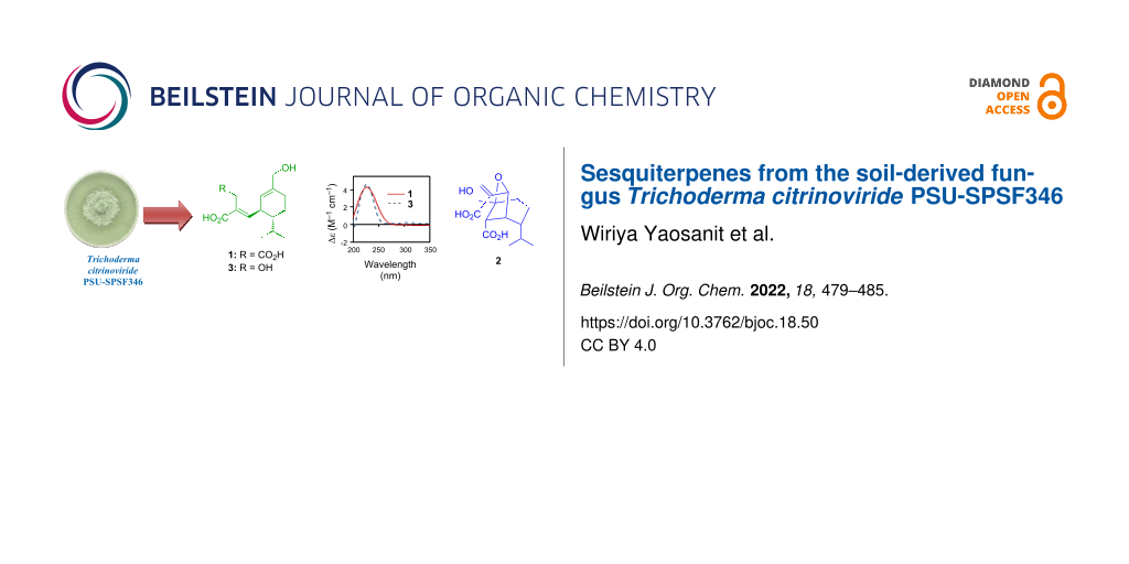
Abstract
Two new sesquiterpenes, trichocitrinovirenes A (1) and B (2), and five known compounds including four structurally related sesquiterpenes and one γ-lactone were isolated from the soil-derived fungus Trichoderma citrinoviride PSU-SPSF346. The structures were identified by analysis of their spectroscopic data. The relative configuration was assigned based on NOEDIFF data. The absolute configuration of compound 1 was established according to specific rotations and ECD data while that of compound 2 was proposed based on biosynthetic considerations. Compound 2 possesses a rare bicyclic sesquiterpene skeleton. The antimicrobial and cytotoxic activities of the isolated compounds were evaluated.

Graphical Abstract
Introduction
The fungus Trichoderma citrinoviride produces structurally diverse secondary metabolites including diterpenes [1,2], alkaloids [3], sorbicillinoids [4,5], long chain alcohols [6], and cyclonerane sesquiterpenes [7]. Some of them display antibacterial [1,2,7], cytotoxic [3], anti-inflammatory [4], and antimicroalgal [7] activities. Based on these data, the investigation of secondary metabolites from this fungus is still limited. In our ongoing search for antimicrobial secondary metabolites from soil-derived fungi, T. citrinoviride PSU-SPSF346 was isolated from a soil sample collected from the Sirindhorn Peat Swamp Forest, Narathiwat Province, Thailand. The crude mycelial extract of T. citrinoviride PSU-SPSF346 displayed antimicrobial activities against Staphylococcus aureus, methicillin-resistant S. aureus, and Cryptococcus neoformans ATCC90113 with MIC values of 128, 200 and 64 μg/mL, respectively. Herein, we report the isolation and structure elucidation as well as antimicrobial and cytotoxic activities of some isolated compounds which were obtained in sufficient amount.
Results and Discussion
Chemical investigation of the broth and mycelial extracts of T. citrinoviride PSU-SPSF346 by various chromatography techniques led to the isolation of two new sesquiterpenes, trichocitrinovirenes A (1) and B (2), four known structurally related sesquiterpenes including gliocladic acid (3), hydroheptelidic acid (4), and xylaric acids B (5) and D (6) [8], as well as one known γ-lactone, fusidilactone A (7) [9] (Figure 1). Their structures were elucidated on the basis of analysis of their spectroscopic data including IR, UV, NMR, and MS. The relative configuration was assigned based on NOEDIFF data. Furthermore, the absolute configuration of compound 1 was determined by comparison of its specific rotation and electronic circular dichroism (ECD) data with those of compound 3, whereas that of compound 2 was proposed based on biosynthetic considerations. The structures of the known compounds were further confirmed by comparison of their 1H and 13C NMR spectroscopic data, specific rotations, and ECD data with those previously reported.
![[1860-5397-18-50-1]](/bjoc/content/figures/1860-5397-18-50-1.svg?scale=2.0&max-width=1024&background=FFFFFF)
Figure 1: Structures of compounds 1–7 isolated from Trichoderma citrinoviride PSU-SPSF346.
Figure 1: Structures of compounds 1–7 isolated from Trichoderma citrinoviride PSU-SPSF346.
Trichocitrinovirene A (1) was isolated as a colorless gum and had the molecular formula C15H22O5 on the basis of the HRESIMS peak at m/z 305.1359 [M + Na]+. The IR spectrum exhibited absorption bands at 3336, 1684, and 1649 cm−1 for hydroxy, conjugated carboxyl carbonyl, and double bond functional groups, respectively [10]. The 1H NMR spectrum (Table 1) displayed characteristic signals for two olefinic protons of two trisubstituted alkenes (δH 6.64, d, J = 10.5 Hz, and 5.31, s, each 1H), two methine protons (δH 3.06 and 1.35, each m, 1H), one set of equivalent oxymethylene protons (δH 3.92, s, 2H), three sets of nonequivalent methylene protons (δH 3.40 and 3.30, each d, J = 16.8 Hz, 1H; δH 2.13 and 2.04, each m, 1H; δH 1.81 and 1.39, each m, 1H), and an isopropyl group (δH 1.71, m, 1H, and δH 0.95 and 0.82, each d, J = 6.9 Hz, 3H). The 13C NMR spectrum (Table 1) consisted of signals for two carboxyl carbonyl carbons (δC 175.1 and 171.1), two olefinic quaternary carbons (δC 141.1 and 128.1), two olefinic methine carbons (δC 149.7 and 123.5) and three methine carbons (δC 46.8, 40.7 and 29.8), one oxymethylene carbon (δC 67.0), three methylene carbons (δC 33.6, 26.7 and 22.4), and two methyl carbons (δC 21.7 and 17.3). These NMR spectroscopic data were similar to those of compound 3 except for the replacement of signals for the nonequivalent oxymethylene protons (δH 4.37 and 4.32, Hab-3; δC 57.4, C-3) in compound 3 with signals of the nonequivalent methylene protons resonating at higher field (δH 3.40 and 3.30, each d, J = 16.8 Hz, 1H; δC 33.6, C-3), and an additional signal for a carboxyl carbonyl carbon (δC 175.1) in compound 1. The HMBC cross peaks of these nonequivalent methylene protons with C-1 (δC 171.1), C-2 (δC 128.1), C-4 (δC 175.1), and C-5 (δC 149.7) (Figure 2) together with the chemical shift of C-3 indicated that the 3-OH group in compound 3 was replaced by a carboxyl group in compound 1. The relative configuration was determined by the NOEDIFF data (Figure 2). A signal enhancement of H-7, but not H-5, after irradiation of Hab-3, indicated an E-configuration of the trisubstituted α,β-unsaturated carboxylic acid. In addition, a trans relationship between H-6 and H-11 was established according to signal enhancement of H-12 (δH 1.71), H3-13, and H3-14 after irradiation of H-6. The absolute configuration at C-6 was assigned as R based on the experimental ECD spectrum of compound 1 which showed a positive Cotton effect at 227 nm (Δε +4.3, c 0.0008, MeOH), the same sign as that of compound 3, (λmax: 227 nm, Δε +4.8, c 0.0008 M, MeOH) [8] (Figure 3). Accordingly, C-11 had an R configuration. The observed specific rotation of compound 1, ![[Graphic 1]](/bjoc/content/inline/1860-5397-18-50-i1.svg?max-width=637&scale=1.18182) +46.1 (c 0.67, MeOH), was similar to that of compound 3,
+46.1 (c 0.67, MeOH), was similar to that of compound 3, ![[Graphic 2]](/bjoc/content/inline/1860-5397-18-50-i2.svg?max-width=637&scale=1.18182) +41.4 (c 0.67, MeOH) [8], thus supporting the assigned absolute configurations at both C-6 and C-11 of compound 1. Therefore, 1 was a 3-carboxyl derivative of compound 3.
+41.4 (c 0.67, MeOH) [8], thus supporting the assigned absolute configurations at both C-6 and C-11 of compound 1. Therefore, 1 was a 3-carboxyl derivative of compound 3.
Table 1: The NMR data of compounds 1 and 2 in CD3OD.
| No. | 1 | 2 | |||
|
δC,
C-type |
δH,
mult. (J [Hz]) |
δC,
C-type |
δH,
mult. (J [Hz]) |
||
| 1 | 171.1, C | 174.5, C | |||
| 2 | 128.1, C | 150.2, C | |||
| 3 | 33.6, CH2 | a: 3.40, d (16.8) | 118.7, CH2 | a: 5.93, s | |
| b: 3.30, d (16.8) | b: 5.52, s | ||||
| 4 | 175.1, C | 84.6, CH | 4.72, s | ||
| 5 | 149.7, CH | 6.64, d (10.5) | 47.0, CH | 2.81, brs | |
| 6 | 40.7, CH | 3.06, m | 51.4, CH | 2.63, d (3.0) | |
| 7 | 123.5, CH | 5.31, s | 83.4, C | ||
| 8 | 141.1, C | 29.3, CH2 |
a: 2.45, ddd (14.5, 9.5, 6.5)
b: 1.37, m |
||
| 9 | 26.7, CH2 |
a: 2.13, m
b: 2.04, m |
22.5, CH2 |
a: 1.84, m
b: 1.64, m |
|
| 10 | 22.4, CH2 |
a: 1.81, m
b: 1.39, m |
49.0, CH | 1.35, m | |
| 11 | 46.8, CH | 1.35, m | 30.6, CH | 1.83, m | |
| 12 | 29.8, CH | 1.71, m | 23.0, CH3 | 1.06, d (6.5) | |
| 13 | 17.3, CH3 | 0.82, d (6.9) | 22.5, CH3 | 0.88, d (6.5) | |
| 14 | 21.7, CH3 | 0.95, d (6.9) | 179.6, C | ||
| 15 | 67.0, CH2 | 3.92, s | 69.2, CH2 |
a: 3.69, d (10.5)
b: 3.65, d (10.5) |
|
![[1860-5397-18-50-2]](/bjoc/content/figures/1860-5397-18-50-2.svg?scale=2.0&max-width=1024&background=FFFFFF)
Figure 2: 1H-1H COSY, key HMBC, and NOEDIFF data of compounds 1 and 2.
Figure 2: 1H-1H COSY, key HMBC, and NOEDIFF data of compounds 1 and 2.
![[1860-5397-18-50-3]](/bjoc/content/figures/1860-5397-18-50-3.png?scale=2.0&max-width=1024&background=FFFFFF)
Figure 3: ECD spectra of compounds 1 and 3 in MeOH.
Figure 3: ECD spectra of compounds 1 and 3 in MeOH.
Trichocitrinovirene B (2) was isolated as a colorless gum. The molecular formula C15H22O6 was determined on the basis of the HRESIMS peak at m/z 321.1320 [M + Na]+. The IR spectrum was similar to that of compound 1, indicating that they possessed similar functional groups. The 1H NMR spectrum (Table 1) displayed characteristic signals for two geminal olefinic protons (δH 5.93 and 5.52, each s, 1H), one oxymethine proton (δH 4.72, s, 1H), three methine protons (δH 2.81, brs, 1H, 2.63, d, J = 3.0 Hz, 1H and 1.35, m, 1H), one set of nonequivalent oxymethylene protons (δH 3.69 and 3.65, each d, J = 10.5 Hz, 1H), two sets of nonequivalent methylene protons (δH 2.45, ddd, J = 14.5, 9.5 and 6.5 Hz, 1H and 1.37, m, 1H; δH 1.84 and 1.64, each m, 1H), and an isopropyl group (δH 1.83, m, 1H, and δH 1.06 and 0.88, each d, J = 6.5 Hz, 3H). The 13C NMR spectrum (Table 1) consisted of signals for two carboxyl carbonyl carbons (δC 179.6 and 174.5), two quaternary carbons (one olefinic carbon, δC 150.2, and one oxycarbon, δC 83.4), one oxymethine carbon (δC 84.6), four methine carbons (δC 51.4, 49.0, 47.0 and 30.6), one oxymethylene carbon (δC 69.2), three methylene carbons (δC 118.7, 29.3 and 22.5), and two methyl carbons (δC 23.0 and 22.5). The 1H-1H COSY correlations (Figure 2) of H-6 (δH 2.63)/H-5 (δH 2.81), H-5/H-10 (δH 1.35), and Hab-9 (δH 1.84 and 1.64)/Hab-8 (δH 2.45 and 1.37) and H-10, and the HMBC correlations (Figure 2) from H3-12 (δH 1.06) and H3-13 (δH 0.88) of the isopropyl group to C-10 (δC 49.0) and Hab-15 (δH 3.69 and 3.65) to C-6 (δC 51.4), C-7 (δC 83.4), and C-8 (δC 29.3) as well as the chemical shifts of C-7 and C-15 (δC 69.2) constructed a cyclohexane ring with both a hydroxymethyl group and an oxy substituent at C-7, the isopropyl group at C-10 and other substituents at C-5 (δC 47.0) and C-6. The substituent at C-6 was assigned as a carboxyl group on the basis of the HMBC correlation of H-6 with the carboxyl carbonyl carbon (C-14, δC 179.6). The HMBC correlations of the geminal olefinic protons (Hab-3, δH 5.93 and 5.52) to the remaining carboxyl carbonyl carbon (C-1, δC 174.5), C-2 (δC 150.2) and C-4 (δC 84.6) and H-4 (δH 4.72) to C-2 and C-3 (δC 118.7) and the chemical shift of C-4 constructed a propenoic acid unit with a 1,1-disubstituted oxymethyl substituent at C-2. The HMBC correlations of H-4 of this unit with C-5, C-6, C-7, and C-10 as well as the chemical shifts of C-4 and C-7 established an ether bond between C-4 and C-7 and a C–C bond between C-4 and C-5 to form a bicyclic skeleton. The relative configuration was determined by the NOEDIFF data (Figure 2). The signal enhancement of H-5 and H-10 upon irradiation of H-4 indicated their close proximity and the orientation of the isopropyl group at an α-position. Irradiation of H-6 enhanced the signal intensities of Hb-3, H-5, and Hab-15, indicating that the carboxyl moiety was α-orientated. Biosynthetically, compound 2 might be derived from compound 4 or compound 5 by oxa-Michael reaction of 7-OH to the α,β-unsaturated carboxylic acid moiety to form a tetrahydrofuran unit followed by ring opening of the lactone moiety and demethylation, respectively (Figure 4). Subsequent dehydration would afford compound 2 with an α,β-unsaturated carboxylic acid moiety. Alternatively, the ring opening of compound 4 and demethylation of compound 5 would occur prior to the oxa-Michael reaction. Accordingly, the absolute configurations at C-5, C-6, C-7, and C-10 of compound 2 were proposed to be 5S, 6S, 7S, and 10R identical to those of the co-metabolites 4 and 5. The absolute configuration at C-4 was thus assigned to be R. Therefore, compound 2 is a rare bicyclic sesquiterpene.
![[1860-5397-18-50-4]](/bjoc/content/figures/1860-5397-18-50-4.svg?scale=2.0&max-width=1024&background=FFFFFF)
Figure 4: Proposed biosynthetic pathway for compound 2.
Figure 4: Proposed biosynthetic pathway for compound 2.
The isolated compounds 1 and 3–6 with sufficient amount were evaluated for their antibacterial activity against S. aureus ATCC25923 and methicillin-resistant S. aureus SK1, antifungal activity against C. neoformans ATCC90113 as well as cytotoxic activity against KB, MCF-7, and noncancerous Vero (African green monkey kidney fibroblast) cells. None of them showed antimicrobial activity against the tested pathogenic microorganisms at the concentration of 200 μg/mL. These results indicate either a synergistic effect and/or that the active components were not isolated. In addition, these compounds were inactive against the tested cell lines at the concentration of 50 μg/mL.
Conclusion
The investigation of the crude extracts of the soil-derived fungus T. citrinoviride PSU-SPSF346 resulted in the isolation of seven compounds including two new (1 and 2) and four known sesquiterpenes (3–6), and one known γ-lactone (7). Sesquiterpenes of this type were previously isolated from T. virens [11,12], T. reesei [10], and Xylaria sp. [8,13] whereas compound 7 was obtained from Fusidium sp. [9] and T. hypoxylon [14]. Therefore, this is the first report on the isolation of these types of compounds from the fungus T. citrinoviride. In addition, compound 2 is a rare bicyclic sesquiterpene. Unfortunately, none of the tested compounds 1 and 3–6 displayed antimicrobial and cytotoxic activities.
Experimental
General experimental procedures
Infrared (IR) spectra were recorded with a Perkin-Elmer spectrum BX FTIR spectrometer. Ultraviolet (UV) spectra were obtained using a Shimadzu UV-2600 UV–vis spectrophotometer in MeOH. ECD spectra were recorded on a JASCO J-815 polarimeter. 1H and 13C nuclear magnetic resonance (1H and 13C NMR) spectra were recorded on 300 and 500 MHz Bruker FTNMR Ultra ShieldTM spectrometers. Specific rotations were measured with a JASCO P-2000 polarimeter. ESI-TOF mass spectra were obtained using a TOF/Q-TOF Mass spectrometer. Thin-layer chromatography (TLC) and preparative TLC were performed on silica gel 60 GF254 (Merck). Column chromatography (CC) was conducted on silica gel (Merck) type 100 (70–230 mesh ASTM) and type 60 (230–400 mesh ASTM), Sephadex LH-20, or reversed-phase C18 silica gel.
Fungal material
The fungus PSU-SPSF346 was isolated from a soil sample collected from the Sirindhorn Peat Swamp Forest, Narathiwat Province, Thailand. This fungus was deposited as BCC88125 at BIOTEC Culture Collection (BCC), National Center for Genetic Engineering and Biotechnology (BIOTEC), Thailand. The fungus SPSF346 was identified based on its morphological and molecular characteristics. The molecular analysis of the internal transcribed spacers (ITS) (GenBank accession number MH997885) and partial large subunit (LSU) (GenBank accession number MH997897) ribosomal RNA gene revealed that the fungus PSU-SPSF346 had close relationships with several strains of Trichoderma citrinoviride with 99% nucleotide identity for both DNA regions. Therefore, this fungus can be identified as Trichoderma citrinoviride.
Fermentation, extraction, and isolation
The crude broth ethyl acetate (BE, 16.5 g) and the mycelial ethyl acetate (CE, 2.6 g) extracts were obtained as a dark brown gum and yellow-brown gum, respectively, using the same procedure as previously described [15]. The broth extract was separated by CC over Sephadex LH-20 using a mixture of MeOH/CH2Cl2 1:1 to afford five fractions (A1–A5). Fraction A4 (7.0 g) was purified by CC over Sephadex LH-20 using a mixture of MeOH/CH2Cl2 1:3 to obtain seven subfractions (A4A–A4G). Subfraction A4F (3.0 g) was purified using the same procedure as fraction A4 to afford eight subfractions (A4F1–A4F8). Subfraction A4F6 (770.3 mg) was separated by CC over silica gel using a mixture of EtOAc/CH2Cl2/MeOH 18:1:1 to give 12 subfractions (A4F6A–A4F6L). Subfraction A4F6H contained compound 3 (91.3 mg). Subfraction A4F6J (532.4 mg) afforded compound 4 (178.3 mg) after purification by CC over reversed-phase C18 silica gel using a mixture of MeOH/H2O 1:1. Subfraction A4F6K (30.1 mg) was purified using the same procedure as subfraction A4F6J followed by CC over Sephadex LH-20 using a mixture of MeOH/CH2Cl2 3:1 to afford compound 1 (4.1 mg). Subfraction A4G (80.0 mg) was subjected to CC over reversed-phase C18 silica gel using a mixture of MeOH/H2O 3:2 to yield six subfractions (A4G1–A4G6). Subfraction A4G2 (18.2 mg) was separated by CC over silica gel using a mixture of CH2Cl2/hexane/MeOH 17:2:1 followed by CC over Sephadex LH-20 using MeOH to obtain compound 2 (2.6 mg). The mycelial EtOAc extract (2.6 g) was fractionated by CC over Sephadex LH-20 using a mixture of MeOH/CH2Cl2 1:1 to give five fractions (B1–B5). Fraction B3 (892.5 mg) was purified by CC over silica gel using a mixture of MeOH/CH2Cl2 3:97 to give ten subfractions (B3A–B3J). Subfraction B3E (81.5 mg) was separated by CC over silica gel using a mixture of acetone/hexane 1:4 to afford five subfractions (B3E1–B3E5). Subfraction B3E2 (5.1 mg) was purified using the same procedure as fraction A4 to afford compound 7 (2.8 mg). Subfraction B3I (454.1 mg) was separated using the same procedure as subfraction A4F6J to provide seven subfractions (B3I1–B3I7). Subfraction B3I3 (39.1 mg) was purified by CC over reversed-phase C18 silica gel using a mixture of MeOH/H2O 2:3 to give compound 6 (5.3 mg). Subfraction B3J (89.9 mg) was further purified using the same procedure as subfraction A4F6J to give seven subfractions (B3J1–B3J7). Subfraction B3J3 (6.0 mg) was then washed with acetone to afford compound 5 (3.7 mg).
Trichocitrinovirene A (1): Colorless gum; ![[Graphic 3]](/bjoc/content/inline/1860-5397-18-50-i3.svg?max-width=637&scale=1.18182) +46.1 (c 0.67, MeOH); UV (MeOH) λmax, nm (log ε): 210 (3.32); ECD (MeOH, c 0.0008) λmax, nm (Δε): 227 (+4.3); IR (neat) νmax: 3336, 1684, 1649 cm−1; 1H and 13C NMR (CD3OD) see Table 1; HRMS–ESI (m/z): [M + Na]+ calcd for C15H22O5Na, 305.1356; found, 305.1359.
+46.1 (c 0.67, MeOH); UV (MeOH) λmax, nm (log ε): 210 (3.32); ECD (MeOH, c 0.0008) λmax, nm (Δε): 227 (+4.3); IR (neat) νmax: 3336, 1684, 1649 cm−1; 1H and 13C NMR (CD3OD) see Table 1; HRMS–ESI (m/z): [M + Na]+ calcd for C15H22O5Na, 305.1356; found, 305.1359.
Trichocitrinovirene B (2): Colorless gum; ![[Graphic 4]](/bjoc/content/inline/1860-5397-18-50-i4.svg?max-width=637&scale=1.18182) +44.6 (c 0.67, MeOH); UV (MeOH) λmax, nm (log ε): 210 (3.67); IR (neat) νmax: 3386, 1683, 1645 cm−1; 1H and 13C NMR (CD3OD) see Table 1; HRMS–ESI (m/z): [M + Na]+ calcd for C15H22O6Na, 321.1309; found, 321.1320.
+44.6 (c 0.67, MeOH); UV (MeOH) λmax, nm (log ε): 210 (3.67); IR (neat) νmax: 3386, 1683, 1645 cm−1; 1H and 13C NMR (CD3OD) see Table 1; HRMS–ESI (m/z): [M + Na]+ calcd for C15H22O6Na, 321.1309; found, 321.1320.
Antimicrobial assay
Antimicrobial activity was evaluated according to the Clinical and Laboratory Standards Institute [16]. Vancomycin was used as a positive control for S. aureus and methicillin-resistant S. aureus and exhibited MIC values of 0.25 and 1.0 μg/mL, respectively. Amphotericin B was used as a positive control for C. neoformans ATCC90113 and displayed a MIC value of 0.25 μg/mL.
Cytotoxicity assay
The activity assay against African green monkey kidney fibroblast (Vero) cells was performed in triplicate employing the method described by Hunt and co-workers [17]. Ellipticine, the standard drug, displayed an IC50 value of 4.06 μM. The activities against KB and MCF-7 cell lines were evaluated using the resazurin microplate assay [18]. Doxorubicin was used as a standard drug for KB and MCF-7 cell lines and displayed IC50 values of 1.21 and 15.84 μM, respectively.
Supporting Information
| Supporting Information File 1: HRESIMS profiles for compounds 1 and 2 and copies of NMR spectra for compounds 1–7. | ||
| Format: PDF | Size: 2.1 MB | Download |
Acknowledgements
V. Rukachaisirikul thanks the NSTDA Chair Professor grant (the fourth Grant) of the Crown Property Bureau and the National Science and Technology Development Agency. W. Yaosanit acknowledges the Faculty of Science, Prince of Songkla University for the research assistantship (contract no. 1-2562-02-004), and the Center of Excellence for Innovation in Chemistry (PERCH-CIC), the Graduate School and Division of Physical Science, Faculty of Science, Prince of Songkla University for partial support. Finally, National Center for Genetic Engineering and Biotechnology (BIOTEC) is acknowledged for evaluation of cytotoxic activity.
References
-
Liang, X.-R.; Miao, F.-P.; Song, Y.-P.; Guo, Z.-Y.; Ji, N.-Y. Nat. Prod. Res. 2016, 30, 1605–1610. doi:10.1080/14786419.2015.1126264
Return to citation in text: [1] [2] -
Liang, X.-R.; Miao, F.-P.; Song, Y.-P.; Liu, X.-H.; Ji, N.-Y. Bioorg. Med. Chem. Lett. 2016, 26, 5029–5031. doi:10.1016/j.bmcl.2016.08.093
Return to citation in text: [1] [2] -
Hu, X.; Gong, M.-W.; Zhang, W.-W.; Zheng, Q.-H.; Liu, Q.-Y.; Chen, L.; Zhang, Q.-Q. Heterocycles 2014, 89, 189–196. doi:10.3987/com-13-12874
Return to citation in text: [1] [2] -
Marra, R.; Nicoletti, R.; Pagano, E.; DellaGreca, M.; Salvatore, M. M.; Borrelli, F.; Lombardi, N.; Vinale, F.; Woo, S. L.; Andolfi, A. Nat. Prod. Res. 2019, 33, 3389–3397. doi:10.1080/14786419.2018.1479702
Return to citation in text: [1] [2] -
Evidente, A.; Andolfi, A.; Cimmino, A.; Ganassi, S.; Altomare, C.; Favilla, M.; De Cristofaro, A.; Vitagliano, S.; Agnese Sabatini, M. J. Chem. Ecol. 2009, 35, 533–541. doi:10.1007/s10886-009-9632-6
Return to citation in text: [1] -
Ganassi, S.; Grazioso, P.; De Cristofaro, A.; Fiorentini, F.; Sabatini, M. A.; Evidente, A.; Altomare, C. Front. Microbiol. 2016, 7, 297. doi:10.3389/fmicb.2016.00297
Return to citation in text: [1] -
Liu, X.-H.; Hou, X.-L.; Song, Y.-P.; Wang, B.-G.; Ji, N.-Y. Fitoterapia 2020, 141, 104469. doi:10.1016/j.fitote.2020.104469
Return to citation in text: [1] [2] [3] -
Yan, S.; Li, S.; Wu, W.; Zhao, F.; Bao, L.; Ding, R.; Gao, H.; Wen, H.-A.; Song, F.; Liu, H.-W. Chem. Biodiversity 2011, 8, 1689–1700. doi:10.1002/cbdv.201100026
Return to citation in text: [1] [2] [3] [4] -
Krohn, K.; Biele, C.; Drogies, K.-H.; Steingröver, K.; Aust, H.-J.; Draeger, S.; Schulz, B. Eur. J. Org. Chem. 2002, 2331–2336. doi:10.1002/1099-0690(200207)2002:14<2331::aid-ejoc2331>3.0.co;2-p
Return to citation in text: [1] [2] -
Rukachaisirikul, V.; Chinpha, S.; Phongpaichit, S.; Saikhwan, N.; Sakayaroj, J.; Preedanon, S. Phytochem. Lett. 2019, 30, 124–129. doi:10.1016/j.phytol.2019.01.023
Return to citation in text: [1] [2] -
Hu, Z.; Tao, Y.; Tao, X.; Su, Q.; Cai, J.; Qin, C.; Ding, W.; Li, C. J. Agric. Food Chem. 2019, 67, 10646–10652. doi:10.1021/acs.jafc.9b04053
Return to citation in text: [1] -
Jiao, W.-H.; Salim, A. A.; Khalil, Z. G.; Dewapriya, P.; Lin, H.-W.; Butler, M. S.; Capon, R. J. J. Nat. Prod. 2019, 82, 3165–3175. doi:10.1021/acs.jnatprod.9b00760
Return to citation in text: [1] -
Wei, H.; Xu, Y.-M.; Espinosa-Artiles, P.; Liu, M. X.; Luo, J.-G.; U'Ren, J. M.; Arnold, A. E.; Gunatilaka, A. A. L. Phytochemistry 2015, 118, 102–108. doi:10.1016/j.phytochem.2015.08.010
Return to citation in text: [1] -
Liu, H.; Pu, Y.-H.; Ren, J.-W.; Li, E.-W.; Guo, L.-X.; Yin, W.-B. Org. Biomol. Chem. 2020, 18, 5344–5348. doi:10.1039/d0ob01202e
Return to citation in text: [1] -
Phainuphong, P.; Rukachaisirikul, V.; Saithong, S.; Phongpaichit, S.; Bowornwiriyapan, K.; Muanprasat, C.; Srimaroeng, C.; Duangjai, A.; Sakayaroj, J. J. Nat. Prod. 2016, 79, 1500–1507. doi:10.1021/acs.jnatprod.5b00961
Return to citation in text: [1] -
Phongpaichit, S.; Rungjindamai, N.; Rukachaisirikul, V.; Sakayaroj, J. FEMS Immunol. Med. Microbiol. 2006, 48, 367–372. doi:10.1111/j.1574-695x.2006.00155.x
Return to citation in text: [1] -
Hunt, L.; Jordan, M.; De Jesus, M.; Wurm, F. M. Biotechnol. Bioeng. 1999, 65, 201–205. doi:10.1002/(sici)1097-0290(19991020)65:2<201::aid-bit10>3.0.co;2-h
Return to citation in text: [1] -
O'Brien, J.; Wilson, I.; Orton, T.; Pognan, F. Eur. J. Biochem. 2000, 267, 5421–5426. doi:10.1046/j.1432-1327.2000.01606.x
Return to citation in text: [1]
| 14. | Liu, H.; Pu, Y.-H.; Ren, J.-W.; Li, E.-W.; Guo, L.-X.; Yin, W.-B. Org. Biomol. Chem. 2020, 18, 5344–5348. doi:10.1039/d0ob01202e |
| 8. | Yan, S.; Li, S.; Wu, W.; Zhao, F.; Bao, L.; Ding, R.; Gao, H.; Wen, H.-A.; Song, F.; Liu, H.-W. Chem. Biodiversity 2011, 8, 1689–1700. doi:10.1002/cbdv.201100026 |
| 13. | Wei, H.; Xu, Y.-M.; Espinosa-Artiles, P.; Liu, M. X.; Luo, J.-G.; U'Ren, J. M.; Arnold, A. E.; Gunatilaka, A. A. L. Phytochemistry 2015, 118, 102–108. doi:10.1016/j.phytochem.2015.08.010 |
| 9. | Krohn, K.; Biele, C.; Drogies, K.-H.; Steingröver, K.; Aust, H.-J.; Draeger, S.; Schulz, B. Eur. J. Org. Chem. 2002, 2331–2336. doi:10.1002/1099-0690(200207)2002:14<2331::aid-ejoc2331>3.0.co;2-p |
| 1. | Liang, X.-R.; Miao, F.-P.; Song, Y.-P.; Guo, Z.-Y.; Ji, N.-Y. Nat. Prod. Res. 2016, 30, 1605–1610. doi:10.1080/14786419.2015.1126264 |
| 2. | Liang, X.-R.; Miao, F.-P.; Song, Y.-P.; Liu, X.-H.; Ji, N.-Y. Bioorg. Med. Chem. Lett. 2016, 26, 5029–5031. doi:10.1016/j.bmcl.2016.08.093 |
| 7. | Liu, X.-H.; Hou, X.-L.; Song, Y.-P.; Wang, B.-G.; Ji, N.-Y. Fitoterapia 2020, 141, 104469. doi:10.1016/j.fitote.2020.104469 |
| 11. | Hu, Z.; Tao, Y.; Tao, X.; Su, Q.; Cai, J.; Qin, C.; Ding, W.; Li, C. J. Agric. Food Chem. 2019, 67, 10646–10652. doi:10.1021/acs.jafc.9b04053 |
| 12. | Jiao, W.-H.; Salim, A. A.; Khalil, Z. G.; Dewapriya, P.; Lin, H.-W.; Butler, M. S.; Capon, R. J. J. Nat. Prod. 2019, 82, 3165–3175. doi:10.1021/acs.jnatprod.9b00760 |
| 6. | Ganassi, S.; Grazioso, P.; De Cristofaro, A.; Fiorentini, F.; Sabatini, M. A.; Evidente, A.; Altomare, C. Front. Microbiol. 2016, 7, 297. doi:10.3389/fmicb.2016.00297 |
| 10. | Rukachaisirikul, V.; Chinpha, S.; Phongpaichit, S.; Saikhwan, N.; Sakayaroj, J.; Preedanon, S. Phytochem. Lett. 2019, 30, 124–129. doi:10.1016/j.phytol.2019.01.023 |
| 4. | Marra, R.; Nicoletti, R.; Pagano, E.; DellaGreca, M.; Salvatore, M. M.; Borrelli, F.; Lombardi, N.; Vinale, F.; Woo, S. L.; Andolfi, A. Nat. Prod. Res. 2019, 33, 3389–3397. doi:10.1080/14786419.2018.1479702 |
| 5. | Evidente, A.; Andolfi, A.; Cimmino, A.; Ganassi, S.; Altomare, C.; Favilla, M.; De Cristofaro, A.; Vitagliano, S.; Agnese Sabatini, M. J. Chem. Ecol. 2009, 35, 533–541. doi:10.1007/s10886-009-9632-6 |
| 8. | Yan, S.; Li, S.; Wu, W.; Zhao, F.; Bao, L.; Ding, R.; Gao, H.; Wen, H.-A.; Song, F.; Liu, H.-W. Chem. Biodiversity 2011, 8, 1689–1700. doi:10.1002/cbdv.201100026 |
| 3. | Hu, X.; Gong, M.-W.; Zhang, W.-W.; Zheng, Q.-H.; Liu, Q.-Y.; Chen, L.; Zhang, Q.-Q. Heterocycles 2014, 89, 189–196. doi:10.3987/com-13-12874 |
| 8. | Yan, S.; Li, S.; Wu, W.; Zhao, F.; Bao, L.; Ding, R.; Gao, H.; Wen, H.-A.; Song, F.; Liu, H.-W. Chem. Biodiversity 2011, 8, 1689–1700. doi:10.1002/cbdv.201100026 |
| 7. | Liu, X.-H.; Hou, X.-L.; Song, Y.-P.; Wang, B.-G.; Ji, N.-Y. Fitoterapia 2020, 141, 104469. doi:10.1016/j.fitote.2020.104469 |
| 9. | Krohn, K.; Biele, C.; Drogies, K.-H.; Steingröver, K.; Aust, H.-J.; Draeger, S.; Schulz, B. Eur. J. Org. Chem. 2002, 2331–2336. doi:10.1002/1099-0690(200207)2002:14<2331::aid-ejoc2331>3.0.co;2-p |
| 17. | Hunt, L.; Jordan, M.; De Jesus, M.; Wurm, F. M. Biotechnol. Bioeng. 1999, 65, 201–205. doi:10.1002/(sici)1097-0290(19991020)65:2<201::aid-bit10>3.0.co;2-h |
| 4. | Marra, R.; Nicoletti, R.; Pagano, E.; DellaGreca, M.; Salvatore, M. M.; Borrelli, F.; Lombardi, N.; Vinale, F.; Woo, S. L.; Andolfi, A. Nat. Prod. Res. 2019, 33, 3389–3397. doi:10.1080/14786419.2018.1479702 |
| 10. | Rukachaisirikul, V.; Chinpha, S.; Phongpaichit, S.; Saikhwan, N.; Sakayaroj, J.; Preedanon, S. Phytochem. Lett. 2019, 30, 124–129. doi:10.1016/j.phytol.2019.01.023 |
| 18. | O'Brien, J.; Wilson, I.; Orton, T.; Pognan, F. Eur. J. Biochem. 2000, 267, 5421–5426. doi:10.1046/j.1432-1327.2000.01606.x |
| 3. | Hu, X.; Gong, M.-W.; Zhang, W.-W.; Zheng, Q.-H.; Liu, Q.-Y.; Chen, L.; Zhang, Q.-Q. Heterocycles 2014, 89, 189–196. doi:10.3987/com-13-12874 |
| 15. | Phainuphong, P.; Rukachaisirikul, V.; Saithong, S.; Phongpaichit, S.; Bowornwiriyapan, K.; Muanprasat, C.; Srimaroeng, C.; Duangjai, A.; Sakayaroj, J. J. Nat. Prod. 2016, 79, 1500–1507. doi:10.1021/acs.jnatprod.5b00961 |
| 1. | Liang, X.-R.; Miao, F.-P.; Song, Y.-P.; Guo, Z.-Y.; Ji, N.-Y. Nat. Prod. Res. 2016, 30, 1605–1610. doi:10.1080/14786419.2015.1126264 |
| 2. | Liang, X.-R.; Miao, F.-P.; Song, Y.-P.; Liu, X.-H.; Ji, N.-Y. Bioorg. Med. Chem. Lett. 2016, 26, 5029–5031. doi:10.1016/j.bmcl.2016.08.093 |
| 7. | Liu, X.-H.; Hou, X.-L.; Song, Y.-P.; Wang, B.-G.; Ji, N.-Y. Fitoterapia 2020, 141, 104469. doi:10.1016/j.fitote.2020.104469 |
| 8. | Yan, S.; Li, S.; Wu, W.; Zhao, F.; Bao, L.; Ding, R.; Gao, H.; Wen, H.-A.; Song, F.; Liu, H.-W. Chem. Biodiversity 2011, 8, 1689–1700. doi:10.1002/cbdv.201100026 |
| 16. | Phongpaichit, S.; Rungjindamai, N.; Rukachaisirikul, V.; Sakayaroj, J. FEMS Immunol. Med. Microbiol. 2006, 48, 367–372. doi:10.1111/j.1574-695x.2006.00155.x |
© 2022 Yaosanit et al.; licensee Beilstein-Institut.
This is an open access article licensed under the terms of the Beilstein-Institut Open Access License Agreement (https://www.beilstein-journals.org/bjoc/terms), which is identical to the Creative Commons Attribution 4.0 International License (https://creativecommons.org/licenses/by/4.0). The reuse of material under this license requires that the author(s), source and license are credited. Third-party material in this article could be subject to other licenses (typically indicated in the credit line), and in this case, users are required to obtain permission from the license holder to reuse the material.