Intermediates and shunt products of massiliachelin biosynthesis in Massilia sp. NR 4-1
,
and
Till Steinmetz
Department of Biochemical and Chemical Engineering, Laboratory of Technical Biology, TU Dortmund University, Emil-Figge-Strasse 66, 44227 Dortmund, Germany
Department of Biochemical and Chemical Engineering, Laboratory of Technical Biology, TU Dortmund University, Emil-Figge-Strasse 66, 44227 Dortmund, Germany
Department of Biochemical and Chemical Engineering, Laboratory of Technical Biology, TU Dortmund University, Emil-Figge-Strasse 66, 44227 Dortmund, Germany
Department of Biochemical and Chemical Engineering, Laboratory of Technical Biology, TU Dortmund University, Emil-Figge-Strasse 66, 44227 Dortmund, Germany
Corresponding author email
Associate Editor: S. Bräse Beilstein J. Org. Chem.2023,19, 909–917.https://doi.org/10.3762/bjoc.19.69 Received 13 Apr 2023,
Accepted 16 Jun 2023,
Published 23 Jun 2023
Siderophores are small molecules secreted by microorganisms in order to scavenge iron from the environment. An example is the thiazoline-containing natural product massiliachelin, which is produced by Massilia sp. NR 4-1 under iron-deficient conditions. Based on experimental evidence and genome analysis, it was suspected that this bacterium synthesizes further iron-chelating molecules. After a thorough inspection of its metabolic profile, six previously overlooked compounds were isolated that were active in the chrome azurol S (CAS) assay. Mass spectrometric measurements and nuclear magnetic resonance spectroscopic analyses identified these compounds as possible biosynthetic intermediates or shunt products of massiliachelin. Their bioactivity was tested against one Gram-positive and three Gram-negative bacteria.
Iron is crucial for many important biological processes, such as photosynthesis, respiration or nitrogen fixation, in which iron-containing proteins are engaged in electron transfer reactions. In fact, the transition metal is perfectly suited for shifting electrons due to its ability to easily interconvert between a reduced ferrous (Fe2+) and an oxidized ferric state (Fe3+) [1]. To maintain iron homeostasis, all living organisms need to regulate the intake of this essential element from the environment. In bacteria, this is typically achieved through the use of siderophores [2], which are small molecules that are secreted under iron-limiting conditions to solubilize and chelate environmental Fe3+. Ligand groups, such as hydroxamate, phenolate, catecholate, carboxylate, or oxazoline/thiazoline residues, confer siderophores their high affinity for the binding of Fe3+[3-5]. Following the coordination of the metal, the Fe3+-loaded siderophore complex is transported back into the cell through membrane receptors and transporters. Eventually, the bound metal is released through reductive or hydrolytic mechanisms [2].
In the past years, β-proteobacteria have received increasing attention as producers of siderophores with interesting chemical features. For instance, Burkholderia thailandensis produces malleobactins (Figure 1), which possess nitro, nitroso, and azoxy groups [6]. Although the malleobactins are weaker iron chelators than related siderophores featuring hydroxamate groups [7], recent evidence suggests that their structural peculiarities might be of relevance for microbial communication processes [8]. Another unusual siderophore, gramibactin, is released by the rhizosphere bacterium Paraburkholderia graminis[9]. Gramibactin features an extremely rare diazeniumdiolate ligand with potent complexing properties [9,10]. Noteworthy is also bolagladin from Burkholderia gladioli, which possesses an unprecedented citrate-derived fatty acid moiety [11]. Furthermore, lipopeptide siderophores with photocleavable moieties, like taiwachelin, were reported from bacteria of the genera Cupriavidus and Variovorax[12-14].
Figure 1:
Selected siderophores from β-proteobacteria.
Figure 1:
Selected siderophores from β-proteobacteria.
The β-proteobacterial genus Massilia was recently identified as an untapped reservoir of novel natural products [15]. In particular, the strain Massilia sp. NR 4-1 was subject of several chemical investigations. In these studies, the bacterium was demonstrated to produce a diverse array of secondary metabolites, namely the pigment violacein [16], the cyclic guanidine alkaloid massinidine [17], and the siderophore massiliachelin [18]. An antiSMASH analysis [19] of the genome of Massilia sp. NR 4-1 revealed the presence of additional biosynthetic gene clusters, including another putative metallophore gene cluster (ACZ75_RS05545–ACZ75_RS06020). Although the architecture of the corresponding locus suggested an involvement in the production of a lipopeptide siderophore, the gene cluster could not be unequivocally associated with a known compound. The assumption that Massilia sp. NR 4-1 synthesizes iron-chelating molecules in addition to massiliachelin was further supported by laboratory experiments involving the chrome azurol S (CAS) assay [20]. For these reasons, Massilia sp. NR 4-1 appeared to be a promising candidate for the discovery of further siderophores. The renewed analysis led to the identification of six previously overlooked iron-chelating molecules, which are structurally and likely also biosynthetically linked to massiliachelin. However, these compounds are not related to the predicted lipopeptide siderophore. Herein, we describe their isolation, characterization and antibacterial activities, and we discuss their biosynthetic relationship to massiliachelin.
Results and Discussion
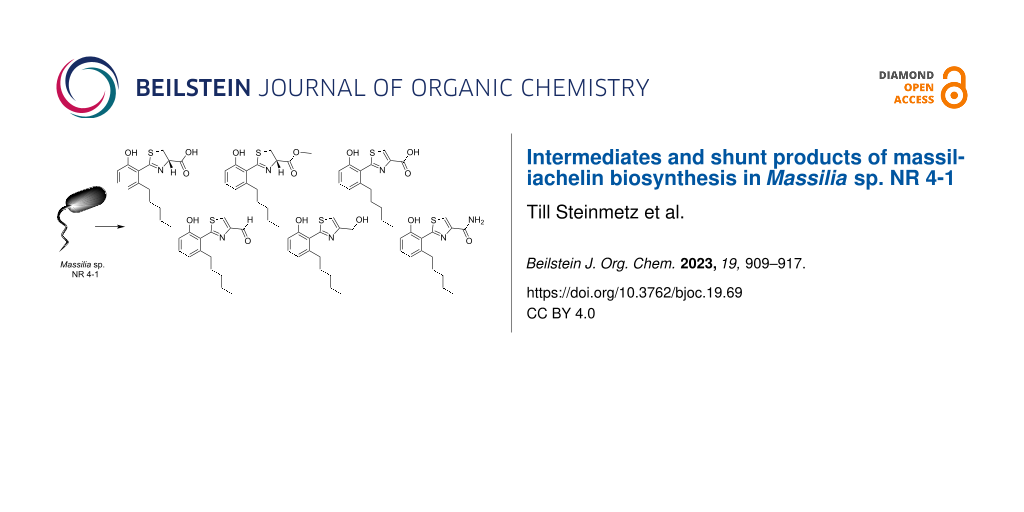
Massilia sp. NR 4-1 was cultivated in modified R2A medium under iron-replete conditions. As opposed to our former study [18], sodium pyruvate was not included in the medium, as this ingredient had been observed to enhance the production of violacein (Supporting Information File 1, Figure S1). In the absence of sodium pyruvate, the identification and isolation of minor metabolites from Massilia sp. NR 4-1 is facilitated, as already observed in the discovery of massinidine [17]. In the present study, several batch fermentations with a total culture volume of 12 L were carried out to secure sufficient material for structure elucidation. The metabolites secreted into the culture broth were recovered post-fermentation with the adsorber resin XAD-7. After the removal of the culture supernatant by filtration, the adsorbed compounds were eluted from the resin with methanol. The resulting extract was concentrated to dryness. For an initial separation of its components, the raw extract was resuspended in water and extracted three times with ethyl acetate. The organic phases were pooled. A testing in the CAS assay indicated the presence of iron-chelating molecules only in the organic phase but not in the aqueous phase. Therefore, the organic phase was concentrated under reduced pressure and the resulting residue was subjected to reversed-phase HPLC. Those fractions that showed a color change from blue to pink in the CAS assay were collected and purified in a second reversed-phase HPLC run. This led to the isolation of six compounds (1–6, Figure 2).
Figure 2:
Chemical structures of compounds 1–6 isolated in this study and of the structurally related siderophores massiliachelin (7) and (S)-dihydroaeruginoic acid (8).
Figure 2:
Chemical structures of compounds 1–6 isolated in this study and of the structurally related siderop...
The NMR spectroscopic data of compound 1 (brownish oil, 1.7 mg) were found to be very similar to those of the previously reported massiliachelin [18], suggesting a structural relatedness. The empirical formula of 1 was assigned to be C15H19NO3S by high-resolution (HR) ESIMS (m/z 294.1180 [M + H]+; calcd. 294.1169 for C15H20NO3S), which indicated the presence of seven double bond equivalents (DBEs). Upon analyzing the 13C NMR spectrum, the DBEs were assigned to two ring structures, two carbon–heteroatom double bonds (δC 171.9, 167.1 ppm; Table 1) and three carbon–carbon double bonds. The latter involved carbon atoms resonating at δC 155.4, 142.3, 130.3, 119.9, 119.8, and 113.2 ppm. The signals in the 1H NMR spectrum of 1 could be distributed among three discrete spin systems according to proton–proton correlation spectroscopy (COSY; Figure 3).
Table 1:1H and 13C NMR spectroscopic data for 1–6 in DMSO-d6.
No.
1
2
3
δH, M (J in Hz)
δC
δH, M (J in Hz)
δC
δH, M (J in Hz)
δC
1
–
155.4
–
155.3
–
155.8
2
–
119.8
–
119.8
–
119.1
3
–
142.3
–
142.2
–
143.4
4
6.69 dd (1.1, 7.8)
119.9
6.70 dd (1.1, 7.8)
119.9
6.79 dd (1.2, 7.7)
120.6
5
7.15 t (7.8)
130.3
7.15 t (7.8)
130.4
7.21 dd (7.7, 8.2)
130.5
6
6.70 dd (1.1, 7.8)
113.2
6.71 dd (1.1, 7.8)
113.2
6.83 dd (1.2, 8.2)
113.4
7
2.54 m
2.62 m
32.7
2.58 m
32.6
2.62 dd (3.8, 7.8)
33.2
8
1.49 m
31.0
1.49 m
30.9
1.38 m
30.2
9
1.25 m
31.2
1.25 m
31.2
1.17 m
31.0
10
1.25 m
21.9
1.29 m
21.9
1.17 m
21.7
11
0.85 t (7.0)
13.9
0.85 t (6.3)
13.9
0.77 t (7.1)
13.8
12
–
167.1
–
167.6
–
163.3
13
3.63 dd (8.1, 11.1)
3.69 dd (9.6, 11.1)
35.4
3.63 dd (8.3, 11.2)
3.72 dd (9.7, 11.2)
35.3
8.55 s
129.5
14
5.25 dd (8.1, 9.6)
77.6
5.33 dd (8.2, 9.7)
77.5
–
146.3
15
–
171.9
–
170.9
–
162.3
16
3.73 s
52.3
No.
4
5
6
δH, M (J in Hz)
δC
δH, M (J in Hz)
δC
δH, M (J in Hz)
δC
1
–
155.7
–
156.5
–
155.7
2
–
118.8
–
119.9
–
119.3
3
–
143.5
–
143.5
–
143.5
4
6.81 dd (1.2, 7.8)
120.8
6.78 dd (1.2, 7.6)
121.0
6.78 dd (1.1, 7.9)
120.5
5
7.23 dd (7.8, 8.2)
130.8
7.19 dd (7.6, 8.2)
130.5
7.20 t (7.9)
130.5
6
6.84 dd (1.2, 8.2)
113.4
6.81 dd (1.2, 8.2)
114.1
6.81 dd (1.1, 7.9)
113.3
7
2.65 m
33.3
2.61 m
33.9
2.59 m
33.2
8
1.39 m
30.3
1.42 m
30.8
1.38 m
30.2
9
1.17 m
31.1
1.20 m
31.7
1.16 m
31.1
10
1.16 m
21.7
1.21 m
22.3
1.15 m
21.7
11
0.77 t (7.1)
13.8
0.81 t (6.9)
14.3
0.76 t (7.1)
13.8
12
–
164.4
–
163.5
–
163.1
13
8.84 s
132.6
7.53 s
115.8
8.36 s
125.3
14
–
153.7
–
157.0
–
149.5
15
9.98 s
184.9
4.63 s
60.2
–
162.4
Figure 3:1H,1H-COSY and selected 1H,13C-HMBC correlations in 1.
Figure 3:1H,1H-COSY and selected 1H,13C-HMBC correlations in 1.
The first spin system is part of a 2,3-substituted phenol moiety featuring proton signals at δH 7.15 (H-5), 6.70 (H-6) and 6.69 ppm (H-4; Table 1). Three aromatic carbon atoms could be assigned due to heteronuclear multiple bond correlation (HMBC) correlations from H-6 to 155.4 ppm (C-1) and 119.8 ppm (C-2) as well as from H-4 to 142.3 ppm (C-3) and 119.8 ppm (C-2). The upfield shifted resonance of H-6 suggested an electron-donating substituent at C-1 and the chemical shift of the latter (δC 155.4 ppm) supported the assignment of an hydroxy function in this position. An HMBC correlation from H-4 to the carbon atom at 32.7 ppm (C-7) allowed the linkage of the phenol moiety with an n-pentyl sidechain in meta position to the hydroxy group. The spin system of the latter includes proton resonances at δH 2.54 (H-7a), 2.62 (H-7b), 1.49 (H-8), 1.25 (H-9), 1.25 (H-10) and 0.85 ppm (H-11). The last spin system is part of a thiazoline moiety with proton resonances at δH 5.25 (H-14), δH 3.69 (H-13a) and δH 3.63 (H-13b). HMBC correlations from H-13 and H-4 to C-12, in combination with characteristic chemical shift values confirmed the presence of the thiazoline substituent at C-2 of the phenol moiety. The final carbon atom at 171.9 ppm (C-15) could be attributed to a carboxylic acid function with HMBC correlations from H-13 and H-14, thereby completing the determination of the planar structure of 1. To determine the configuration of 1, we measured its optical rotation. The obtained value ( = +37°) was consistent with the value of (S)-dihydroaeruginoic acid (8, = +47°) which is structurally almost identical to 1 except for the presence of the n-pentyl side chain [21]. We thus propose that 1 is also S-configured.
Compound 2 (0.4 mg) was obtained as a brown oil. It possesses a molecular ion at m/z 308.1343 [M + H]+, which is consistent with a molecular formula of C16H21NO3S (calcd. for C16H22NO3S, 308.1326) and seven degrees of unsaturation. The NMR data are comparable with compound 1, except for the presence of an additional carbon atom at δC 52.3 ppm (C-16) and a proton resonance at δH 3.73 ppm (H-16). Both H-16 and H-14 show HMBC correlations to the carbonyl C-15. It can therefore be concluded that a methyl ester function replaced the carboxylic acid function of compound 1. Measurements of the optical rotation of 2 were not possible due to the low quantity of isolated material.
Compound 3 (0.8 mg) was obtained as a brown oil. It possesses a molecular ion at m/z 292.1017 [M + H]+, which suggests a molecular formula of C15H17NO3S (calcd. for C15H18NO3S, 292.1013) and corresponds to eight degrees of unsaturation. A key distinction between compounds 1 and 3 is the lack of two proton signals associated with the thiazoline moiety. Instead, the proton spectrum of 3 features a signal at δH 8.55 (H-13). The HMBC correlations linking H-13 to 163.3 ppm (C-12) and 146.3 ppm (C-14) indicate the presence of a thiazole rather than a thiazoline moiety. This aligns well with the observed increase in the degree of unsaturation and the reduction in mass by 2 Da.
Compound 4 (0.5 mg) was obtained as a brown oil. It possesses a molecular ion at m/z 276.0985 [M + H]+, which suggests a molecular formula of C15H17NO2S (calcd. for C15H18NO3S, 276.1002) and is in accordance with eight degrees of unsaturation. The NMR data for 4 is closely related with that of compound 3. The carbon atom at 184.9 ppm (C-15) possesses an HSQC correlation to a proton resonance at δH 9.98 (H-15), which is characteristic of an aldehyde function. This is in full accordance with the loss of 16 Da compared to compound 3, confirming that it is the reduced form of the carboxylic acid.
Compound 5 (0.6 mg) was obtained as a brown oil. It possesses a molecular ion at m/z 278.1231 [M + H]+, which suggests a molecular formula of C15H19NO2S (calcd. for C15H20NO3S, 278.1220) and corresponds to seven degrees of unsaturation. The NMR data of compound 5 are very similar to those of compounds 3 and 4. However, instead of the aldehyde function seen in compound 4, a proton signal at δH 4.63 (H-15) can be observed. Together with its distinct carbon signal at 60.3 ppm (C-15), HMBC correlations to the carbon atoms C-13 and C-14 of the thiazole moiety, the loss of one degree of unsaturation and a loss of 2 Da in mass, it can be deduced that compound 5 possesses an alcohol function.
Compound 6 (0.4 mg) was obtained as a brown oil. It possesses a molecular ion at m/z 291.1165 [M + H]+, which suggests a molecular formula of C15H18N2O2S (calcd. for C15H19N2O2S, 291.1173) and corresponds to eight degrees of unsaturation. In comparison to the other five molecules compound 6 must contain an even number of nitrogen atoms. The NMR data is comparable to compound 3, but includes a deuterium exchangeable proton signal at δH 7.62 ppm characteristic for an amide.
In vitro tests were conducted for all compounds to assess their antibacterial activities against three Gram-negative bacteria (Escherichia coli, Agrobacterium tumefaciens and Pseudomonas fluorescens) and one Gram-positive bacterium (Bacillus subtilis). The filter paper method was used to determine the inhibition zone diameters, which are reported in Supporting Information File 1, Table S1. It turned out that the compounds possess only modest inhibitory activities against B. subtilis. These results are consistent with previous studies [22,23]. Natural products that are structurally related to 1–6 were discovered in Pseudomonas aeruginosa. It was shown that these compounds function as signaling molecules involved in quorum sensing and stress response [24] which might be an explanation for their low bioactivity against the tested bacteria.
Upon analyzing the structures of the isolated metabolites, it became evident that the predicted lipopeptide siderophore [17] had not been produced by Massilia sp. NR 4-1 under the chosen cultivation conditions, as no further extract fraction possessed CAS activity. The reasons for the absence of this compound are not clear. According to the literature, the occurrence of multiple siderophore pathways in a single bacterium is not unusual. Furthermore, it has been repeatedly observed that not all siderophores of such a bacterium must be produced at the same time [25-27]. An illustrative example is the pathogen P. aeruginosa, which is capable of adapting its iron acquisition strategy. In general, P. aeruginosa relies on a comparatively weak iron chelator named pyochelin. During acute infections, however, when iron availability is severely limited, P. aeruginosa switches to the production of a high-affinity siderophore, which further acts as a signal molecule for the production of virulence factors [26]. Similar to P. aeruginosa, the causative agent of splenic fever, Bacillus anthracis, is also known to synthesize two structurally different siderophores. The corresponding compounds were found to underlie discrete regulation mechanisms, which explains their context-dependent production [27]. The examples of P. aeruginosa and B. anthracis suggest that siderophore biosynthesis sometimes requires specific triggers beyond iron deficiency, which may not be met under laboratory conditions.
The six metabolites that were recovered in this study share a phenolic moiety with a thiazole or thiazoline substituent. This motif is present in many siderophores, e.g., in pyochelin [26], yersiniabactin [28], agrochelin [29], micacocidin [30], the Massilia-derived massiliachelin [18], as well as in piscibactin [31] and the photoxenobactins [32]. A unifying theme in the biosynthesis of these natural products is the use of a thiotemplate-based assembly strategy [33]. The molecular building blocks that are needed for the biosynthesis are covalently bound via thioester bonds to multi-domain enzymes. The domains fulfill specific functions, ranging from the selection and linkage of building blocks to their chemical modification. A plausible scenario for the formation of compounds 1–6 involves the enzymatic machinery for massiliachelin biosynthesis, namely the protein RS02200 [18]. According to our proposal (Figure 4), the biosynthesis starts from hexanoic acid which, upon its thioesterification, is elongated by three decarboxylative Claisen condensations with malonyl-CoA to a 6-pentylsalicyl thioester. A condensation with cysteine and a subsequent cyclization generate a 6-pentylsalicyl-thiazolinyl thioester intermediate (1’). In massiliachelin biosynthesis this intermediate is further processed and elongated with another cysteine-derived thiazoline, which is eventually reduced [18]. A premature hydrolytic release of 1’ from the assembly line would give 1, which could be further modified to 2–5. Some corresponding reactions (e.g., hydrolysis, esterification, oxidation) might be due to the isolation conditions or they could be attributed to unspecific enzymatic biotransformations. For compound 1, no spontaneous conversion to the ester 2 was observed, even after storage in methanol for two months. In contrast, the formation of the terminal carboxamide in 6 might be due to a spontaneous C–N bond cleavage, which occurs in 1’’ prior to the cyclization, consistent with a mechanism recently proposed in photoxenobactin biosynthesis [34].
Figure 4:
Proposed origin of the isolated compounds 1–6 as well as massiliachelin (7). Domain notation of the protein RS02200: FAAL: fatty acyl-AMP ligase; ACP: acyl carrier protein; KS: β-ketoacyl synthase; AT: acyltransferase; KR: ketoreductase; C: condensation; A: adenylation; MT: methyltransferase; PCP: peptidyl carrier protein. A discrete enzyme, the thiazolinyl imide reductase RS02195 (Red), catalyzes the conversion of a thiazoline into a thiazolidine ring.
Figure 4:
Proposed origin of the isolated compounds 1–6 as well as massiliachelin (7). Domain notation of the...
Despite the widespread occurrence of siderophores featuring a phenolic moiety with a thiazole or thiazoline substituent, compounds 2–6 have not been reported before according to literature searches. Compound 1 was already described as an intermediate in the chemical total synthesis of micacocidin [35], but has not been isolated from a natural source before.
Conclusion
In summary, six metabolites that are structurally related to the siderophore massiliachelin were recovered from a culture extract of Massilia sp. NR 4-1. The structures of the natural products were verified by high-resolution mass spectrometry as well as 1D and 2D NMR analyses. The newly found compounds are assumed to represent intermediates or shunt products in massiliachelin biosynthesis. A model for their formation is presented.
Experimental
Analytical methods
Preparative HPLC was conducted on a Shimadzu LC-20A system equipped with two pumps (LC-20AD), a photo-diode array detector (SPD-M20A), a degasser unit (DGU-20A), an automatic sampler (SIL-20A), and a column oven (CTO-20AC). LC–MS analysis was performed on a compact quadrupole-time of flight (Q-TOF) mass spectrometer from Bruker Daltonics with an Agilent 1260 Infinity LC system equipped with a Nucleoshell RP18 column (150 × 2.0 mm, 5 µm, Macherey-Nagel). NMR spectra were recorded on a Bruker 600 MHz Avance III HD system with DMSO-d6 as solvent and internal standard. The solvent signal was referenced to δH 2.50 ppm and δC 39.52 ppm, respectively. Optical rotation was measured at 20 °C on a PerkinElmer polarimeter 341 with a sodium lamp (wavelength = 589 nm) using a 1 dm cuvette. For this, samples were dissolved in 1 mL methanol.
Cultivation and extraction of Massilia sp. NR 4-1
For metabolite production, the strain was grown in 5 L Erlenmeyer flasks containing 1.5 L of modified R2A medium: 0.5 g/L yeast extract, 0.5 g/L proteose peptone, 0.5 g/L casamino acids, 0.5 g/L glucose, 0.5 g/L soluble starch, 0.3 g/L KH2PO4, 0.05 g/L MgSO4 × 7 H2O. The pH of the medium was adjusted to pH 7.2. The cultivation was conducted on a rotary shaker at 130 rpm and 30 °C for one week. Afterwards adsorber resin (XAD-7, 20 g/L) was added to the culture broth to bind the secreted metabolites. The resin was separated from the culture broth by filtration, washed with distilled water and exhaustively extracted with methanol.
Isolation of derivatives
The concentrated extract was first fractionated by reversed-phase HPLC using a Nucleodur C18 Isis column (250 × 10 mm, 5 μm, Macherey-Nagel) and a gradient of acetonitrile in water supplemented with 0.1% (v/v) trifluoroacetic acid. The gradient conditions were as follows: from 20% acetonitrile to 90% in 30 minutes and kept at 90% for 10 minutes. The flow rate was set to 5 mL/min. The elution of compounds was monitored with a diode array detector over the range from 190 to 650 nm. Subsequently, the relevant metabolites were further purified by isocratic fractionation, by lowering the concentration of acetonitrile by 10% compared to the elution concentration that was achieved with the gradient method.
Chrome azurol S assay
The CAS assay solution was prepared according to a previously reported protocol [20]. Briefly, chrome azurol S (CAS) and hexadecyltrimethylammonium bromide (HDTMA) were independently dissolved in water to prepare 2 mM and 6.585 M solutions, respectively. In a beaker, 7.5 mL of the CAS solution were mixed with 15 mL of the HDTMA solution under stirring. To this mixture, 50 mL of water, 1.5 mL of an iron chloride solution (1 mM FeCl × 6 H2O in 10 mM HCl), 4.3 g of anhydrous piperazine and 6.25 mL of 12 M hydrochloric acid were added. The resulting mixture was then diluted with water to a final volume of 100 mL, hence yielding the blue CAS assay solution. To perform the assay, 1 mL of the CAS assay solution was pipetted into a cuvette followed by the addition of 0.2 mL of the substance to be tested. A color change from blue to yellow indicated the presence of metal-chelating molecules.
Filter paper assay
The bacterial strains were acquired from the Leibniz Institute DSMZ (German Collection of Microorganisms and Cell Cultures GmbH). To test antibacterial activity, 0.1 mL of an overnight culture of each bacterial strain was plated on agar plates containing their preferred medium. The samples being tested were dissolved in methanol at a concentration of 50 µg/10 µL. Each compound was impregnated onto filter papers at a volume of 10 µL per disc. The inoculated plates were then incubated at 30 °C for 48 hours. The antibacterial activity was evaluated by measuring the zone of inhibition against the test organism. Ampicillin (Roth, Carl Roth GmbH + Co. KG, Germany), tetracycline (Fluka Honeywell International Inc., United States of America) and ciprofloxacin (Sigma, Sigma-Aldrich Chemie GmbH, Germany) were used as positive controls.
Supporting Information
Supporting Information File 1:
UV and total ion chromatograms of culture extracts from Massilia sp. NR 4-1. Copies of MS/MS and NMR spectra for new compounds.
Prof. Wolf Hiller (TU Dortmund University) is gratefully acknowledged for NMR measurements. We thank Professors Stephan Lütz and Oliver Kayser (both TU Dortmund University) for providing access to their LC–MS facilities, Anton Lindig (TU Dortmund University) for HRMS measurements, and Katharina Kuhr (TU Dortmund University) for the antimicrobial bioactivity tests.
References
Hofmann, M.; Retamal-Morales, G.; Tischler, D. Nat. Prod. Rep.2020,37, 1262–1283. doi:10.1039/c9np00058e
Return to citation in text:
[1]
Raymond, K. N.; Allred, B. E.; Sia, A. K. Acc. Chem. Res.2015,48, 2496–2505. doi:10.1021/acs.accounts.5b00301
Return to citation in text:
[1]
[2]
Kurth, C.; Kage, H.; Nett, M. Org. Biomol. Chem.2016,14, 8212–8227. doi:10.1039/c6ob01400c
Return to citation in text:
[1]
Hider, R. C.; Kong, X. Nat. Prod. Rep.2010,27, 637–657. doi:10.1039/b906679a
Return to citation in text:
[1]
Swayambhu, G.; Bruno, M.; Gulick, A. M.; Pfeifer, B. A. Curr. Opin. Biotechnol.2021,69, 242–251. doi:10.1016/j.copbio.2021.01.021
Return to citation in text:
[1]
Franke, J.; Ishida, K.; Ishida-Ito, M.; Hertweck, C. Angew. Chem., Int. Ed.2013,52, 8271–8275. doi:10.1002/anie.201303196
Return to citation in text:
[1]
Franke, J.; Ishida, K.; Hertweck, C. Chem. – Eur. J.2015,21, 8010–8014. doi:10.1002/chem.201500757
Return to citation in text:
[1]
Trottmann, F.; Franke, J.; Ishida, K.; García-Altares, M.; Hertweck, C. Angew. Chem., Int. Ed.2019,58, 200–204. doi:10.1002/anie.201811131
Return to citation in text:
[1]
Hermenau, R.; Ishida, K.; Gama, S.; Hoffmann, B.; Pfeifer-Leeg, M.; Plass, W.; Mohr, J. F.; Wichard, T.; Saluz, H.-P.; Hertweck, C. Nat. Chem. Biol.2018,14, 841–843. doi:10.1038/s41589-018-0101-9
Return to citation in text:
[1]
[2]
Gama, S.; Hermenau, R.; Frontauria, M.; Milea, D.; Sammartano, S.; Hertweck, C.; Plass, W. Chem. – Eur. J.2021,27, 2724–2733. doi:10.1002/chem.202003842
Return to citation in text:
[1]
Dashti, Y.; Nakou, I. T.; Mullins, A. J.; Webster, G.; Jian, X.; Mahenthiralingam, E.; Challis, G. L. Angew. Chem., Int. Ed.2020,59, 21553–21561. doi:10.1002/anie.202009110
Return to citation in text:
[1]
Kreutzer, M. F.; Kage, H.; Nett, M. J. Am. Chem. Soc.2012,134, 5415–5422. doi:10.1021/ja300620z
Return to citation in text:
[1]
Kreutzer, M. F.; Nett, M. Org. Biomol. Chem.2012,10, 9338–9343. doi:10.1039/c2ob26296g
Return to citation in text:
[1]
Kurth, C.; Schieferdecker, S.; Athanasopoulou, K.; Seccareccia, I.; Nett, M. J. Nat. Prod.2016,79, 865–872. doi:10.1021/acs.jnatprod.5b00932
Return to citation in text:
[1]
Miess, H.; Frediansyah, A.; Göker, M.; Gross, H. Microbiol. Resour. Announce.2020,9, e00226–20. doi:10.1128/mra.00226-20
Return to citation in text:
[1]
Agemathu, H.; Suzuki, K.; Tsuya, H. Biosci., Biotechnol., Biochem.2011,75, 2008–2010. doi:10.1271/bbb.100729
Return to citation in text:
[1]
Lombe, B. K.; Winand, L.; Diettrich, J.; Töbermann, M.; Hiller, W.; Kaiser, M.; Nett, M. Org. Lett.2022,24, 2935–2939. doi:10.1021/acs.orglett.2c00963
Return to citation in text:
[1]
[2]
[3]
Diettrich, J.; Kage, H.; Nett, M. Beilstein J. Org. Chem.2019,15, 1298–1303. doi:10.3762/bjoc.15.128
Return to citation in text:
[1]
[2]
[3]
[4]
[5]
[6]
Blin, K.; Shaw, S.; Kloosterman, A. M.; Charlop-Powers, Z.; van Wezel, G. P.; Medema, M. H.; Weber, T. Nucleic Acids Res.2021,49, W29–W35. doi:10.1093/nar/gkab335
Return to citation in text:
[1]
Roberts, A. A.; Schultz, A. W.; Kersten, R. D.; Dorrestein, P. C.; Moore, B. S. FEMS Microbiol. Lett.2012,335, 95–103. doi:10.1111/j.1574-6968.2012.02641.x
Return to citation in text:
[1]
Cornelis, P.; Dingemans, J. Front. Cell. Infect. Microbiol.2013,3, 75. doi:10.3389/fcimb.2013.00075
Return to citation in text:
[1]
[2]
[3]
Lee, J. Y.; Passalacqua, K. D.; Hanna, P. C.; Sherman, D. H. PLoS One2011,6, e20777. doi:10.1371/journal.pone.0020777
Return to citation in text:
[1]
[2]
Drechsel, H.; Stephan, H.; Lotz, R.; Haag, H.; Zähner, H.; Hantke, K.; Jung, G. Liebigs Ann.1995, 1727–1733. doi:10.1002/jlac.1995199510243
Return to citation in text:
[1]
Acebal, C.; Cañedo, L. M.; Puentes, J. L. F.; Baz, J. P.; Romero, F.; De La Calle, F.; Grávalos, M. D. G.; Rodriguez, P. J. Antibiot.1999,52, 983–987. doi:10.7164/antibiotics.52.983
Return to citation in text:
[1]
Kobayashi, S.; Nakai, H.; Ikenishi, Y.; Sun, W.-Y.; Ozaki, M.; Hayase, Y.; Takeda, R. J. Antibiot.1998,51, 328–332. doi:10.7164/antibiotics.51.328
Return to citation in text:
[1]
Souto, A.; Montaos, M. A.; Rivas, A. J.; Balado, M.; Osorio, C. R.; Rodríguez, J.; Lemos, M. L.; Jiménez, C. Eur. J. Org. Chem.2012, 5693–5700. doi:10.1002/ejoc.201200818
Return to citation in text:
[1]
Shi, Y.-M.; Hirschmann, M.; Shi, Y.-N.; Ahmed, S.; Abebew, D.; Tobias, N. J.; Grün, P.; Crames, J. J.; Pöschel, L.; Kuttenlochner, W.; Richter, C.; Herrmann, J.; Müller, R.; Thanwisai, A.; Pidot, S. J.; Stinear, T. P.; Groll, M.; Kim, Y.; Bode, H. B. Nat. Chem.2022,14, 701–712. doi:10.1038/s41557-022-00923-2
Return to citation in text:
[1]
Sundaram, S.; Hertweck, C. Curr. Opin. Chem. Biol.2016,31, 82–94. doi:10.1016/j.cbpa.2016.01.012
Return to citation in text:
[1]
Shi, Y.-M.; Hirschmann, M.; Shi, Y.-N.; Bode, H. B. ACS Chem. Biol.2022,17, 2221–2228. doi:10.1021/acschembio.2c00367
Return to citation in text:
[1]
Ino, A.; Hasegawa, Y.; Murabayashi, A. Tetrahedron Lett.1998,39, 3509–3512. doi:10.1016/s0040-4039(98)00518-8
Return to citation in text:
[1]
Acebal, C.; Cañedo, L. M.; Puentes, J. L. F.; Baz, J. P.; Romero, F.; De La Calle, F.; Grávalos, M. D. G.; Rodriguez, P. J. Antibiot.1999,52, 983–987. doi:10.7164/antibiotics.52.983
Blin, K.; Shaw, S.; Kloosterman, A. M.; Charlop-Powers, Z.; van Wezel, G. P.; Medema, M. H.; Weber, T. Nucleic Acids Res.2021,49, W29–W35. doi:10.1093/nar/gkab335
Souto, A.; Montaos, M. A.; Rivas, A. J.; Balado, M.; Osorio, C. R.; Rodríguez, J.; Lemos, M. L.; Jiménez, C. Eur. J. Org. Chem.2012, 5693–5700. doi:10.1002/ejoc.201200818
Roberts, A. A.; Schultz, A. W.; Kersten, R. D.; Dorrestein, P. C.; Moore, B. S. FEMS Microbiol. Lett.2012,335, 95–103. doi:10.1111/j.1574-6968.2012.02641.x


![[1860-5397-19-69-1]](/bjoc/content/figures/1860-5397-19-69-1.svg?scale=2.0&max-width=1024&background=FFFFFF)
![[1860-5397-19-69-2]](/bjoc/content/figures/1860-5397-19-69-2.svg?scale=2.0&max-width=1024&background=FFFFFF)
![[1860-5397-19-69-3]](/bjoc/content/figures/1860-5397-19-69-3.svg?scale=2.0&max-width=1024&background=FFFFFF)
![[Graphic 1]](/bjoc/content/inline/1860-5397-19-69-i1.svg?max-width=637&scale=1.18182) = +37°) was consistent with the value of (S)-dihydroaeruginoic acid (8,
= +37°) was consistent with the value of (S)-dihydroaeruginoic acid (8, ![[Graphic 2]](/bjoc/content/inline/1860-5397-19-69-i2.svg?max-width=637&scale=1.18182) = +47°) which is structurally almost identical to 1 except for the presence of the n-pentyl side chain [21]. We thus propose that 1 is also S-configured.
= +47°) which is structurally almost identical to 1 except for the presence of the n-pentyl side chain [21]. We thus propose that 1 is also S-configured.![[1860-5397-19-69-4]](/bjoc/content/figures/1860-5397-19-69-4.svg?scale=2.0&max-width=1024&background=FFFFFF)