

Search results
Search for "binding site" in Full Text gives 186 result(s) in Beilstein Journal of Organic Chemistry.
Catalysis: transition-state molecular recognition?
Beilstein J. Org. Chem. 2010, 6, 1026–1034, doi:10.3762/bjoc.6.117
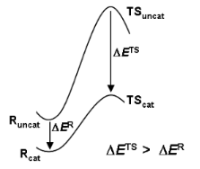
- concept. It is shown that reactant binding is intrinsically inhibitory, and that attempts to design catalysts that focus simply upon attractive interactions in a binding site may fail. Free-energy changes along the reaction coordinate for SN2 methyl transfer catalysed by the enzyme catechol-O-methyl


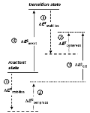
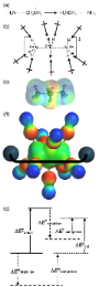

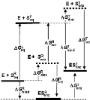


En route to photoaffinity labeling of the bacterial lectin FimH
Beilstein J. Org. Chem. 2010, 6, 810–822, doi:10.3762/bjoc.6.91
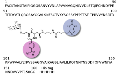
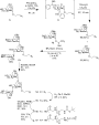
- . It requires a photoactive ligand derivative, which can form a reactive species upon photo-excitation. Thus, incubation of the photoprobe with a protein followed by irradiation can result in a photo-crosslinked product, that provides structural information on the binding site of the protein (Figure 1
- just one monovalent carbohydrate binding site on the FimH protein [9][10][11][12][13]. Thus, we decided to employ photoactive ligands to probe carbohydrate binding to the known CRD in solution and to identify possibly auxiliary, so far unknown, binding sites on bacterial lectin FimH [2]. The known FimH



A bivalent glycopeptide to target two putative carbohydrate binding sites on FimH
Beilstein J. Org. Chem. 2010, 6, 801–809, doi:10.3762/bjoc.6.90
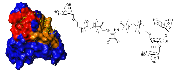
- 1 fimbriae with a monovalent carbohydrate recognition domain (CRD) that is known from X-ray studies. However, binding studies with multivalent ligands have suggested an additional carbohydrate-binding site on this protein. In order to prove this hypothesis, a bivalent glycopeptide ligand with the
- and a pilin domain, which is required for fimbriae assembly [7]. The FimH adhesin domain features a carbohydrate binding site at its tip, called the carbohydrate recognition domain (CRD), which is known from X-ray studies [8][9][10][11]. It is a monovalent binding site, which can accommodate one α-D
- trisaccharide substructures, mainly α-D-Man-(1→3)-[α-D-Man-(1→6)]-D-Man. By employing site directed mutagenesis, it was found that mutations in one of these cavities significantly reduces binding, indicating that this could be a second carbohydrate binding site, relevant for ligand binding [21]. Thus, it was



RAFT polymers for protein recognition
Beilstein J. Org. Chem. 2010, 6, No. 66, doi:10.3762/bjoc.6.66
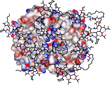
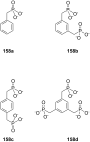
- tetramethyl ester was subjected to LiBr-assisted nucleophilic cleavage to furnish the free bisphosphonate dianion binding site. This procedure has two major drawbacks. First, if the functional groups on the polymer backbone become restricted in their accessability, the final deprotection step will suffer from




Anthracene coupled adenine for the selective sensing of copper ions
Beilstein J. Org. Chem. 2010, 6, No. 44, doi:10.3762/bjoc.6.44
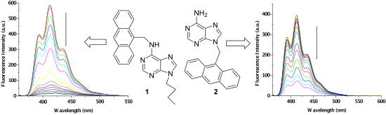
- sensor, shown in Figure 1. In both 1 and 2, adenine is defined as the binding site, which is connected to the anthracene probe via –CH2– spacer at the different regions of adenine. In order to find out the possible site for binding of metal ions, we optimized the geometries of both 1 and 2 at AM1 level
- compared to the cases of 1 and 2 with Cu2+. During the interaction process no other changes were observed. This finding thus indicates that the adenine moiety in both 1 and 2 acts as a metal ion binding site for which participation of anthracene in cation-π interaction is facilitated upon binding of metal




Molecular recognition of organic ammonium ions in solution using synthetic receptors
Beilstein J. Org. Chem. 2010, 6, No. 32, doi:10.3762/bjoc.6.32
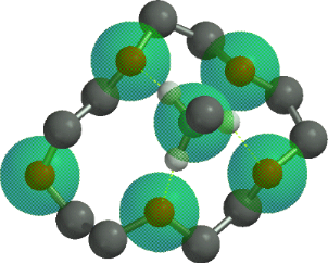
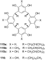
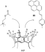
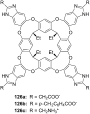
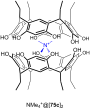
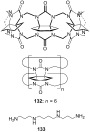
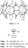
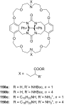
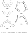
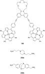
- stoichiometry between a specific host and guest – or dynamic, where the binding of the first guest to the first binding site of a receptor affects the association constant of a second guest with a second binding site. Either positive allosteric binding – the first guest increases the association constant of the
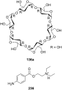
- “key” in the complementary binding site or an inclusion compound. This host pre-organization leads to a major enhancement of the overall energy of guest complexation. The binding is energetically favored: Both enthalpic – a less solvent accessible area leads to a less strongly solvated guest with fewer
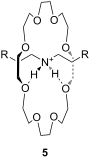
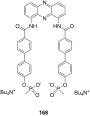
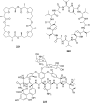
- has been attributed to the tetrahedral binding site geometry that favors complexation of the tetrahedral ammonium ion over that of the spherically symmetrical potassium ion, underlining the particular importance of hydrogen bonding and symmetry considerations in the design of ammonium ion recognition












































Dipyridodiazepinone derivatives; synthesis and anti HIV-1 activity
Beilstein J. Org. Chem. 2009, 5, No. 36, doi:10.3762/bjoc.5.36
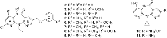
- groups in 7 and 8 led to diminished activity relative to that of 5 and 6. 10 and 11, 8-amino analogues of nevirapine, were found to be ineffective inhibitors. Molecular docking To understand the binding mode of the new potent derivatives 5, 6 and 9 were docked into the HIV-1 RT binding site by using the
- program. The docked conformations of 5, 6, 9 and nevirapine are shown in Figure 3, and their GoldScores are presented in Table 2. In the wild-type and K103N binding sites, the docked orientations of 5, 6 and 9 are similar to that of nevirapine. In the Y181C binding site, except for 5, the orientations of


Synthesis and enzymatic evaluation of 2- and 4-aminothiazole- based inhibitors of neuronal nitric oxide synthase
Beilstein J. Org. Chem. 2009, 5, No. 28, doi:10.3762/bjoc.5.28

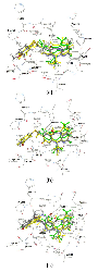
- -groups at the 5-position of 4 should allow us to probe the hydrophobic binding pocket defined by P565, A566, V567, and F584 in the substrate binding site and optimize this interaction. Figure 2 shows the docking mode for 3 and 4a in rat nNOS. Initially, attempts were made to construct 3 and 4 using


Synthesis of rigidified flavin–guanidinium ion conjugates and investigation of their photocatalytic properties
Beilstein J. Org. Chem. 2009, 5, No. 26, doi:10.3762/bjoc.5.26
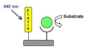
- catalytic efficacy, flavin derivatives bearing a guanidinium ion as oxoanion binding site were prepared. Chromophore and substrate binding site are linked by a rigid Kemp’s acid structure. The molecular structure of the new flavins was confirmed by an X-ray structure analysis and their photocatalytic
- activity was investigated in benzyl ester cleavage, nitroarene reduction and a Diels–Alder reaction. The modified flavins photocatalyze the reactions, but the introduced substrate binding site does not enhance their performance. Keywords: flavin; guanidine; Kemp’s acid; photocatalysis; template
- flavin chromophore, the guanidinium substrate binding site and a Kemp’s acid derived rigid linker, starts from Kemp’s acid anhydride (5) [50][51][52]. The anhydride 5 was allowed to react with previously prepared flavins 4 and 8 [21] in the presence of DMAP as catalyst. The amide formation of the


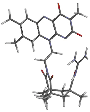


The first salen- type ligands derived from 3',5'-diamino- 3',5'-dideoxythymidine and -dideoxyxylothymidine and their corresponding copper(II) complexes
Beilstein J. Org. Chem. 2006, 2, No. 17, doi:10.1186/1860-5397-2-17
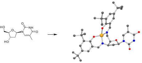
- binding site and the N-glycosidic bound thymine provides an additional chiral information and steric shielding. After the ligands had been added to a mixture of copper(II) acetate in THF the complexes were formed within a few minutes resulting in a dark green solution (Scheme 2). All four ligands 8–11
- 15 (384 nm). The complexes of the diastereomeric ligands could be obtained in a straightforward synthesis. They possess interesting features especially with regard to chiral catalysts or DNA strand formation. Although located off the metal ion the thymine base may act as a substrate binding site in


Colchitaxel, a coupled compound made from microtubule inhibitors colchicine and paclitaxel
Beilstein J. Org. Chem. 2006, 2, No. 13, doi:10.1186/1860-5397-2-13
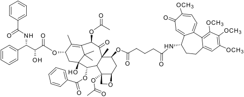
- stacked on it in such a way that the nucleotide-binding site is embedded in the interface [8]. Ravelli and coworkers found colchicine binding site to be buried in the β subunit, boxed in by β-strands of the second domain and helices #7 and #8. The A ring of colchicine contacts residue 241[9], consistent
- binding. However, the acetamide linkage on ring B could be replaced by other alkyl amides with little change in potency [32]. Moreover, colchicine with an altered B ring still bound tubulin [33]. The binding site of paclitaxel is situated on the inner face of the polymerized microtubule, tucked into a
- bend formed by helix #6 and helix #7 near the + end of the microtubule and adjacent to strand #7. Here, the taxane may affect both the structure of the M loop and that of helix #6 which contacts the GTP binding site, so that it may both stabilize GTP against hydrolysis and stabilize the M loop against