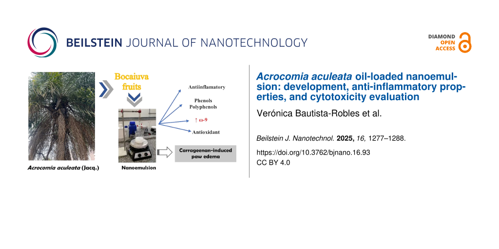
Abstract
The oil from the pulp of the bocaiúva fruit may have several medical applications. However, little is known about its pharmacological activity. Therefore, this study aimed to develop and evaluate the anti-inflammatory activity of a nanoemulsion loaded with the oil extracted from the pulp of the fruit of Acrocomia aculeata. Griffin’s method determined the hydrophilic–lipophilic equilibrium ratio of the nanoemulsion. It was shown to have an adequate droplet size (173.60 nm) with excellent homogeneity (polydispersity index 0.200). The anti-inflammatory activity of the nanoemulsion was evaluated by the carrageenan-induced paw edema method. Finally, the non-hemolytic and cytotoxic activity of the nanoformulation was determined to assess its safety. The nanoemulsion loaded with Acrocomia aculeata fruit pulp oil was shown to have parameters suitable for its characterization, impressive anti-inflammatory activity, and a safe profile.
Introduction
Acrocomia aculeata Jacq is a palm of the Arecaceae family, commonly known as bocaiúva or macaúba. It is widespread in South America and is particularly abundant in Mato Grosso do Sul, located in the Center-West region of Brazil. A. aculeata stands out for its wide geographic distribution, being native to tropical forests [1,2]. Its rounded fruits present sensory attractions, such as color, distinctive and intense flavor, and aroma. They are traditionally consumed by the native population, occupying an important place in the regional culture [3,4].
Bocaiúva oil contains several antioxidant compounds such as phenols, terpenes, β-carotenes, and compounds that present antioxidant properties [5,6]. It contains free fatty acids, monoglycerides, triglycerides, sterols [6-8], and saturated and unsaturated fatty acids predominantly [9]. These compounds have the potential to enhance immune response, reduce the risk of degenerative diseases, and contribute to anti-inflammatory activity [10,11], reducing the indiscriminate use of non-steroidal anti-inflammatory drugs (NSAIDs) and corticosteroids in the population [12,13]. There are several reports about the severe adverse reactions in patients taking these drugs [14,15]. In addition, the economic impact of these degenerative and inflammatory diseases, such as rheumatoid arthritis, is significant, as demonstrated by heightened healthcare resource utilization and substantially increased annual aggregate costs. This underscores the need to find effective alternative treatments to prevent and treat these pathologies [16-18].
On a global scale, vegetable oils play a fundamental role not only in human nutrition but also as strategic inputs in the chemical, pharmaceutical, and food industries. In recent decades, there has been a growing imbalance between the demand and supply of these oils, which has generated challenges in terms of sustainability and supply. In this scenario, several oil palm species have been identified as potentially efficient sources of vegetable oil production, given their high yield rates per hectare and their ability to adapt to different agroecological environments [19]. In the state of Mato Grosso do Sul, it is possible to find virgin, refined, or conventional bocaiúva oil in the market for cosmetic, food, pharmaceutical, nutraceutical, and industrial applications. However, due to the chemical and physicochemical characteristics as well as the solubility and stability of this oil, it was decided to make a nanoemulsion to enhance its already known therapeutic benefits.
Nanoemulsions are nanoemulsified systems, either oil-in-water (O/W) or water-in-oil (W/O), which are isotropic, homogeneous, and thermodynamically unstable, with droplet sizes ranging from 20 to 200 nm [20]. They present properties such as high surface area per unit volume, robust kinetic stability, and tunable rheology [21]. It has been demonstrated that emulsified systems loaded with plant extracts have better pharmacological activity than extracts when used naturally [22]. For example, plant oil-loaded nanoemulsions exhibit high water solubility, improved permeability, and enhanced bioavailability [23]. This contrasts with the limited solubility and poor bioavailability of natural oil through different routes of administration [24-27].
The possibility of developing nanotechnological products with potent pharmacological activity was considered to add more value to the oil obtained from bocaiúva pulp, which contains phenols and carotenoids. Therefore, the objective of this study was to develop, characterize, and evaluate the anti-inflammatory activity of the nanoemulsion loaded with Acrocomia aculeata oil.
Results and Discussion
Vegetable oils are known for their high content of fatty acids, which possess a diverse range of biological activities, including hypoglycemic [28], cholesterol-lowering, anti-inflammatory, and antioxidant effects [11,22-30]. Bocaiúva oil is widely used to treat cardiovascular, inflammatory, and renal diseases [31,32].
In addition, one of the main characteristics of this oil is its orange color due to the presence of phenols and carotenoids, which were characterized in this study. These secondary metabolites are considered to have high antioxidant activity and provide high stability to the oil [5]. These metabolites have been shown to possess anti-inflammatory and immunostimulant properties [1,33].
Physicochemical characterization of Acrocomia aculeata fruit pulp oil
The physicochemical parameters of bocaiúva oil, such as acidity index, iodine index, and refractive index, were analyzed. The acidity index indicates the state of conservation of oils and fats and is related to the oxidation process. Our results showed an acid index of 0.92 ± 0.10. The iodine index determines the amount of unsaturation in fatty acids [28]. Our results showed an iodine index of 74.50 ± 1.50 g I2/100 g, values that are within the range allowed (58–75 g I2/100 g) by OMS/FAO for oils with high oleic acid content [34].
Also, quality indicators such as refractive index, solubility in different organic solvents, and relative density showed that the bocaiúva oil used in that study had good purity [35]. Coimbra and Jorge analyzed Acrocomia aculeata oil and found refractive index values similar to those in this study (1.46 ± 0.01) [19]. These results were found within the reference values established for oils rich in oleic acids, such as extra virgin olive oil, palm oil, and almond oil [34]. The presence of polyphenols and carotenoids was also identified in this oil (see Table 1).
Table 1: Physicochemical properties of Acrocomia aculeata fruit pulp oil.
| Property | Value |
| relative density | 0.9000 ± 0.0001 |
| iodine value (g I2/100 g) | 74.50 ± 1.50 |
| refractive index (30 °C) | 1.456 ± 0.001 |
| peroxide value (mEq/kg oil) | 4.50 ± 0.40 |
| saponification index (mg KOH/g) | 133.00 ± 4.50 |
| acidity | 0.92 ± 0.10 |
| total carotenoids (µg/g) | 266.00 ± 12.00 |
| polyphenols (mg/g) | 12.60 ± 0.30 |
Table 2 shows the profile of fatty acids present in Acrocomia aculeata fruit pulp oil. Oleic acid is the major component (71.25%) among monounsaturated fatty acids (73.79%). Therefore, bocaiúva oil can be considered an oil with a cardioprotective effect due to its high oleic acid content [36,37]. In addition, its levels of monounsaturated fatty acids are higher than those found in extra virgin olive, soybean, corn, sunflower, and flaxseed oils [37,38].
Table 2: Lipid profile of Acrocomia aculeata fruit pulp oil.
| Fatty acid | Content (%) | RE Index | LRa Index |
| saturated | |||
| hexanoic acid | 0.22 ± 0.02 | 974 | 975 |
| octanoic acid | 0.25 ± 0.02 | 1169 | 1170 |
| decanoic acid | 0.13 ± 0.01 | 1365 | 1365 |
| dodecanoic acid | 0.85 ± 0.01 | 1548 | 1547 |
| tetradecanoic acid | 0.70 ± 0.01 | 1747 | 1749 |
| hexadecanoic acid | 16.52 ± 0.15 | 1970 | 1969 |
| octadecanoic acid | 4.11 ± 0.15 | 2164 | 2165 |
| docosanoic acid | 0.06 ± 0.03 | 2562 | 2564 |
| subtotal | 22.84 ± 0.05 | — | — |
| monounsaturated | |||
| 9-hexadecenoic acid | 2.54 ± 0.01 | 1939 | 1938 |
| 9-octadecenoic acid | 71.25 ± 2.21 | 2241 | 2142 |
| subtotal | 73.79 ± 1.11 | — | — |
| polyunsaturated | |||
| 9,12,15-octadecatrienoic acid | 0.80 ± 0.04 | 2154 | 2155 |
| 9,12-octadecadienoic acid | 2.20 ± 0.33 | 2176 | 2175 |
| eicosanoic acid | 0.20 ± 0.03 | 2369 | 2370 |
| subtotal | 3.20 ± 0.13 | — | — |
| total fatty acids | >99.00% | — | — |
aLiterate retention rate (from NIST chemistry webbook, SRD 69).
The bocaiúva oil utilized in this study demonstrated excellent quality, as assessed by established parameters for evaluating vegetable oils reported in the literature [11,39]. The results are consistent with findings by Hiane and collaborators [40] and Lieb and collaborators [7], who also observed a high concentration of monounsaturated fatty acids in the fruit pulp. Amaral et al. further identified a notable oleic acid content of 69.07% in Acrocomia aculeata pulp oil [41]. Minor discrepancies in composition may be attributed to variations in environmental conditions, such as climatic conditions, temperature, and pulp drying duration before oil extraction. Additionally, the specific extraction technique employed can influence lipid degradation and promote the formation of free fatty acids [19]. Despite these variations, the compositional profiles remain comparable, underscoring the distinctive chemical characteristics of the oil studied.
Preparation of nanoemulsions, required hydrophilic–lipophilic balance, droplet size, zeta potential, and shelf stability
The development of a nanoemulsion requires the determination of key formulation parameters, including the required hydrophilic–lipophilic balance (HLBr), droplet size, and polydispersity index (PDI) [42-44]. In this study, the Acrocomia aculeata oil nanoemulsion (AANE) exhibited a uniform droplet size distribution and a stable PDI upon formulation with the surfactant system characterized by a hydrophilic–lipophilic balance (HLB) value of 12. These physicochemical parameters are critical for defining the kinetic stability and structural integrity of the nanoemulsion system.
Surfactants or emulsifiers are characterized by their HLB values, which reflect their affinity for either aqueous or lipid phases. Hydrophilic emulsifiers typically exhibit a high HLB value, while lipophilic emulsifiers possess lower values. The HLB scale generally ranges from 1 to 20, with an approximate midpoint of 10, distinguishing emulsifiers suited for oil-in-water versus water-in-oil systems [43] (Figure 1).
![[2190-4286-16-93-1]](/bjnano/content/figures/2190-4286-16-93-1.png?scale=2.0&max-width=1024&background=FFFFFF)
Figure 1: Droplet size and polydispersity index of nanoemulsions versus the hydrophilic–lipophilic balance of the surfactant system used for the preparation. HLBr: required hydrophilic-lipophilic balance.
Figure 1: Droplet size and polydispersity index of nanoemulsions versus the hydrophilic–lipophilic balance of...
A surfactant system characterized by an HLB value of 12 (Figure 1) was employed to formulate the bocaiúva oil nanoemulsion, resulting in a satisfactory polydispersity index of 0.200. The formulation exhibited excellent physical stability, maintaining consistent zeta potential and droplet size parameters over a 180-day storage period at 25 ± 2 °C. Dynamic light scattering analysis revealed a mean nanodroplet size (by intensity) of 173.6 ± 0.70 nm (Figure 2A). The nanoemulsion, composed of 0.28 parts of Span 80® and 0.72 parts of Tween 80®, exhibited a zeta potential of −14.10 ± 1.06 mV (Figure 2B), indicative of sufficient electrostatic repulsion for colloidal stability.
![[2190-4286-16-93-2]](/bjnano/content/figures/2190-4286-16-93-2.png?scale=2.0&max-width=1024&background=FFFFFF)
Figure 2: Droplet size distribution and zeta potential of the nanoemulsion prepared with 0.28 parts Span 80® and 0.72 parts Tween 80® (HLB = 12). Droplet size: 173.6 ± 0.70 nm. Zeta potential: 14.0 ± 1.06 mV.
Figure 2: Droplet size distribution and zeta potential of the nanoemulsion prepared with 0.28 parts Span 80® ...
It should be noted that the phenolic compounds and carotenoids contained in this oil are considered potent antioxidants, which may contribute to the stability of the nanoemulsion [45,46].
Figure 3 shows the behavior of zeta potential and droplet size in Bocaiúva oil-loaded nanoemulsion over 180 days. The droplet size remained stable at around 170 nm, and no statistical differences were found at any point in time over 180 days (Ftest = 0.18, pvalue = 0.0804). In contrast, the zeta potential underwent a significant decrease from approximately −10 to −20 mV within the first 45 days and then stabilized for the remainder of the 180 days. The analysis of variance found statistically significant differences among the zeta potential values (Ftest = 2.4258, pvalue = 0.0021). The Tukey test suggests that the zeta potential values at zero and 15 days were not statistically different, forming a homogenous group statistically different from the rest of the time points (45, 90, and 180 days), which formed another homogeneous group.
![[2190-4286-16-93-3]](/bjnano/content/figures/2190-4286-16-93-3.png?scale=2.0&max-width=1024&background=FFFFFF)
Figure 3: Stability of bocaiúva oil-loaded nanoemulsion prepared with 0.28 parts of Span 80® and 0.72 parts of Tween 80® (HLB = 12).
Figure 3: Stability of bocaiúva oil-loaded nanoemulsion prepared with 0.28 parts of Span 80® and 0.72 parts o...
The progressive increase in the absolute value of zeta potential over time suggests enhanced system stability, attributed to the intensification of electrostatic repulsion among nanodroplets. Although this phenomenon is atypical for systems stabilized by nonionic surfactants (Span 80® and Tween 80®), it may result from the presence of ionizable bioactive compounds such as phenolics and carotenoids, which can associate with the droplet interface. These compounds likely expand the diffuse electrical double layer surrounding the nanodroplets, thereby increasing the magnitude of the zeta potential, enhancing stability by preventing droplet aggregation [47].
Figure 4 presents the impact of temperature (ranging from 10 to 80 °C) on the nanodroplet size of the AANE formulation over a 180-day period. Between 10 and 60 °C, the nanodroplet size remained relatively stable, ranging from 171 to 181 nm. However, temperatures exceeding 60 °C led to a marked reduction in droplet size, stabilizing between 110 and 120 nm across all time points. This reduction can be attributed to several interrelated physicochemical mechanisms. Primarily, the nonionic surfactants Span 80® and Tween 80® reduce interfacial tension between oil and aqueous phases, a phenomenon that becomes increasingly efficient at elevated temperatures, promoting the formation of smaller droplets. Additionally, elevated temperatures enhance molecular mobility and solubility, facilitating droplet disruption and dispersion effects observed in bocaiúva oil nanoemulsions above 60 °C [48]. Moreover, the surfactants exhibit temperature-responsive behavior, reorganizing at higher temperatures to stabilize finer dispersions. The concurrent decline in oil phase viscosity with increasing temperature also improves emulsification efficiency by promoting shear-induced droplet breakup, particularly under mechanical agitation [48]. Nevertheless, while these conditions favor the formation of smaller nanodroplets, temperatures above 60 °C may compromise emulsion stability, potentially triggering phase separation or degradation of labile components. This highlights the necessity for stringent temperature control during both formulation and storage to ensure the long-term stability of the nanoemulsion [49].
![[2190-4286-16-93-4]](/bjnano/content/figures/2190-4286-16-93-4.png?scale=2.0&max-width=1024&background=FFFFFF)
Figure 4: Effect of temperature on droplet size of nanoemulsion loaded with bocaiúva oil.
Figure 4: Effect of temperature on droplet size of nanoemulsion loaded with bocaiúva oil.
Hemolytic and cytotoxic activity of Acrocomia aculeata oil-based nanoemulsion
The hemolytic and cytotoxic activities of the nanoemulsion were assessed to evaluate its potential therapeutic use, with particular focus on its interaction with erythrocyte membranes. The nanoemulsion demonstrated no hemolytic activity against murine erythrocytes at concentrations of 1, 10, 100, and 1000 μg/mL. These findings were benchmarked against Triton X-100, a well-established positive control known for its potent hemolytic effect [50]. As shown in Figure 5A, AANE maintained erythrocyte membrane integrity across all tested concentrations, reinforcing its biocompatibility.
![[2190-4286-16-93-5]](/bjnano/content/figures/2190-4286-16-93-5.png?scale=2.0&max-width=1024&background=FFFFFF)
Figure 5: Hemolytic effect (A) and cell viability assay (B) of nanoemulsion loaded with Acrocomia aculeata fruit oil.
Figure 5: Hemolytic effect (A) and cell viability assay (B) of nanoemulsion loaded with Acrocomia aculeata fr...
Furthermore, cytotoxicity evaluation showed that AANE did not inhibit cell growth, as cellular viability remained at 100% across all tested concentrations (1, 10, 100, and 1000 μg/mL, Figure 5B). These findings underscore the nanoemulsion’s biocompatibility and support its potential for safe therapeutic applications. This is consistent with the traditional use of bocaiúva oil, which is commonly taken in or applied topically by traditional populations for managing joint inflammation and some infections [51]. Nevertheless, further comprehensive studies are necessary to confirm the long-term safety and therapeutic viability of the bocaiúva oil loaded nanoemulsions.
Anti-inflammatory activity of Acrocomia aculeata oil-based nanoemulsion
After confirmation of the nonhemolytic effect of the nanoemulsion, the anti-inflammatory effect was evaluated. In inflammatory processes, therapeutic interventions aim primarily to attenuate the productive phase of inflammation, particularly by inhibiting leukocyte infiltration at the injury site [52].
In this study, acute inflammation was induced via subplantar injection of carrageenan, a sulfated polysaccharide known to stimulate edema through the release of pro-inflammatory mediators associated with hyperalgesia and vascular alterations [53,54]. The paw edema model provides a reliable assessment of two key inflammatory parameters, namely, leukocyte migration and protein extravasation [55].
The assay demonstrated that Acrocomia aculeata nanoemulsion at a dose of 50 mg/kg has a pharmacological effect approximately two-fold greater than that of the pristine oil at 100 mg/kg (Figure 6). This finding reinforces the premise that the nanoformulation of drugs potentially enhances the biological activity of natural drugs [22]. Similar outcomes were reported by Silva et al., who observed enhanced anti-inflammatory efficacy of a Copaifera spp. essential oil nanoemulsion compared to its unformulated counterpart [47].
![[2190-4286-16-93-6]](/bjnano/content/figures/2190-4286-16-93-6.png?scale=2.0&max-width=1024&background=FFFFFF)
Figure 6: Anti-inflammatory effect of Acrocomia aculeata fruit oil and oil-loaded nanoemulsion. Different letters indicate statistically significant differences at p ≤ 0.05.
Figure 6: Anti-inflammatory effect of Acrocomia aculeata fruit oil and oil-loaded nanoemulsion. Different let...
The superior pharmacological response of the nanoemulsion may be attributed to the nanoscale droplet size, which increases the surface area-to-volume ratio, enhances solubility and stability, and promotes rapid absorption and cellular uptake [56]. The nanometric scale facilitates more efficient interaction with cellular receptors, contributing to its heightened bioactivity [57]. Collectively, these findings suggest that bocaiúva oil-based nanoemulsions hold promising potential as anti-inflammatory agents.
Further analysis using the carrageenan-induced paw edema model revealed that the oil at 100 mg/kg exerted a modest anti-inflammatory effect up to 3 h post-treatment, which was considerably lower than that of both diclofenac and the nanoemulsion at 5, 10, 20, and 50 mg/kg body weight. Notably, at the 3 h time point, the nanoemulsion at 20 and 50 mg/kg elicited a more potent anti-inflammatory response than diclofenac. At 6 h post-treatment, the nanoemulsion maintained a comparable effect to diclofenac at the same dose levels (Figure 6). These results further substantiate the enhanced efficacy of the oil in the form of nanoemulsion in modulating acute inflammation in rats.
Conclusion
The Acrocomia aculeata oil-loaded nanoemulsion exhibited a homogeneously distributed droplet size (173.60 nm) within the nanometric range and demonstrated excellent physicochemical stability, maintaining its structural integrity and key parameters over a six-month period of shelf storage. Nanoemulsion showed a markedly greater anti-inflammatory effect compared to unformulated bocaiúva oil with an efficacy comparable to that of diclofenac. In addition, this nanoemulsion showed no cytotoxicity or hemolytic activity, indicating a favorable safety profile. These findings underscore the potential of the Acrocomia aculeata oil-loaded nanoemulsion as a nanotechnological innovative product that enhances the therapeutic value of A. aculeata oil and supports its development as a promising anti-inflammatory agent.
Experimental
Materials
Bocaiúva oil was used as the lipid core (relative density 0.9000, iodine value 74.50 g I2/100 g, refractive index (30 °C): 1.456, peroxide value 4.50 mEq/kg, saponification index: 133.00 mg KOH/g ± 4.50) characterized in this study. The food-grade surfactant was Tween® 80 (nonionic polyoxyethylene (20) sorbitan monooleate; C64H124O26; HLB = 15.0), Span 80® (2R)-2-[(2R,3R,4S)-3,4-dihydroxyoxyoxolan-2-yl]-2-hydroxyethyl (9Z)-octadec-9-enoate; C24H44O6 HLB 4.3). Deionized water was used in the preparation of all experiments throughout the study.
Plant material
The fruits of Acrocomia aculeata were collected in Campo Grande, Mato Grosso do Sul, Brazil (20°50'00.1" S 54°36'45.7" W) after the natural fall of the first ripe fruits. The fruit pulp was manually separated from the seeds and preserved until oil extraction.
Bocaiúva oil extraction
One kilogram of fresh fruit pulp was placed in an Erlenmeyer flask and extracted with n-hexane (1000 mL) by mechanical agitation for 24 h. The n-hexane solution was separated from the pulp and preserved. Another 500 mL of n-hexane was added to the pulp for a second extraction under the same conditions. The two extractions were combined in a rotary evaporator system (Ika Werke, Germany). It was subjected to a slow stream of nitrogen for 24 h to obtain the solvent-free oil.
Physicochemical characterization of Acrocomia aculeata oil
Relative density and refractive index of Acrocomia aculeata oil (AAO) were evaluated according to the American Pharmacopoeia [58]. Iodine value, peroxide value, acid value, and saponification index were also evaluated following the protocols of the Brazilian Pharmacopoeia [58].
Determination of phenolic content of Acrocomia aculeata oil
The total phenols present in AAO were evaluated using the Folin–Ciocalteu spectrophotometric method. In this method, 3 mL of bocaiúva oil are mixed with 10 mL of a 75% ethanol solution. The mixture was stirred on a mechanical shaker for 2 h and allowed to stand in the dark for 24 h. The liquid was then centrifuged at 5000 rpm (LKP, Brazil). Aliquots of 1 mL of the ethanolic phase were used for analysis. The calibration curve was constructed using the standard addition method and a standard reference material (Sigma, USA). The results were expressed as gallic acid equivalent.
Determination of carotenoid content
Carotenoid content was evaluated spectrophotometrically (Shimatsu, Japan) following the procedure described by Rodriguez-Amaya. The molar extinction coefficient of β-carotene (β-C) in n-hexane at 453 nm (2592 mol−1·cm−1) was used. The carotenoid content (CT), expressed as β-carotene, was calculated by the formula:
![[Graphic 1]](/bjnano/content/inline/2190-4286-16-93-i1.svg?max-width=637&scale=1.18182)
where A is the absorbance of the sample, V is the volume of the sample, ε is the molar absorbance of β-carotene in n-hexane at 453 nm, and m is the mass of the sample [59].
Lipid profile
A derivatization process was carried out to improve the stability of bocaiúva samples. One gram of bocaiúva oil was dissolved in n-hexane and vortexed for 5 min. The hexane phase was separated by centrifugation, transferred to a derivatization tube, and dried under a stream of nitrogen for 24 h. Then, 3 mL of a 2% methanolic NaOH solution was added to the tube. The tube was hermetically sealed and heated at 85 °C for 3 min. After cooling to room temperature, 2 mL of a BF3/methanol solution was added. The tube was resealed and heated for 25 min.
Once cooled, the solution was extracted with 5 mL of n-hexane and centrifuged. 20 μL of supernatant (hexane phase) were injected directly into the GC-MS system (Mega 2 series gas chromatograph coupled to a SHIMADZU GC-MS-QP500 mass spectrometer (GC-MS) (Japan)) [60]. A 30 m × 0.32 mm capillary column with a 0.25 mm thick layer (66DB-5MS, Agilent Technologies, USA) was used as the stationary phase. Helium gas was used as carrier gas at a flow rate of 1.0 mL/min with a split ratio of 1:10. The injector temperature was set to 250 °C. The oven temperature was set to 130 °C for 10 min and then increased to 250 °C at a rate of 5 K/min, maintaining the final temperature for 10 min.
Mass spectra were acquired using a mass range of m/z 40–500, an interface temperature of 250 °C, and an ion source temperature of 220 °C. The solvent cutoff time was 3 min, and the event time was 0.20 min. The sweep speed was set at 2,500 mL/min. The composition (in percent) was calculated using the peak normalization method.
Preparation of nanoemulsions
Acrocomia aculeata oil nanoemulsions were prepared using the phase inversion method [61,62]. The formulations comprised 5% w/w bocaiúva oil, 5% surfactants (Span 80®: Tween 80®), and 90% deionized water. The organic phase, composed of bocaiúva oil and surfactants, was stirred at 400 rpm at 35 °C for 20 min. The aqueous phase (deionized water with conductivity below 0.4 μS and pH 6.5) was added to the organic phase at 1 mL/min under continuous magnetic stirring (400 rpm). Stirring was maintained for 20 min after adding the total volume of water. Finally, the initial volume of the nanoemulsion (50 mL) was restored with deionized water [42].
Required hydrophilic–lipophilic balance (HLBr)
Griffin's method determined the hydrophilic–lipophilic balance (HLBr) necessary to emulsify bocaiúva oil [63]. A set of nanoemulsions was prepared using HLB values from 4.3 to 15, obtained by mixing different proportions of Span 80® (HLB 4.3) and Tween 80® (HLB 15). The temperature was maintained at 25 ± 1 °C. The surfactant mixture that produced the stable nanoemulsion with the smallest droplet size was selected as the (HLBr) to emulsify bocaiúva oil [63].
Droplet size and zeta potential
Droplet size and polydispersity index (PDI) were measured by dynamic light scattering (DLS) with a Zetasizer Nano-ZS instrument (Malvern, UK). Zeta potential was determined by electrophoretic light scattering with a Zetasizer Nano-ZS instrument (Malvern, UK). AANE was diluted with Milli-Q water (1:25, v/v). All measurements were performed in triplicate, and results were presented as the mean ± standard deviation [42].
Shelf stability
The selected AANE was transferred to an amber vial and stored at 25 ± 2 °C for 180 days. Droplet size, polydispersity index, and zeta potential were measured at 0, 15, 45, 90, and 180 days. Measurements were performed in triplicate, and results were presented as the mean ± standard deviation.
The effect of temperature on droplet size was also evaluated, from 10 to 70 °C, at the same time intervals mentioned above. Measurements were performed with the Zetasizer instrument (Malvern, UK). The nanoemulsion was equilibrated at temperatures of 10, 20, 30 40, 40, 50, 60, and 70 °C for 5 min prior to measurement [42].
Hemolytic activity
Hemolytic activity was assessed using a murine erythrocyte suspension as described by Amado et al. Briefly, 190 μL of erythrocyte suspension was added to the wells of a 96-well polycarbonate plate. Then, 10 μL of nanoemulsion solution at different concentrations in PBS buffer (0, 5, 10, 20, and 50 μg/mL) was added to each well [41]. The plates were incubated for 1 h at 37 °C and were centrifuged at 3000 rpm at 5 °C for 15 min. After centrifugation, the concentration was suspended in 50 mL of phosphate-buffered saline (PBS, pH 7.4). The amount of hemoglobin was determined at 540 nm using a docetaxel (DTX) 880 multi-mode detector (Beckman, UK). A solution of 10 μg/mL Triton X-100 was used as a positive control, and 10 μL of PBS was used as a negative control. The assay was performed in triplicate.
The hemolytic activity (% of Hemolysis) was calculated using:
![[Graphic 2]](/bjnano/content/inline/2190-4286-16-93-i2.svg?max-width=637&scale=1.18182)
where AM is the absorbance of the sample; AS is the absorbance of the solvent, and AC is the absorbance of the positive control [61,62].
Cytotoxic activity
The cytotoxic activity of AANE was evaluated in murine macrophages of the J774 strain (ATCC USA) according to the technique described by Nakayama and colleagues [64]. Staurosporine (5 μg/mL) was used as a positive control, whereas cells from the culture without the nanoemulsion served as a negative control. Different concentrations of the nanoemulsion (1.65, 3.30, 6.60, 12.5, 25, 50, and 100 μg/mL) were added to the cultured cells and kept in contact for 24 h. Assays were performed in triplicate, and cell viability was expressed as a percentage according to International Organization for Standardization ISO 10993-5 guidelines [65].
Anti-inflammatory activity of Acrocomia aculeata oil-based nanoemulsion
Animals
The anti-inflammatory effect was evaluated using carrageenan-induced paw edema. Six- to eight-week-old female Swiss mice weighing 22 to 28 g were used [66]. Animals were acclimatized under laboratory conditions (25 ± 3 °C, 65 ± 5% humidity) with a 12/12 h light/dark cycle. Animals had free access to food and water at all times and were deprived of food 6 h before the experiment.
Formation of experimental groups and induction of paw edema
Eight experimental groups were randomly formed, with five animals per group (n = 5). 30 min before edema induction, groups 3, 4, 5, 6, 7, and 8 received the test substances (diclofenac sodium, AAO, or AANE). Group 1 received a blank (obtained under the same conditions, but without AAO) and group 2 received only carrageenan as it is explained in Table 3.
Therefore, the experimental groups were organized as follows:
- Group 1 recieved a blank (i.e., no carrageenan) but AANE treatment.
- Group 2 received only water before carrageenan administration.
- Group 3 received diclofenac sodium before carrageenan administration.
- Group 4 received AAO before carrageenan administration.
- Groups 5, 6, 7, and 8 received AANE before carrageenan administration.
Table 3 details how the experiment (Figure 7) was conducted with the corresponding doses of each treatment.
Table 3: Experimental groups of the paw edema protocol induced by carrageenan.
| Groups | Treatment orally | Induction of edema |
| Group 1 (blank) (mg/kg body weight) | 50 | —a |
| Group 2 (D.W.b) (µL) | 200 | 50 µL carrageenan |
| Group 3 (D.S.c) (mg/kg body weight) | 50 | 50 µL carrageenan |
| Group 4 (AAOd) (mg/kg body weight) | 100 | 50 µL carrageenan |
| Group 5 (AANEe) (mg/kg body weight) | 5 | 50 µL carrageenan |
| Group 6 (AANEe) (mg/kg body weight) | 10 | 50 µL carrageenan |
| Group 7 (AANEe) (mg/kg body weight) | 20 | 50 µL carrageenan |
| Group 8 (AANEe) (mg/kg body weight) | 50 | 50 µL carrageenan |
a50 µL injected saline; bD.W. = distilled water; cD.S. = diclofenac sodium; dAAO = Acrocomia aculeata oil; eAANE: Acrocomia aculeata oil nanoemulsions.
![[2190-4286-16-93-7]](/bjnano/content/figures/2190-4286-16-93-7.png?scale=2.0&max-width=1024&background=FFFFFF)
Figure 7: Representative scheme of the experimental protocol of paw edema induced by carrageenan. D.W. = distilled water; D.S. = diclofenac sodium; AAO = Acrocomia aculeata oil; AANE = Acrocomia aculeata oil nanoemulsions.
Figure 7: Representative scheme of the experimental protocol of paw edema induced by carrageenan. D.W. = dist...
Edema volume was measured by plethysmometry (NovaLab, Brazil) at 3 and 6 h after carrageenan injection [67]. Inhibition of paw edema was expressed (in percentage) as the difference between the control value (paw volume of each animal before carrageenan injection) and the volumes measured at each time point after the treatments [63].
The expression of the results obtained, which is calculated using the formula:
![[Graphic 3]](/bjnano/content/inline/2190-4286-16-93-i3.svg?max-width=637&scale=1.18182)
where a = mean hind paw volume of test/standard group animals after carageenan injection, b = mean hind paw volume of positive control animals after carageenan injection, x = mean hind paw volume of test/standard group animals before carageenan injection, y = mean hind paw volume of positive control animals before carageenan injection [51].
Statistical analysis
A one-way ANOVA followed by Tukey’s HSD test was performed to determine statistical differences between experimental groups. A statistically significant difference was considered at p ≤ 0.05. StatGraphics® Centurion XV.1 software (StatEase, USA) was used for the analyses.
Ethical approval
All experiments were performed in accordance with the Ethics Committee for the Experimental Use of Animals of the Federal University of Mato Grosso do Sul, Brazil (Reference number: 1.250/2022).
Data Availability Statement
Data generated and analyzed during this study is available from the corresponding author upon reasonable request.
References
-
Dario, M. F.; Oliveira, F. F.; Marins, D. S. S.; Baby, A. R.; Velasco, M. V. R.; Löbenberg, R.; Bou-Chacra, N. A. Ind. Crops Prod. 2018, 112, 305–312. doi:10.1016/j.indcrop.2017.12.021
Return to citation in text: [1] [2] -
Borges, C. E.; dos Santos, J. C. B.; Evaristo, A. B.; da Cunha, T. G.; Von dos Santos Veloso, R.; Barroso, G. M.; Souza, P. G. C.; da Silva, R. S. Theor. Appl. Climatology 2021, 146, 1069–1078. doi:10.1007/s00704-021-03788-6
Return to citation in text: [1] -
Andrade, A. C.; Marinho, J. F. U.; de Souza, A. C.; de Sousa Tavares, T.; Dias, D. R.; Schwan, R. F.; Nunes, C. A.; Bastos, S. C. Food Res. Int. 2020, 136, 109595. doi:10.1016/j.foodres.2020.109595
Return to citation in text: [1] -
Sanjinez-Argandoña, E. J.; Chuba, C. A. M. Rev. Bras. Frutic. 2011, 33, 1023–1028. doi:10.1590/s0100-29452011000300040
Return to citation in text: [1] -
del Río, J. C.; Evaristo, A. B.; Marques, G.; Martín-Ramos, P.; Martín-Gil, J.; Gutiérrez, A. Ind. Crops Prod. 2016, 84, 294–304. doi:10.1016/j.indcrop.2016.02.018
Return to citation in text: [1] [2] -
Bressan, J.; Hermsdorff, H. H. M.; Zulet, M. Á.; Martínez, J. A. Arq. Bras. Endocrinol. Metabol. 2009, 53, 572–581. doi:10.1590/s0004-27302009000500010
Return to citation in text: [1] [2] -
Lieb, V. M.; Schex, R.; Esquivel, P.; Jiménez, V. M.; Schmarr, H.-G.; Carle, R.; Steingass, C. B. NFS J. 2019, 14–15, 6–13. doi:10.1016/j.nfs.2019.02.002
Return to citation in text: [1] [2] -
Souza, L. T. A.; Oliveira, J. S.; Rodrigues, M. Q. R. B.; dos Santos, V. L.; Pessela, B. C.; Resende, R. R. Microb. Cell Fact. 2015, 14, 87. doi:10.1186/s12934-015-0266-9
Return to citation in text: [1] -
César, A. d. S.; Almeida, F. d. A.; de Souza, R. P.; Silva, G. C.; Atabani, A. E. Renewable Sustainable Energy Rev. 2015, 49, 1213–1220. doi:10.1016/j.rser.2015.04.125
Return to citation in text: [1] -
Oliveira, U. F.; Costa, A. M.; Roque, J. V.; Cardoso, W.; Motoike, S. Y.; Barbosa, M. H. P.; Teofilo, R. F. Food Chem. 2021, 351, 129314. doi:10.1016/j.foodchem.2021.129314
Return to citation in text: [1] -
Lescano, C. H.; Iwamoto, R. D.; Sanjinez-Argandoña, E. J.; Kassuya, C. A. L. J. Med. Food 2015, 18, 656–662. doi:10.1089/jmf.2014.0077
Return to citation in text: [1] [2] [3] -
Abduljaleel, Y. R.; Jwaid, A. H.; Hussein, A. K. J. Med. Life 2023, 16, 1519–1526. doi:10.25122/jml-2023-0107
Return to citation in text: [1] -
Hussain, S.; Ur-Rehman, M.; Arif, A.; Cailleau, C.; Gillet, C.; Saleem, R.; Noor, H.; Naqvi, F.; Jabeen, A.; Atta-ur-Rahman; Iqbal Choudhary, M.; Fattal, E.; Tsapis, N. Int. J. Pharm. 2023, 643, 123227. doi:10.1016/j.ijpharm.2023.123227
Return to citation in text: [1] -
Temporelli, P. L.; Zito, G. B.; Pedretti, R. F.; Belisarii, F. I.; Putortì, G.; Faggiano, P. Monaldi Arch. Chest Dis. 2014, 82, 165–170. doi:10.4081/monaldi.2014.58
Return to citation in text: [1] -
Ventura, M. T.; Cenci, L.; Giuliano, G.; Di Corato, R.; Matino, M. G.; Tursi, A. Immunopharmacol. Immunotoxicol. 1999, 21, 455–468. doi:10.3109/08923979909007120
Return to citation in text: [1] -
Hsieh, P.-H.; Wu, O.; Geue, C.; McIntosh, E.; McInnes, I. B.; Siebert, S. Ann. Rheum. Dis. 2020, 79, 771–777. doi:10.1136/annrheumdis-2019-216243
Return to citation in text: [1] -
Roodenrijs, N. M. T.; Welsing, P. M. J.; van der Goes, M. C.; Tekstra, J.; Lafeber, F. P. J. G.; Jacobs, J. W. G.; van Laar, J. M. Rheumatology (Oxford, U. K.) 2021, 60, 4681–4690. doi:10.1093/rheumatology/keab078
Return to citation in text: [1] -
Verstappen, S. M. M. Rheumatology (Oxford, U. K.) 2017, 56, 1051–1052. doi:10.1093/rheumatology/kew428
Return to citation in text: [1] -
Coimbra, M. C.; Jorge, N. J. Food Sci. 2011, 76, C1156–C1161. doi:10.1111/j.1750-3841.2011.02358.x
Return to citation in text: [1] [2] [3] -
Choi, S. J.; McClements, D. J. Food Sci. Biotechnol. 2020, 29, 149–168. doi:10.1007/s10068-019-00731-4
Return to citation in text: [1] -
Patel, R. B.; Patel, M. R.; Thakore, S. D.; Patel, B. G. Nanoemulsion as a Valuable Nanostructure Platform for Pharmaceutical Drug Delivery. In Nano- and Microscale Drug Delivery Systems; Grumezescu, A. M., Ed.; Elsevier: Amsterdam, Netherlands, 2017; pp 321–341. doi:10.1016/b978-0-323-52727-9.00017-0
Return to citation in text: [1] -
Prada, A. L.; Amado, J. R. R.; Keita, H.; Zapata, E. P.; Carvalho, H.; Lima, E. S.; de Sousa, T. P.; Carvalho, J. C. T. Biomed. Pharmacother. 2018, 103, 421–428. doi:10.1016/j.biopha.2018.04.059
Return to citation in text: [1] [2] [3] -
Salvia-Trujillo, L.; Soliva-Fortuny, R.; Rojas-Graü, M. A.; McClements, D. J.; Martín-Belloso, O. Annu. Rev. Food Sci. Technol. 2017, 8, 439–466. doi:10.1146/annurev-food-030216-025908
Return to citation in text: [1] [2] -
Ansari, S. H.; Islam, F.; Sameem, M. J. Adv. Pharm. Technol. Res. 2012, 3, 142–146. doi:10.4103/2231-4040.101006
Return to citation in text: [1] [2] -
Gupta, A.; Eral, H. B.; Hatton, T. A.; Doyle, P. S. Soft Matter 2016, 12, 2826–2841. doi:10.1039/c5sm02958a
Return to citation in text: [1] [2] -
Kumari, A.; Kumar, V.; Yadav, S. K. Trends Med. Res. 2012, 7, 34–42. doi:10.3923/tmr.2012.34.42
Return to citation in text: [1] [2] -
McClements, D. J. Curr. Opin. Colloid Interface Sci. 2012, 17, 235–245. doi:10.1016/j.cocis.2012.06.002
Return to citation in text: [1] [2] -
Nunes, Â. A.; Buccini, D. F.; dos Santos Jaques, J. A.; Portugal, L. C.; Guimarães, R. C. A.; Favaro, S. P.; de Araújo Caldas, R.; Carvalho, C. M. E. J. Funct. Foods 2020, 75, 104295. doi:10.1016/j.jff.2020.104295
Return to citation in text: [1] [2] [3] -
Duarte, J. L.; Amado, J. R. R.; Oliveira, A. E. M. F. M.; Cruz, R. A. S.; Ferreira, A. M.; Souto, R. N. P.; Falcão, D. Q.; Carvalho, J. C. T.; Fernandes, C. P. Rev. Bras. Farmacogn. 2015, 25, 189–192. doi:10.1016/j.bjp.2015.02.010
Return to citation in text: [1] -
Silva, P. V. B. Caracterização química e avaliação do potencial antidiabético e citotóxico de óleo extraído de Acrocomia aculeata (macaúba). Dissertação (Mestrado). Dourados-MS. Brasil, 2012.
Return to citation in text: [1] -
da Silva, P. V. B.; Ramiro, M. M.; Iriguchi, E. K. K.; Corrêa, W. A.; Lowe, J.; Cardoso, C. A. L.; Arena, A. C.; Kassuya, C. A. L.; Muzzi, R. M. Nat. Prod. Res. 2019, 33, 2413–2416. doi:10.1080/14786419.2018.1446006
Return to citation in text: [1] -
Monteiro-Alfredo, T.; Oliveira, S.; Amaro, A.; Rosendo-Silva, D.; Antunes, K.; Pires, A. S.; Teixo, R.; Abrantes, A. M.; Botelho, M. F.; Castelo-Branco, M.; Seiça, R.; Silva, S.; de Picoli Souza, K.; Matafome, P. Nutrients 2021, 13, 2856. doi:10.3390/nu13082856
Return to citation in text: [1] -
Belmonte-Herrera, B. H.; Domínguez-Avila, J. A.; Wall-Medrano, A.; Ayala-Zavala, J. F.; Preciado-Saldaña, A. M.; Salazar-López, N. J.; López-Martínez, L. X.; Yahia, E. M.; Robles-Sánchez, R. M.; González-Aguilar, G. A. Nutrients 2022, 14, 3663. doi:10.3390/nu14173663
Return to citation in text: [1] -
OMS/FAO. Organización de las Naciones Unidas para la Alimentación y la Agricultura, 1999. https://www.fao.org/fao-who-codexalimentarius/sh-proxy/en/?lnk=1&url=https%253A%252F%252Fworkspace.fao.org%252Fsites%252Fcodex%252FStandards%252FCXS%2B210-1999%252FCXS_210s.pdf.
Return to citation in text: [1] [2] -
Barros Gomes, P. R.; Mouchrek Filho, V. E.; Ferreira Rabêlo, W.; Albuquerque do Nascimento, A.; Costa Louzeiro, H.; Da Silva Lyra, W.; Alves Fontenele, M. Rev. Colomb. Cienc. Quim.-Farm. 2018, 47, 37–52. doi:10.15446/rcciquifa.v47n1.70657
Return to citation in text: [1] -
Faria e Souza, B. S.; Carvalho, H. O.; Taglialegna, T.; Barros, A. S. A.; da Cunha, E. L.; Ferreira, I. M.; Keita, H.; Navarrete, A.; Carvalho, J. C. T. J. Med. Food 2017, 20, 830–837. doi:10.1089/jmf.2017.0027
Return to citation in text: [1] -
Keita, H.; Ramírez-San Juan, E.; Paniagua-Castro, N.; Garduño-Siciliano, L.; Quevedo, L. Diabetol. Metab. Syndr. 2013, 5, 53. doi:10.1186/1758-5996-5-53
Return to citation in text: [1] [2] -
Brito Mariano, R. G.; Da Silva, C. M.; Couri, S.; Nogueira, R. I.; Freitas, S. P. Defect Diffus. Forum 2011, 312–315, 554–559. doi:10.4028/www.scientific.net/ddf.312-315.554
Return to citation in text: [1] -
ANVISA. Agencia Nacional de Vigilancia. Agencia Nacional de Vigilancia Sanitaría, ed. 2019, 1. 2019; https://bibliotecadigital.anvisa.gov.br/jspui/bitstream/anvisa/12396/1/Manual%20de%20reda%c3%a7%c3%a3o%20da%20Farmacopeia%20Brasileira%20-%20vers%c3%a3o%20final%20-%20formato%20ANVISA%20.pdf.
Return to citation in text: [1] -
Hiane, P. A.; Ramos Filho, M. M.; Ramos, M. I. L.; Macedo, M. L. Braz. J. Food Technol. 2005, 8, 256–259.
Return to citation in text: [1] -
Amaral, F. P. d.; Broetto, F.; Batistella, C. B.; Jorge, S. M. A. Energy Agric. 2011, 26, 12–20. doi:10.17224/energagric.2011v26n1p12-20
Return to citation in text: [1] [2] -
Amado, J. R. R.; Prada, A. L.; Diaz, J. G.; Souto, R. N. P.; Arranz, J. C. E.; de Souza, T. P. Environ. Sci. Pollut. Res. 2020, 27, 9410–9423. doi:10.1007/s11356-020-07608-8
Return to citation in text: [1] [2] [3] [4] -
Gadhave, A. Int. J. Sci. Res. 2014, 3, 573–575.
Return to citation in text: [1] [2] -
Silva, G. C. R.; Andrade, M. H. C. J. Food Process Eng. 2013, 36, 134–145. doi:10.1111/j.1745-4530.2011.00657.x
Return to citation in text: [1] -
Liu, Q.; Huang, H.; Chen, H.; Lin, J.; Wang, Q. Molecules 2019, 24, 4242. doi:10.3390/molecules24234242
Return to citation in text: [1] -
Garavand, F.; Jalai-Jivan, M.; Assadpour, E.; Jafari, S. M. Food Chem. 2021, 364, 130376. doi:10.1016/j.foodchem.2021.130376
Return to citation in text: [1] -
Silva, E. A.; Prada, A. L.; Boechat, A. L.; Lima, E. S.; Simplício, F. G.; de Souza, T. P.; Rodriguez-Amado, J. R. Bol. Latinoam. Caribe Plant. Med. Aromat. 2022, 21, 323–342.
Return to citation in text: [1] [2] -
Astaraki, A. M.; Shirvani, F. Russ. J. Appl. Chem. 2020, 93, 916–919. doi:10.1134/s1070427220060191
Return to citation in text: [1] [2] -
Jintapattanakit, A. Pharm. Sci. Asia 2018, 45, 1–12.
Return to citation in text: [1] -
Unver, Y.; Yildiz, S.; Acar, M. Bioprocess Biosyst. Eng. 2022, 3, 553–561. doi:10.1007/s00449-021-02678-5
Return to citation in text: [1] -
Preté, P. S. C.; Gomes, K.; Malheiros, S. V. P.; Meirelles, N. C.; de Paula, E. Biophys. Chem. 2002, 97, 45–54. doi:10.1016/s0301-4622(02)00042-x
Return to citation in text: [1] [2] -
de Souza, F. G.; de Araújo, F. F.; de Paulo Farias, D.; Zanotto, A. W.; Neri-Numa, I. A.; Pastore, G. M. Food Res. Int. 2020, 138, 109690. doi:10.1016/j.foodres.2020.109690
Return to citation in text: [1] -
Sousa, L. P.; Alessandri, A. L.; Pinho, V.; Teixeira, M. M. Curr. Opin. Pharmacol. 2013, 13, 625–631. doi:10.1016/j.coph.2013.03.007
Return to citation in text: [1] -
Morris, C. J. Carrageenan-Induced Paw Edema in the Rat and Mouse. In Inflammation Protocols. Methods in Molecular Biology; Winyard, P. G.; Willoughby, D. A., Eds.; Humana Press: Totowa, NJ, USA, 2003; pp 115–121. doi:10.1385/1-59259-374-7:115
Return to citation in text: [1] -
Doğan, A.; Yanılmaz, E. M. B.; Karakoc, G.; Parlar, A.; Annaç, E.; Lolak, N.; Akocak, S. J. Biochem. Mol. Toxicol. 2025, 39, e70217. doi:10.1002/jbt.70217
Return to citation in text: [1] -
Sandri, G.; Bonferoni, C.; Ferrari, F.; Rossi, S.; Caramella, C. M. The Role of Particle Size in Drug Release and Absorption. In Particulate Products; Merkus, H. G.; Meesters, G. M. H., Eds.; Particle Technology Series, Vol. 19; Springer International Publishing: Cham, Switzerland. doi:10.1007/978-3-319-00714-4_11
Return to citation in text: [1] -
Gertsch, J.; Leonti, M.; Raduner, S.; Racz, I.; Chen, J.-Z.; Xie, X.-Q.; Altmann, K.-H.; Karsak, M.; Zimmer, A. Proc. Natl. Acad. Sci. U. S. A. 2008, 105, 9099–9104. doi:10.1073/pnas.0803601105
Return to citation in text: [1] -
United States Pharmacopeia (USP). United States Pharmacopeial Convention, 42nd ed. 2020. http://www.usp.org (accessed Aug 25, 2024).
Return to citation in text: [1] [2] -
Rodriguez-Amaya, D. B. A guide to carotenoids analysis in foods; ILSI Press: Washington, D.C., USA, 2001.
Return to citation in text: [1] -
Moigradean, D.; Poiana, M. A.; Alda, L. M.; Gogoasa, I. J. Agroaliment. Processes Technol. 2013, 19, 459–463.
Return to citation in text: [1] -
Ostertag, F.; Weiss, J.; McClements, D. J. J. Colloid Interface Sci. 2012, 388, 95–102. doi:10.1016/j.jcis.2012.07.089
Return to citation in text: [1] [2] -
Prada, A. L.; Amado, J. R. R.; Reis, E. T. d. N.; Bicudo, G.; Perdomo, R. T.; Martines, M. A. U. Biotechnol. Res. Innovation 2023, 7, e2023014. doi:10.4322/biori.0142023
Return to citation in text: [1] [2] -
Griffin, W. C. J. Soc. Cosmet. Chem. 1954, 5, 249–256.
Return to citation in text: [1] [2] [3] -
Nakayama, G. R.; Caton, M. C.; Nova, M. P.; Parandoosh, Z. J. Immunol. Methods 1997, 204, 205–208. doi:10.1016/s0022-1759(97)00043-4
Return to citation in text: [1] -
International Organization for Standardization “(ISO) 10993-5:2009 2009”. https://www.iso.org/obp/ui/en/#iso:std:iso:10993:-5:ed-3:v1:en (accessed Sept 7, 2024).
Return to citation in text: [1] -
Pitsillides, A. A. Rheumatology (Oxford, U. K.) 2004, 43, 814. doi:10.1093/rheumatology/keh165
Return to citation in text: [1] -
Winter, C. A.; Risley, E. A.; Nuss, G. W. Proc. Soc. Exp. Biol. Med. 1962, 111, 544–547. doi:10.3181/00379727-111-27849
Return to citation in text: [1]
| 11. | Lescano, C. H.; Iwamoto, R. D.; Sanjinez-Argandoña, E. J.; Kassuya, C. A. L. J. Med. Food 2015, 18, 656–662. doi:10.1089/jmf.2014.0077 |
| 39. | ANVISA. Agencia Nacional de Vigilancia. Agencia Nacional de Vigilancia Sanitaría, ed. 2019, 1. 2019; https://bibliotecadigital.anvisa.gov.br/jspui/bitstream/anvisa/12396/1/Manual%20de%20reda%c3%a7%c3%a3o%20da%20Farmacopeia%20Brasileira%20-%20vers%c3%a3o%20final%20-%20formato%20ANVISA%20.pdf. |
| 40. | Hiane, P. A.; Ramos Filho, M. M.; Ramos, M. I. L.; Macedo, M. L. Braz. J. Food Technol. 2005, 8, 256–259. |
| 7. | Lieb, V. M.; Schex, R.; Esquivel, P.; Jiménez, V. M.; Schmarr, H.-G.; Carle, R.; Steingass, C. B. NFS J. 2019, 14–15, 6–13. doi:10.1016/j.nfs.2019.02.002 |
| 48. | Astaraki, A. M.; Shirvani, F. Russ. J. Appl. Chem. 2020, 93, 916–919. doi:10.1134/s1070427220060191 |
| 48. | Astaraki, A. M.; Shirvani, F. Russ. J. Appl. Chem. 2020, 93, 916–919. doi:10.1134/s1070427220060191 |
| 45. | Liu, Q.; Huang, H.; Chen, H.; Lin, J.; Wang, Q. Molecules 2019, 24, 4242. doi:10.3390/molecules24234242 |
| 46. | Garavand, F.; Jalai-Jivan, M.; Assadpour, E.; Jafari, S. M. Food Chem. 2021, 364, 130376. doi:10.1016/j.foodchem.2021.130376 |
| 47. | Silva, E. A.; Prada, A. L.; Boechat, A. L.; Lima, E. S.; Simplício, F. G.; de Souza, T. P.; Rodriguez-Amado, J. R. Bol. Latinoam. Caribe Plant. Med. Aromat. 2022, 21, 323–342. |
| 42. | Amado, J. R. R.; Prada, A. L.; Diaz, J. G.; Souto, R. N. P.; Arranz, J. C. E.; de Souza, T. P. Environ. Sci. Pollut. Res. 2020, 27, 9410–9423. doi:10.1007/s11356-020-07608-8 |
| 43. | Gadhave, A. Int. J. Sci. Res. 2014, 3, 573–575. |
| 44. | Silva, G. C. R.; Andrade, M. H. C. J. Food Process Eng. 2013, 36, 134–145. doi:10.1111/j.1745-4530.2011.00657.x |
| 41. | Amaral, F. P. d.; Broetto, F.; Batistella, C. B.; Jorge, S. M. A. Energy Agric. 2011, 26, 12–20. doi:10.17224/energagric.2011v26n1p12-20 |
| 19. | Coimbra, M. C.; Jorge, N. J. Food Sci. 2011, 76, C1156–C1161. doi:10.1111/j.1750-3841.2011.02358.x |
| 50. | Unver, Y.; Yildiz, S.; Acar, M. Bioprocess Biosyst. Eng. 2022, 3, 553–561. doi:10.1007/s00449-021-02678-5 |
| 51. | Preté, P. S. C.; Gomes, K.; Malheiros, S. V. P.; Meirelles, N. C.; de Paula, E. Biophys. Chem. 2002, 97, 45–54. doi:10.1016/s0301-4622(02)00042-x |
| 57. | Gertsch, J.; Leonti, M.; Raduner, S.; Racz, I.; Chen, J.-Z.; Xie, X.-Q.; Altmann, K.-H.; Karsak, M.; Zimmer, A. Proc. Natl. Acad. Sci. U. S. A. 2008, 105, 9099–9104. doi:10.1073/pnas.0803601105 |
| 58. | United States Pharmacopeia (USP). United States Pharmacopeial Convention, 42nd ed. 2020. http://www.usp.org (accessed Aug 25, 2024). |
| 47. | Silva, E. A.; Prada, A. L.; Boechat, A. L.; Lima, E. S.; Simplício, F. G.; de Souza, T. P.; Rodriguez-Amado, J. R. Bol. Latinoam. Caribe Plant. Med. Aromat. 2022, 21, 323–342. |
| 56. | Sandri, G.; Bonferoni, C.; Ferrari, F.; Rossi, S.; Caramella, C. M. The Role of Particle Size in Drug Release and Absorption. In Particulate Products; Merkus, H. G.; Meesters, G. M. H., Eds.; Particle Technology Series, Vol. 19; Springer International Publishing: Cham, Switzerland. doi:10.1007/978-3-319-00714-4_11 |
| 55. | Doğan, A.; Yanılmaz, E. M. B.; Karakoc, G.; Parlar, A.; Annaç, E.; Lolak, N.; Akocak, S. J. Biochem. Mol. Toxicol. 2025, 39, e70217. doi:10.1002/jbt.70217 |
| 22. | Prada, A. L.; Amado, J. R. R.; Keita, H.; Zapata, E. P.; Carvalho, H.; Lima, E. S.; de Sousa, T. P.; Carvalho, J. C. T. Biomed. Pharmacother. 2018, 103, 421–428. doi:10.1016/j.biopha.2018.04.059 |
| 52. | de Souza, F. G.; de Araújo, F. F.; de Paulo Farias, D.; Zanotto, A. W.; Neri-Numa, I. A.; Pastore, G. M. Food Res. Int. 2020, 138, 109690. doi:10.1016/j.foodres.2020.109690 |
| 53. | Sousa, L. P.; Alessandri, A. L.; Pinho, V.; Teixeira, M. M. Curr. Opin. Pharmacol. 2013, 13, 625–631. doi:10.1016/j.coph.2013.03.007 |
| 54. | Morris, C. J. Carrageenan-Induced Paw Edema in the Rat and Mouse. In Inflammation Protocols. Methods in Molecular Biology; Winyard, P. G.; Willoughby, D. A., Eds.; Humana Press: Totowa, NJ, USA, 2003; pp 115–121. doi:10.1385/1-59259-374-7:115 |
| 59. | Rodriguez-Amaya, D. B. A guide to carotenoids analysis in foods; ILSI Press: Washington, D.C., USA, 2001. |
| 60. | Moigradean, D.; Poiana, M. A.; Alda, L. M.; Gogoasa, I. J. Agroaliment. Processes Technol. 2013, 19, 459–463. |
| 58. | United States Pharmacopeia (USP). United States Pharmacopeial Convention, 42nd ed. 2020. http://www.usp.org (accessed Aug 25, 2024). |
| 1. | Dario, M. F.; Oliveira, F. F.; Marins, D. S. S.; Baby, A. R.; Velasco, M. V. R.; Löbenberg, R.; Bou-Chacra, N. A. Ind. Crops Prod. 2018, 112, 305–312. doi:10.1016/j.indcrop.2017.12.021 |
| 2. | Borges, C. E.; dos Santos, J. C. B.; Evaristo, A. B.; da Cunha, T. G.; Von dos Santos Veloso, R.; Barroso, G. M.; Souza, P. G. C.; da Silva, R. S. Theor. Appl. Climatology 2021, 146, 1069–1078. doi:10.1007/s00704-021-03788-6 |
| 9. | César, A. d. S.; Almeida, F. d. A.; de Souza, R. P.; Silva, G. C.; Atabani, A. E. Renewable Sustainable Energy Rev. 2015, 49, 1213–1220. doi:10.1016/j.rser.2015.04.125 |
| 24. | Ansari, S. H.; Islam, F.; Sameem, M. J. Adv. Pharm. Technol. Res. 2012, 3, 142–146. doi:10.4103/2231-4040.101006 |
| 25. | Gupta, A.; Eral, H. B.; Hatton, T. A.; Doyle, P. S. Soft Matter 2016, 12, 2826–2841. doi:10.1039/c5sm02958a |
| 26. | Kumari, A.; Kumar, V.; Yadav, S. K. Trends Med. Res. 2012, 7, 34–42. doi:10.3923/tmr.2012.34.42 |
| 27. | McClements, D. J. Curr. Opin. Colloid Interface Sci. 2012, 17, 235–245. doi:10.1016/j.cocis.2012.06.002 |
| 41. | Amaral, F. P. d.; Broetto, F.; Batistella, C. B.; Jorge, S. M. A. Energy Agric. 2011, 26, 12–20. doi:10.17224/energagric.2011v26n1p12-20 |
| 6. | Bressan, J.; Hermsdorff, H. H. M.; Zulet, M. Á.; Martínez, J. A. Arq. Bras. Endocrinol. Metabol. 2009, 53, 572–581. doi:10.1590/s0004-27302009000500010 |
| 7. | Lieb, V. M.; Schex, R.; Esquivel, P.; Jiménez, V. M.; Schmarr, H.-G.; Carle, R.; Steingass, C. B. NFS J. 2019, 14–15, 6–13. doi:10.1016/j.nfs.2019.02.002 |
| 8. | Souza, L. T. A.; Oliveira, J. S.; Rodrigues, M. Q. R. B.; dos Santos, V. L.; Pessela, B. C.; Resende, R. R. Microb. Cell Fact. 2015, 14, 87. doi:10.1186/s12934-015-0266-9 |
| 28. | Nunes, Â. A.; Buccini, D. F.; dos Santos Jaques, J. A.; Portugal, L. C.; Guimarães, R. C. A.; Favaro, S. P.; de Araújo Caldas, R.; Carvalho, C. M. E. J. Funct. Foods 2020, 75, 104295. doi:10.1016/j.jff.2020.104295 |
| 5. | del Río, J. C.; Evaristo, A. B.; Marques, G.; Martín-Ramos, P.; Martín-Gil, J.; Gutiérrez, A. Ind. Crops Prod. 2016, 84, 294–304. doi:10.1016/j.indcrop.2016.02.018 |
| 6. | Bressan, J.; Hermsdorff, H. H. M.; Zulet, M. Á.; Martínez, J. A. Arq. Bras. Endocrinol. Metabol. 2009, 53, 572–581. doi:10.1590/s0004-27302009000500010 |
| 22. | Prada, A. L.; Amado, J. R. R.; Keita, H.; Zapata, E. P.; Carvalho, H.; Lima, E. S.; de Sousa, T. P.; Carvalho, J. C. T. Biomed. Pharmacother. 2018, 103, 421–428. doi:10.1016/j.biopha.2018.04.059 |
| 42. | Amado, J. R. R.; Prada, A. L.; Diaz, J. G.; Souto, R. N. P.; Arranz, J. C. E.; de Souza, T. P. Environ. Sci. Pollut. Res. 2020, 27, 9410–9423. doi:10.1007/s11356-020-07608-8 |
| 3. | Andrade, A. C.; Marinho, J. F. U.; de Souza, A. C.; de Sousa Tavares, T.; Dias, D. R.; Schwan, R. F.; Nunes, C. A.; Bastos, S. C. Food Res. Int. 2020, 136, 109595. doi:10.1016/j.foodres.2020.109595 |
| 4. | Sanjinez-Argandoña, E. J.; Chuba, C. A. M. Rev. Bras. Frutic. 2011, 33, 1023–1028. doi:10.1590/s0100-29452011000300040 |
| 23. | Salvia-Trujillo, L.; Soliva-Fortuny, R.; Rojas-Graü, M. A.; McClements, D. J.; Martín-Belloso, O. Annu. Rev. Food Sci. Technol. 2017, 8, 439–466. doi:10.1146/annurev-food-030216-025908 |
| 42. | Amado, J. R. R.; Prada, A. L.; Diaz, J. G.; Souto, R. N. P.; Arranz, J. C. E.; de Souza, T. P. Environ. Sci. Pollut. Res. 2020, 27, 9410–9423. doi:10.1007/s11356-020-07608-8 |
| 16. | Hsieh, P.-H.; Wu, O.; Geue, C.; McIntosh, E.; McInnes, I. B.; Siebert, S. Ann. Rheum. Dis. 2020, 79, 771–777. doi:10.1136/annrheumdis-2019-216243 |
| 17. | Roodenrijs, N. M. T.; Welsing, P. M. J.; van der Goes, M. C.; Tekstra, J.; Lafeber, F. P. J. G.; Jacobs, J. W. G.; van Laar, J. M. Rheumatology (Oxford, U. K.) 2021, 60, 4681–4690. doi:10.1093/rheumatology/keab078 |
| 18. | Verstappen, S. M. M. Rheumatology (Oxford, U. K.) 2017, 56, 1051–1052. doi:10.1093/rheumatology/kew428 |
| 20. | Choi, S. J.; McClements, D. J. Food Sci. Biotechnol. 2020, 29, 149–168. doi:10.1007/s10068-019-00731-4 |
| 14. | Temporelli, P. L.; Zito, G. B.; Pedretti, R. F.; Belisarii, F. I.; Putortì, G.; Faggiano, P. Monaldi Arch. Chest Dis. 2014, 82, 165–170. doi:10.4081/monaldi.2014.58 |
| 15. | Ventura, M. T.; Cenci, L.; Giuliano, G.; Di Corato, R.; Matino, M. G.; Tursi, A. Immunopharmacol. Immunotoxicol. 1999, 21, 455–468. doi:10.3109/08923979909007120 |
| 21. | Patel, R. B.; Patel, M. R.; Thakore, S. D.; Patel, B. G. Nanoemulsion as a Valuable Nanostructure Platform for Pharmaceutical Drug Delivery. In Nano- and Microscale Drug Delivery Systems; Grumezescu, A. M., Ed.; Elsevier: Amsterdam, Netherlands, 2017; pp 321–341. doi:10.1016/b978-0-323-52727-9.00017-0 |
| 12. | Abduljaleel, Y. R.; Jwaid, A. H.; Hussein, A. K. J. Med. Life 2023, 16, 1519–1526. doi:10.25122/jml-2023-0107 |
| 13. | Hussain, S.; Ur-Rehman, M.; Arif, A.; Cailleau, C.; Gillet, C.; Saleem, R.; Noor, H.; Naqvi, F.; Jabeen, A.; Atta-ur-Rahman; Iqbal Choudhary, M.; Fattal, E.; Tsapis, N. Int. J. Pharm. 2023, 643, 123227. doi:10.1016/j.ijpharm.2023.123227 |
| 61. | Ostertag, F.; Weiss, J.; McClements, D. J. J. Colloid Interface Sci. 2012, 388, 95–102. doi:10.1016/j.jcis.2012.07.089 |
| 62. | Prada, A. L.; Amado, J. R. R.; Reis, E. T. d. N.; Bicudo, G.; Perdomo, R. T.; Martines, M. A. U. Biotechnol. Res. Innovation 2023, 7, e2023014. doi:10.4322/biori.0142023 |
| 10. | Oliveira, U. F.; Costa, A. M.; Roque, J. V.; Cardoso, W.; Motoike, S. Y.; Barbosa, M. H. P.; Teofilo, R. F. Food Chem. 2021, 351, 129314. doi:10.1016/j.foodchem.2021.129314 |
| 11. | Lescano, C. H.; Iwamoto, R. D.; Sanjinez-Argandoña, E. J.; Kassuya, C. A. L. J. Med. Food 2015, 18, 656–662. doi:10.1089/jmf.2014.0077 |
| 19. | Coimbra, M. C.; Jorge, N. J. Food Sci. 2011, 76, C1156–C1161. doi:10.1111/j.1750-3841.2011.02358.x |
| 42. | Amado, J. R. R.; Prada, A. L.; Diaz, J. G.; Souto, R. N. P.; Arranz, J. C. E.; de Souza, T. P. Environ. Sci. Pollut. Res. 2020, 27, 9410–9423. doi:10.1007/s11356-020-07608-8 |
| 5. | del Río, J. C.; Evaristo, A. B.; Marques, G.; Martín-Ramos, P.; Martín-Gil, J.; Gutiérrez, A. Ind. Crops Prod. 2016, 84, 294–304. doi:10.1016/j.indcrop.2016.02.018 |
| 11. | Lescano, C. H.; Iwamoto, R. D.; Sanjinez-Argandoña, E. J.; Kassuya, C. A. L. J. Med. Food 2015, 18, 656–662. doi:10.1089/jmf.2014.0077 |
| 22. | Prada, A. L.; Amado, J. R. R.; Keita, H.; Zapata, E. P.; Carvalho, H.; Lima, E. S.; de Sousa, T. P.; Carvalho, J. C. T. Biomed. Pharmacother. 2018, 103, 421–428. doi:10.1016/j.biopha.2018.04.059 |
| 23. | Salvia-Trujillo, L.; Soliva-Fortuny, R.; Rojas-Graü, M. A.; McClements, D. J.; Martín-Belloso, O. Annu. Rev. Food Sci. Technol. 2017, 8, 439–466. doi:10.1146/annurev-food-030216-025908 |
| 24. | Ansari, S. H.; Islam, F.; Sameem, M. J. Adv. Pharm. Technol. Res. 2012, 3, 142–146. doi:10.4103/2231-4040.101006 |
| 25. | Gupta, A.; Eral, H. B.; Hatton, T. A.; Doyle, P. S. Soft Matter 2016, 12, 2826–2841. doi:10.1039/c5sm02958a |
| 26. | Kumari, A.; Kumar, V.; Yadav, S. K. Trends Med. Res. 2012, 7, 34–42. doi:10.3923/tmr.2012.34.42 |
| 27. | McClements, D. J. Curr. Opin. Colloid Interface Sci. 2012, 17, 235–245. doi:10.1016/j.cocis.2012.06.002 |
| 28. | Nunes, Â. A.; Buccini, D. F.; dos Santos Jaques, J. A.; Portugal, L. C.; Guimarães, R. C. A.; Favaro, S. P.; de Araújo Caldas, R.; Carvalho, C. M. E. J. Funct. Foods 2020, 75, 104295. doi:10.1016/j.jff.2020.104295 |
| 29. | Duarte, J. L.; Amado, J. R. R.; Oliveira, A. E. M. F. M.; Cruz, R. A. S.; Ferreira, A. M.; Souto, R. N. P.; Falcão, D. Q.; Carvalho, J. C. T.; Fernandes, C. P. Rev. Bras. Farmacogn. 2015, 25, 189–192. doi:10.1016/j.bjp.2015.02.010 |
| 30. | Silva, P. V. B. Caracterização química e avaliação do potencial antidiabético e citotóxico de óleo extraído de Acrocomia aculeata (macaúba). Dissertação (Mestrado). Dourados-MS. Brasil, 2012. |
| 31. | da Silva, P. V. B.; Ramiro, M. M.; Iriguchi, E. K. K.; Corrêa, W. A.; Lowe, J.; Cardoso, C. A. L.; Arena, A. C.; Kassuya, C. A. L.; Muzzi, R. M. Nat. Prod. Res. 2019, 33, 2413–2416. doi:10.1080/14786419.2018.1446006 |
| 32. | Monteiro-Alfredo, T.; Oliveira, S.; Amaro, A.; Rosendo-Silva, D.; Antunes, K.; Pires, A. S.; Teixo, R.; Abrantes, A. M.; Botelho, M. F.; Castelo-Branco, M.; Seiça, R.; Silva, S.; de Picoli Souza, K.; Matafome, P. Nutrients 2021, 13, 2856. doi:10.3390/nu13082856 |
| 65. | International Organization for Standardization “(ISO) 10993-5:2009 2009”. https://www.iso.org/obp/ui/en/#iso:std:iso:10993:-5:ed-3:v1:en (accessed Sept 7, 2024). |
| 66. | Pitsillides, A. A. Rheumatology (Oxford, U. K.) 2004, 43, 814. doi:10.1093/rheumatology/keh165 |
| 61. | Ostertag, F.; Weiss, J.; McClements, D. J. J. Colloid Interface Sci. 2012, 388, 95–102. doi:10.1016/j.jcis.2012.07.089 |
| 62. | Prada, A. L.; Amado, J. R. R.; Reis, E. T. d. N.; Bicudo, G.; Perdomo, R. T.; Martines, M. A. U. Biotechnol. Res. Innovation 2023, 7, e2023014. doi:10.4322/biori.0142023 |
| 64. | Nakayama, G. R.; Caton, M. C.; Nova, M. P.; Parandoosh, Z. J. Immunol. Methods 1997, 204, 205–208. doi:10.1016/s0022-1759(97)00043-4 |
| 36. | Faria e Souza, B. S.; Carvalho, H. O.; Taglialegna, T.; Barros, A. S. A.; da Cunha, E. L.; Ferreira, I. M.; Keita, H.; Navarrete, A.; Carvalho, J. C. T. J. Med. Food 2017, 20, 830–837. doi:10.1089/jmf.2017.0027 |
| 37. | Keita, H.; Ramírez-San Juan, E.; Paniagua-Castro, N.; Garduño-Siciliano, L.; Quevedo, L. Diabetol. Metab. Syndr. 2013, 5, 53. doi:10.1186/1758-5996-5-53 |
| 37. | Keita, H.; Ramírez-San Juan, E.; Paniagua-Castro, N.; Garduño-Siciliano, L.; Quevedo, L. Diabetol. Metab. Syndr. 2013, 5, 53. doi:10.1186/1758-5996-5-53 |
| 38. | Brito Mariano, R. G.; Da Silva, C. M.; Couri, S.; Nogueira, R. I.; Freitas, S. P. Defect Diffus. Forum 2011, 312–315, 554–559. doi:10.4028/www.scientific.net/ddf.312-315.554 |
| 19. | Coimbra, M. C.; Jorge, N. J. Food Sci. 2011, 76, C1156–C1161. doi:10.1111/j.1750-3841.2011.02358.x |
| 34. | OMS/FAO. Organización de las Naciones Unidas para la Alimentación y la Agricultura, 1999. https://www.fao.org/fao-who-codexalimentarius/sh-proxy/en/?lnk=1&url=https%253A%252F%252Fworkspace.fao.org%252Fsites%252Fcodex%252FStandards%252FCXS%2B210-1999%252FCXS_210s.pdf. |
| 34. | OMS/FAO. Organización de las Naciones Unidas para la Alimentación y la Agricultura, 1999. https://www.fao.org/fao-who-codexalimentarius/sh-proxy/en/?lnk=1&url=https%253A%252F%252Fworkspace.fao.org%252Fsites%252Fcodex%252FStandards%252FCXS%2B210-1999%252FCXS_210s.pdf. |
| 51. | Preté, P. S. C.; Gomes, K.; Malheiros, S. V. P.; Meirelles, N. C.; de Paula, E. Biophys. Chem. 2002, 97, 45–54. doi:10.1016/s0301-4622(02)00042-x |
| 35. | Barros Gomes, P. R.; Mouchrek Filho, V. E.; Ferreira Rabêlo, W.; Albuquerque do Nascimento, A.; Costa Louzeiro, H.; Da Silva Lyra, W.; Alves Fontenele, M. Rev. Colomb. Cienc. Quim.-Farm. 2018, 47, 37–52. doi:10.15446/rcciquifa.v47n1.70657 |
| 1. | Dario, M. F.; Oliveira, F. F.; Marins, D. S. S.; Baby, A. R.; Velasco, M. V. R.; Löbenberg, R.; Bou-Chacra, N. A. Ind. Crops Prod. 2018, 112, 305–312. doi:10.1016/j.indcrop.2017.12.021 |
| 33. | Belmonte-Herrera, B. H.; Domínguez-Avila, J. A.; Wall-Medrano, A.; Ayala-Zavala, J. F.; Preciado-Saldaña, A. M.; Salazar-López, N. J.; López-Martínez, L. X.; Yahia, E. M.; Robles-Sánchez, R. M.; González-Aguilar, G. A. Nutrients 2022, 14, 3663. doi:10.3390/nu14173663 |
| 67. | Winter, C. A.; Risley, E. A.; Nuss, G. W. Proc. Soc. Exp. Biol. Med. 1962, 111, 544–547. doi:10.3181/00379727-111-27849 |
| 28. | Nunes, Â. A.; Buccini, D. F.; dos Santos Jaques, J. A.; Portugal, L. C.; Guimarães, R. C. A.; Favaro, S. P.; de Araújo Caldas, R.; Carvalho, C. M. E. J. Funct. Foods 2020, 75, 104295. doi:10.1016/j.jff.2020.104295 |
© 2025 Bautista-Robles et al.; licensee Beilstein-Institut.
This is an open access article licensed under the terms of the Beilstein-Institut Open Access License Agreement (https://www.beilstein-journals.org/bjnano/terms), which is identical to the Creative Commons Attribution 4.0 International License (https://creativecommons.org/licenses/by/4.0). The reuse of material under this license requires that the author(s), source and license are credited. Third-party material in this article could be subject to other licenses (typically indicated in the credit line), and in this case, users are required to obtain permission from the license holder to reuse the material.