


Abstract
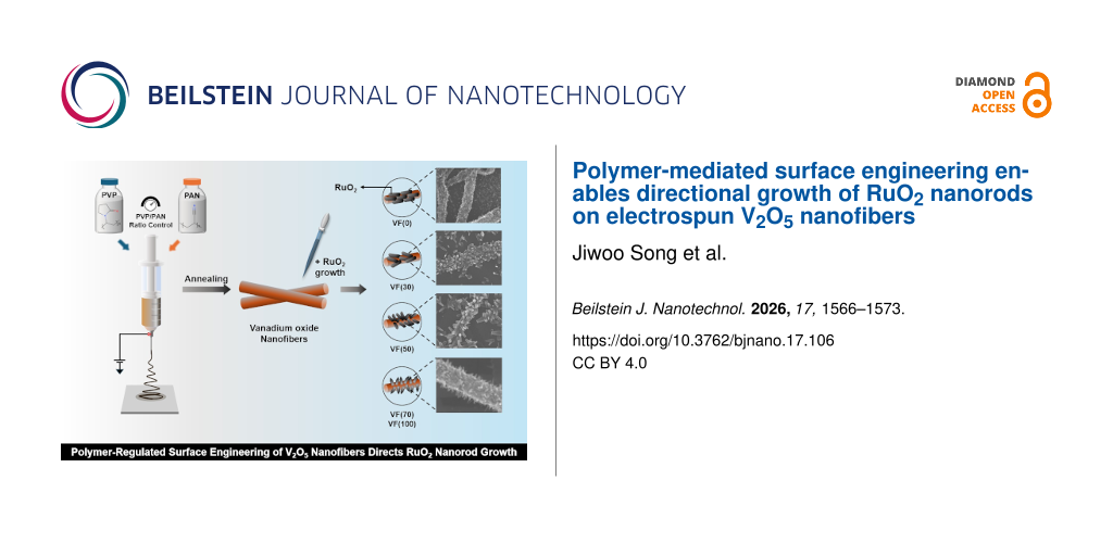
Controlling the morphology of metal oxide nanostructures through simple and scalable synthetic strategies remains challenging because crystal growth is strongly governed by interfacial surface chemistry. Herein, we present a polymer-mediated strategy for directing the growth of RuO2 nanorods on electrospun V2O5 nanofibers by tuning the polyvinylpyrrolidone (PVP)/polyacrylonitrile (PAN) ratio in the precursor solution. PAN incorporation preserves the one-dimensional V2O5 framework while progressively promoting the formation of vertically aligned RuO2 nanorods. Structural and spectroscopic analyses reveal that PAN-induced thermal evolution modifies the surface chemistry of V2O5 by increasing pyridinic nitrogen functionalities and oxygen-deficient surface sites. These surface characteristics are proposed to facilitate preferential Ru precursor nucleation, leading to anisotropic crystal growth without altering the crystal structures of either V2O5 or RuO2. The optimized composition containing 50 wt % PAN exhibits the most homogeneous and vertically aligned RuO2 nanorod arrays.
Introduction
Controlling crystal growth at heterogeneous oxide interfaces remains a fundamental challenge because nucleation and morphological evolution are highly sensitive to the local chemical environment during thermal processing [1]. Although numerous synthetic approaches have been developed to regulate metal oxide nanostructures, precise control over anisotropic crystal growth generally relies on surfactants, templates, or complex solution chemistries that limit scalability and synthetic simplicity [2]. Vanadium oxides (VOx, x = 1.0–2.5) exhibit multiple accessible oxidation states (V3+, V4+, and V5+), enabling reversible redox reactions for a wide range of electrochemical energy-storage and conversion applications [3-7]. Despite these advantages, their relatively poor electrical conductivity often limits reaction kinetics and overall electrochemical performance. Hybridization with highly conductive noble metal oxides provides an attractive strategy to overcome these limitations by simultaneously improving charge transport and interfacial electron transfer. Among various candidates, RuO2 is particularly attractive owing to its metallic conductivity, excellent chemical stability and fast surface redox kinetics, making it one of the most efficient oxide materials for electrocatalysis [8,9], electrochemical sensing [10-12], and heterogeneous catalytic processes [13,14].
Existing synthetic approaches predominantly rely on surfactants, templates, or ligands to manipulate crystal growth [15,16]. While these methods effectively control morphology, they often increase synthetic complexity and provide limited control over the coupled evolution of oxide supports and secondary crystal nucleation during thermal processing. Consequently, developing a simple and scalable strategy that regulates crystal growth through intrinsic interfacial chemistry, rather than external structure-directing agents, remains highly desirable. In this regard, polymer-assisted electrospinning provides a unique opportunity to regulate interfacial chemistry during oxide formation [17,18]. Beyond controlling the morphology of precursor fibers, polymer composition governs the thermal evolution of the precursor matrix, thereby determining the formation of residual surface functionalities and defect structures after calcination. These polymer-derived surface characteristics can significantly influence heterogeneous nucleation and subsequent crystal growth; however, their role in directing the morphology of secondary metal oxides remains largely unexplored [19].
Herein, we demonstrate that polymer composition can be exploited to direct the anisotropic growth of RuO2 nanorods on electrospun V2O5 nanofibers. By systematically tuning the polyvinylpyrrolidone (PVP)/polyacrylonitrile (PAN) ratio, the thermal evolution of the precursor fibers is regulated while preserving the one-dimensional V2O5 framework after calcination. The resulting polymer-derived surface chemistry, characterized by increased pyridinic nitrogen functionalities and oxygen-deficient surface sites, is proposed to facilitate preferential Ru precursor nucleation and anisotropic crystal growth, leading to uniformly aligned nanorods without altering the crystal structures of either V2O5 or RuO2.
Experimental
Materials
Vanadium(III) chloride (VCl3, 97%), ruthenium(III) chloride hydrate (RuCl3·xH2O), polyvinylpyrrolidone (PVP, MW ≈ 1 300 000) and polyacrylonitrile (PAN, MW ≈ 150 000) were purchased from Sigma-Aldrich (St. Louis, MO, USA). N,N-dimethylformamide (DMF, extra pure grade), ethanol (94.5%, extra pure grade), and sodium hydroxide (NaOH) beads were purchased from Ducksan (Korea). All other chemicals used were of analytical grade without further purification, and all solutions used for experiments were prepared with deionized water (resistivity ≥ 18 MΩ·cm).
Synthesis
Vanadium oxide nanofibers were synthesized via electrospinning followed by post-annealing, and the resulting samples were denoted as VF(x), where x represents the PAN weight percentage (wt %) in the precursor polymer composition. For VF(0), total of 50 mg of metal precursors were dissolved in a 1:1 (v/v) mixture of ethanol and deionized water. The resulting solution was ultrasonicated for 10 min, and then 300 mg of PVP was added. The mixture was stirred at 25 °C for 24 h to ensure complete homogenization and viscosity optimization for electrospinning. The as-prepared solution was then loaded into 12 mL plastic syringe fitted with a 25-gauge needle. Electrospinning (NanoNC ESR200R2) was performed under the following conditions: flow rate of 5 μL·min−1, applied voltage of 15.0 kV, and needle-to-collector distance of 11 cm. VF(30), VF(50), VF(70), and VF(100) were prepared by incorporating PAN at 30, 50, 70, and 100 wt %, respectively. All the resulting as-spun nanofibers were calcined at 350 °C under a gas flow of 20 sccm O2 and 80 sccm He, with ramping rate of 0.5 °C·min−1. After synthesizing the VF(x) samples, amorphous ruthenium hydroxide (i.e., Ru(OH)y) was prepared by the dropwise addition of 0.1 M NaOH to an aqueous RuCl3·xH2O solution until the pH reached 10 at 25 °C. The resulting precipitate was thoroughly washed with deionized water and redispersed in ethanol to form a homogeneous suspension. The Ru(OH)y suspension was then drop-cast onto the VF(x) substrates, followed by drying for 30 min. Subsequently, the samples were annealed in air at 200 °C for 3 h to obtain RuO2. The resulting composites were denoted as RuO2–VF(x).
Physical characterization
Morphological and elemental analyses were conducted via field-emission scanning electron microscopy (FESEM, JEOL JSM-7610F). High-resolution structural features were examined using high-resolution transmission electron microscopy (HRTEM, FEI Tecnai F20) and transmission electron microscopy (TEM, JEOL JEM-ARM200F NEOARM). Surface chemical states were investigated by angle-resolved X-ray photoelectron spectroscopy (ARXPS, Thermo Fisher Scientific K-ALPHA XPS, Al Kα radiation at 12 kV) and vibrational properties were probed using Raman microscope (HORIBA LabRAM HR Evolution 800). Thermal analysis was conducted with simultaneous thermogravimetric and differential thermal analysis (TGA-DSC, TA instruments STD 650) with a heating rate of 10 °C·min−1 up to 800 °C in air. Crystal structures were analyzed by X-ray diffraction (XRD; Malvern Panalytical) using Cu Kα radiation (λ = 1.5406 Å).
Results and Discussion
To elucidate the influence of polymer composition on the growth behavior of RuO2, the morphology and microstructure of the RuO2–VF(x) composites were systematically characterized. The corresponding fabrication scheme is shown in Figure 1. SEM images (Figure 2a–e) reveal that the electrospun V2O5 nanofiber framework is well preserved over the entire PAN composition range, indicating that the incorporation of PAN effectively suppresses structural collapse during thermal processing. In the absence of PAN (RuO2–VF(0), Figure 2a), RuO2 is randomly deposited as discontinuous particulate aggregates on the nanofiber surface, suggesting that crystal nucleation occurs isotropically without a preferred growth direction [20,21]. Upon increasing the PAN content, however, the RuO2 growth mode gradually transitions from randomly distributed nanoparticles to vertically aligned nanorods: At 30 wt % PAN, elongated RuO2 nanorods become discernible, while increasing the PAN content to 50 wt % results in densely packed and uniformly aligned nanorod arrays covering the V2O5 nanofiber surface. Further increasing the PAN content to 70 and 100 wt % produces no significant changes in nanorod morphology, indicating that the conditions favoring anisotropic RuO2 growth are achieved at approximately 50 wt % PAN.
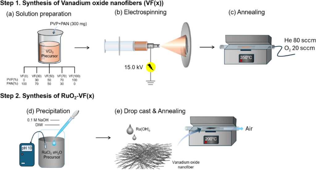
Figure 1: Schematic illustration of the synthetic methodology for RuO2–VF(x) nanostructures.
Figure 1: Schematic illustration of the synthetic methodology for RuO2–VF(x) nanostructures.
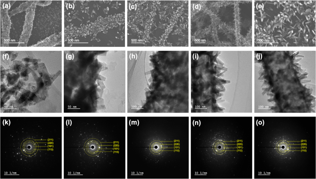
Figure 2: Morphological evolution of RuO2 nanorods on electrospun V2O5 nanofibers with increasing PAN content (from left to right: x = 0, 30, 50, 70, and 100 wt %): (a–e) SEM images, (f–j) corresponding TEM images, and (k–o) SAED patterns of RuO2–VF(x). The diffraction rings are indexed to the (110), (101), (220), and (211) planes of rutile RuO2 (JCPDS No. 65-2824).
Figure 2: Morphological evolution of RuO2 nanorods on electrospun V2O5 nanofibers with increasing PAN content...
TEM observations (Figure 2f–j) further corroborate the SEM results by revealing the microstructural evolution of the RuO2 coating. While RuO2–VF(0) exhibits a discontinuous layer of randomly distributed nanoparticles, PAN incorporation promotes the formation of radially oriented RuO2 nanorods with progressively improved uniformity and alignment. The RuO2–VF(50) sample displays the most homogeneous nanorod architecture, supporting the conclusion that polymer composition governs the transition from isotropic particle deposition to anisotropic crystal growth. The crystal structure of the deposited RuO2 was further examined by selected-area electron diffraction (SAED) (Figure 2k–o). All samples exhibit well-defined diffraction rings indexed to the (110), (101), (220), and (211) planes of rutile RuO2 (JCPDS No. 65-2824), confirming the formation of crystalline RuO2 regardless of PAN content. Despite the significant morphological evolution observed by SEM and TEM, the diffraction patterns remain essentially unchanged, indicating that polymer modulation regulates the nucleation and growth behavior of RuO2 without altering its intrinsic crystal structure. To further investigate the elemental distribution within the RuO2–V2O5 nanostructures, TEM-EDS elemental mapping was performed on RuO2–VF(0) and RuO2–VF(50) as representative samples (Supporting Information File 1, Figure S1 and Figure S2). Consistent with the SEM observations, RuO2–VF(0) exhibits a rough surface, whereas RuO2–VF(50) features distinct RuO2 nanorods grown on V2O5 nanofibers. The detection of C, Ru, N, and V in both samples further supports the formation of RuO2/V2O5 heterostructures. The XRD patterns in Figure S3 (Supporting Information File 1) show characteristic reflections of rutile RuO2, indexed to the (110), (101), (220), and (211) planes (JCPDS No. 65-2824), consistent with the TEM-SAED results. The VF(100) PVP-100 sample exhibits relatively lower peak intensity than the PAN/PVP mixed and PAN-only samples.
To elucidate the structural characteristics of the V2O5 substrate governing the subsequent growth of RuO2 nanorods, Raman spectroscopy was performed on the synthesized VF(x) nanofibers (Figure 3). All samples exhibit the characteristic Raman modes of orthorhombic α-V2O5, confirming the successful formation of the crystalline V2O5 framework regardless of polymer composition. The characteristic bands at approximately 142, 192, 284, 405, 698, and 994 cm−1 are assigned to the B1g/B3g lattice vibration, Ag/B2g vanadium vibration, V–O bending, Ag V–O rocking, B2g V–O stretching, and terminal V=O stretching modes, respectively, in good agreement with previous reports [22-25]. Notably, the positions of these characteristic Raman bands remain essentially unchanged from VF(0) to VF(70), indicating that increasing the PAN content does not alter the crystal structure of the electrospun V2O5 nanofibers. Although VF(100) exhibits reduced peak intensity accompanied by an increased spectral background, the characteristic α-V2O5 Raman modes remain clearly identifiable, suggesting that the crystalline framework is largely preserved even at the highest PAN content. These results demonstrate that modulation of the PVP/PAN ratio primarily influences the surface characteristics of the nanofibers without inducing phase transformation of the V2O5 substrate.
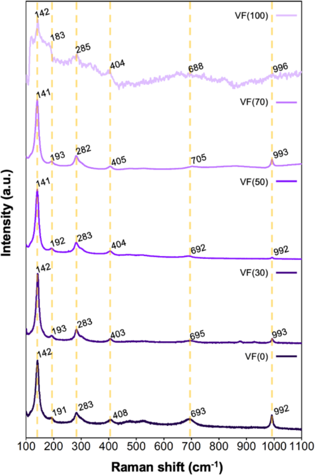
Figure 3: Raman spectra of electrospun V2O5 nanofibers (VF(x)) with increasing PAN content (x = 0, 30, 50, 70 and 100 wt %), demonstrating the preservation of the orthorhombic α-V2O5 structure.
Figure 3: Raman spectra of electrospun V2O5 nanofibers (VF(x)) with increasing PAN content (x = 0, 30, 50, 70...
To understand how polymer composition influences the growth behavior of RuO2 nanorods, the thermal evolution of the as-spun VF(x) nanofibers was investigated by thermogravimetric analysis (TGA) and differential scanning calorimetry (DSC) in air (Figure 4). A notable feature observed in both TGA and DSC is the nearly identical thermal profiles of VF(x) with PAN contents of 50–100 wt %, which coincide with the saturation of the RuO2 morphology observed by SEM and TEM (vide supra). This correlation suggests that the thermal evolution of the polymer matrix plays a critical role in establishing the interfacial chemical environment that governs the nucleation and anisotropic growth of RuO2. As shown in Figure 4a, all samples exhibit an initial weight loss below 100 °C, which is attributed to the evaporation of residual solvent and physically adsorbed moisture. The thermal decomposition behavior thereafter strongly depends on polymer composition. VF(0), consisting solely of PVP, exhibits two major weight-loss events at approximately 330 and 445 °C, accompanied by corresponding exothermic peaks in the DSC profile (Figure 4b), which are associated with PVP side-chain decomposition and backbone combustion [26]. In contrast, PAN-containing samples exhibit distinctly different thermal characteristics. For VF(30), new exothermic events emerge at approximately 300 and 370 °C, corresponding to oxidative cyclization of nitrile groups and subsequent aromatization of the polymer backbone, respectively [27,28]. These reactions progressively dominate the thermal behavior as the PAN content increases, leading to nearly identical thermal profiles for VF(50–100). Importantly, the oxidative stabilization occurring near 300 °C takes place within the calcination temperature range employed for V2O5 nanofiber synthesis. The formation of a thermally stable ladder-like structure during this process is expected to suppress structural relaxation of the precursor fibers while simultaneously modifying the chemical environment of the oxide surface. The close agreement between the thermal evolution and the morphology of the resulting RuO2 suggests that PAN-derived thermal stabilization contributes to the formation of surface conditions favorable for anisotropic RuO2 growth.
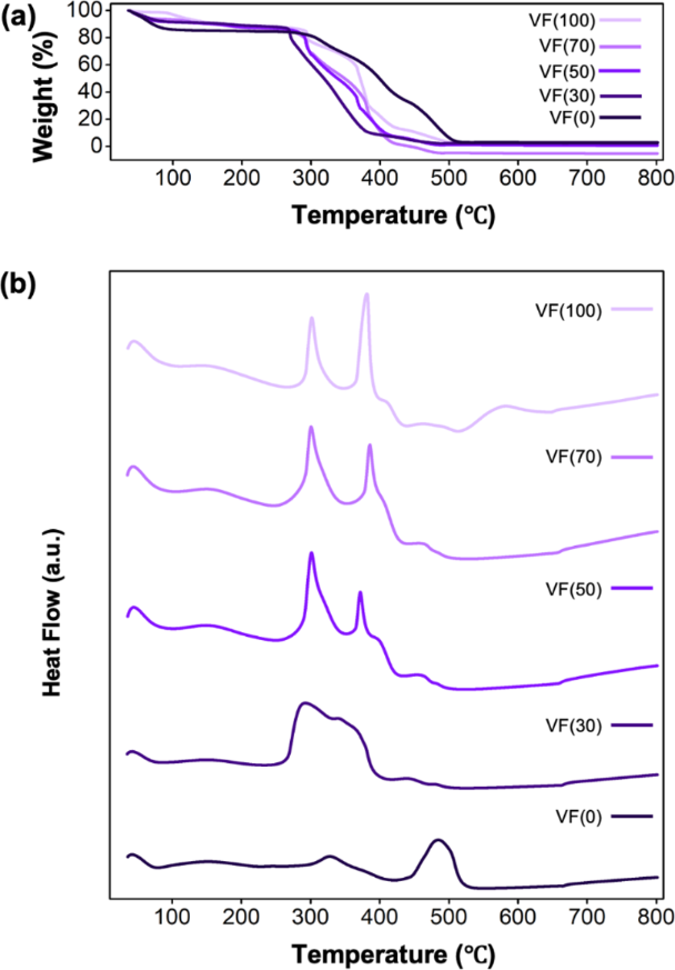
Figure 4: Thermal evolution of electrospun VF(x) nanofibers as a function of PAN content (x = 0, 30, 50, 70 and 100 wt %): (a) TGA curves and (b) DSC thermograms.
Figure 4: Thermal evolution of electrospun VF(x) nanofibers as a function of PAN content (x = 0, 30, 50, 70 a...
To elucidate how polymer composition modifies the surface chemistry of the V2O5 support, X-ray photoelectron spectroscopy (XPS) was performed on the synthesized VF(x) nanofibers (Figure 5). The V 2p spectra (Figure 5a) exhibit characteristic V4+ and V5+ components at 515.9/523.2 and 517.0/524.3 eV, respectively, in good agreement with reported values for orthorhombic V2O5 [29,30]. No appreciable changes in either the binding energies or relative spectral features are observed with increasing PAN content, indicating that polymer modulation does not alter the chemical state of the V2O5 framework, consistent with the Raman results (vide supra). In contrast, the surface carbon and nitrogen chemistries exhibit pronounced changes with polymer composition. The C 1s spectra (Figure 5b) can be deconvoluted into C–C, C–N/C–O, and C=O components centered at 284.6, 286.5, and 288.0 eV, respectively [31,32]. More importantly, the N 1s spectra (Figure 5c) show that increasing the PAN content progressively enriches the V2O5 surface with pyridinic nitrogen functionalities, particularly above 50 wt %. These nitrogen functionalities originate from PAN-derived oxidative stabilization during calcination, in agreement with the thermal analysis. Since pyridinic nitrogen has been reported to serve as an effective anchoring site for Ru precursor species [33], the increased abundance of pyridinic nitrogen is expected to facilitate preferential Ru nucleation during the subsequent deposition process. The evolution of the O 1s spectra (Figure 5d) further supports the polymer-induced surface reconstruction. Deconvolution of the spectra identifies lattice oxygen, oxygen-deficient species, and adsorbed oxygen at approximately 530, 531, and 533 eV, respectively [34,35]. Notably, the relative concentration of oxygen-deficient surface species increases progressively with PAN incorporation, with a pronounced increase observed above 50 wt % PAN. Consistent with this trend, the oxygen-vacancy-to-lattice-oxygen ratio increases from approximately 1.2 for VF(0) and VF(30) to 1.8, 2.6, and 3.5 for VF(50), VF(70), and VF(100), respectively. The evolution of pyridinic nitrogen and oxygen-deficient surface sites closely parallels the morphological transition of RuO2 observed by SEM and TEM, suggesting that polymer-derived surface reconstruction modifies the interfacial chemical environment governing Ru precursor adsorption and anisotropic crystal growth. Furthermore, the V4+ species detected by XPS may facilitate the nucleation of rutile RuO2. Because V4+ can locally form rutile-type VO2-like domains, which are isostructural with RuO2 and have comparable lattice parameters, these domains could serve as low-mismatch templates for RuO2 nucleation [36].
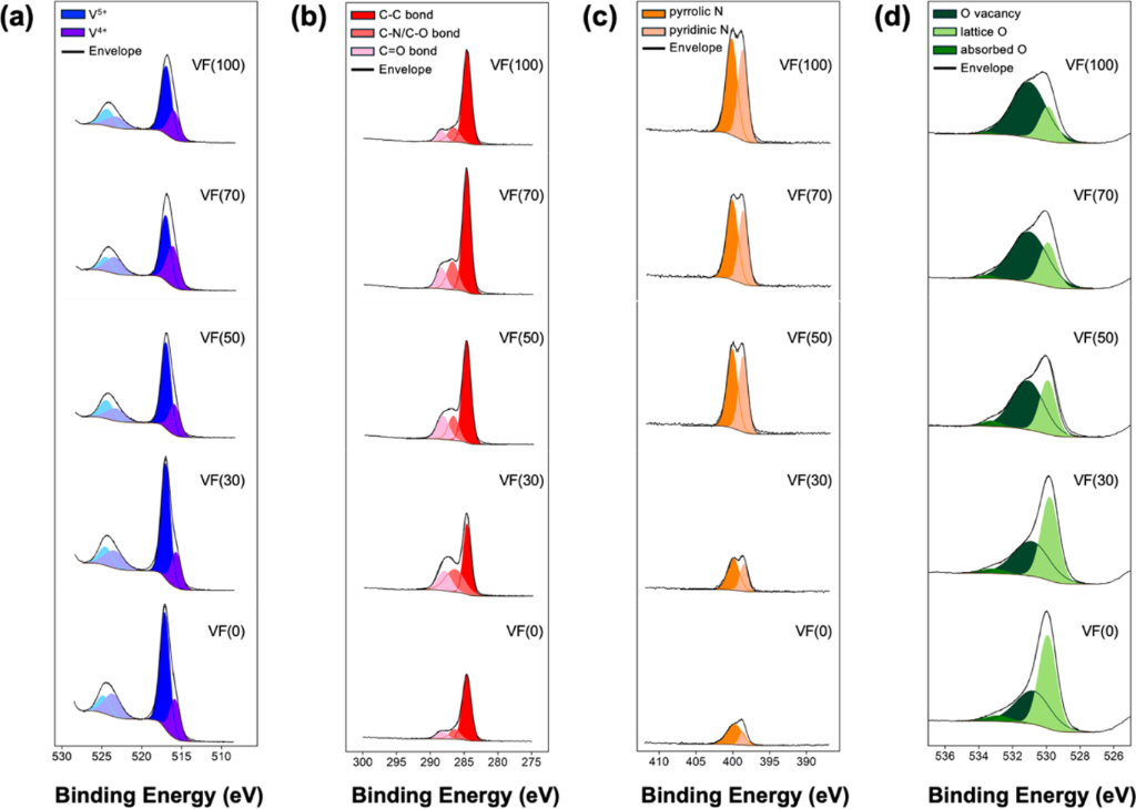
Figure 5: ARXPS spectra of VF(x) with increasing PAN content (x = 0, 30, 50, 70, and 100 wt %), showing the (a) V 2p, (b) C 1s, (c) N 1s, and (d) O 1s core-level regions.
Figure 5: ARXPS spectra of VF(x) with increasing PAN content (x = 0, 30, 50, 70, and 100 wt %), showing the (...
Conclusion
In this study, we developed a polymer-regulated strategy for simultaneously stabilizing electrospun V2O5 nanofibers and directing the anisotropic growth of RuO2 nanorods by tuning the PVP/PAN ratio in the electrospinning precursor. By systematically tuning the PVP/PAN ratio, the thermal evolution of the precursor fibers and the resulting surface chemistry were effectively controlled while preserving the crystalline V2O5 framework. In particular, a PAN content of approximately 50 wt % promotes the formation of pyridinic nitrogen functionalities and oxygen-deficient surface sites, which are proposed to facilitate preferential Ru precursor nucleation and the subsequent growth of uniformly aligned RuO2 nanorods. These findings demonstrate that polymer composition serves as an effective means of regulating interfacial chemistry during thermal processing, providing a simple and scalable strategy for controlling oxide nanostructures without relying on surfactants, templates, or complex synthetic procedures.
Supporting Information
| Supporting Information File 1: Additional figures. | ||
| Format: PDF | Size: 413.8 KB | Download |
Acknowledgements
D.J. gratefully acknowledge support from Department of Chemistry, College of Natural Science at Jeonbuk National University.
Data Availability Statement
Data generated and analyzed during this study is available from the corresponding author upon reasonable request.
References
-
Xu, L.; Xiong, P.; Zeng, L.; Fang, Y.; Liu, R.; Liu, J.; Luo, F.; Chen, Q.; Wei, M.; Qian, Q. Nanoscale 2019, 11, 16308–16316. doi:10.1039/c9nr03574e
Return to citation in text: [1] -
Chamakh, M.; Ayesh, A. I. Mater. Des. 2021, 204, 109678. doi:10.1016/j.matdes.2021.109678
Return to citation in text: [1] -
Sohaimi, K.; Jaafar, J.; Rosman, N. Malays. J. Fundam. Appl. Sci. 2023, 19, 901–914.
Return to citation in text: [1] -
Deng, R.; Wang, Z.; Tan, S.; Huang, X.; Gao, Z.; Fang, Y.; Chen, C.; Wang, R.; Xu, C.; Huang, G.; Wang, J.; Pan, F. Chem. Eng. J. 2024, 489, 151095. doi:10.1016/j.cej.2024.151095
Return to citation in text: [1] -
Zhu, C.; Shu, J.; Wu, X.; Li, P.; Li, X. J. Electroanal. Chem. 2015, 759, 184–189. doi:10.1016/j.jelechem.2015.11.013
Return to citation in text: [1] -
Cheah, Y. L.; Gupta, N.; Pramana, S. S.; Aravindan, V.; Wee, G.; Srinivasan, M. J. Power Sources 2011, 196, 6465–6472. doi:10.1016/j.jpowsour.2011.03.039
Return to citation in text: [1] -
Li, S.; Qin, L.; Li, L.; Cheng, H.; Fang, G.; Zhu, Q. Int. J. Electrochem. Sci. 2021, 16, 210349. doi:10.20964/2021.03.26
Return to citation in text: [1] -
Hou, L.; Li, Z.; Jang, H.; Wang, Y.; Cui, X.; Gu, X.; Kim, M. G.; Feng, L.; Liu, S.; Liu, X. Adv. Energy Mater. 2023, 13, 2300177. doi:10.1002/aenm.202300177
Return to citation in text: [1] -
Liu, M.; Su, B.; Tang, Y.; Jiang, X.; Yu, A. Adv. Energy Mater. 2017, 7, 1700885. doi:10.1002/aenm.201700885
Return to citation in text: [1] -
Chun, S. H.; Choi, H.-A.; Kang, M.; Koh, M.; Lee, N.-S.; Lee, S. C.; Lee, M.; Lee, Y.; Lee, C.; Kim, M. H. ACS Appl. Mater. Interfaces 2013, 5, 8401–8406. doi:10.1021/am4016445
Return to citation in text: [1] -
Shahzad, U.; Saeed, M.; Marwani, H. M.; Al-Humaidi, J. Y.; Rehman, S. u.; Althomali, R. H.; Awual, M. R.; Rahman, M. M. Crit. Rev. Anal. Chem. 2025, 55, 1081–1098. doi:10.1080/10408347.2024.2337876
Return to citation in text: [1] -
Li, T.; Yin, W.; Gao, S.; Sun, Y.; Xu, P.; Wu, S.; Kong, H.; Yang, G.; Wei, G. Nanomaterials 2022, 12, 982. doi:10.3390/nano12060982
Return to citation in text: [1] -
Over, H. Chem. Rev. 2012, 112, 3356–3426. doi:10.1021/cr200247n
Return to citation in text: [1] -
Hodnik, N.; Jovanovič, P.; Pavlišič, A.; Jozinović, B.; Zorko, M.; Bele, M.; Šelih, V. S.; Šala, M.; Hočevar, S.; Gaberšček, M. J. Phys. Chem. C 2015, 119, 10140–10147. doi:10.1021/acs.jpcc.5b01832
Return to citation in text: [1] -
Wang, Y.; Cao, G. Chem. Mater. 2006, 18, 2787–2804. doi:10.1021/cm052765h
Return to citation in text: [1] -
Inoue, Y.; Ohtsuka, S.; Torikai, T.; Watari, T.; Yada, M. Cryst. Growth Des. 2009, 9, 5092–5100. doi:10.1021/cg900261s
Return to citation in text: [1] -
Wu, X.; Bai, W.; Yuan, L.; Li, J.; Qi, L.; Weng, X.; Gu, C.; Bi, M. ACS Appl. Mater. Interfaces 2025, 17, 19848–19855. doi:10.1021/acsami.4c10823
Return to citation in text: [1] -
Walls, B.; Murtagh, O.; Bozhko, S. I.; Ionov, A.; Mazilkin, A. A.; Mullarkey, D.; Zhussupbekova, A.; Shulyatev, D. A.; Zhussupbekov, K.; Andreev, N.; Tabachkova, N.; Shvets, I. V. Materials 2022, 15, 7652. doi:10.3390/ma15217652
Return to citation in text: [1] -
Park, J.; Lee, J. W.; Ye, B. U.; Chun, S. H.; Joo, S. H.; Park, H.; Lee, H.; Jeong, H. Y.; Kim, M. H.; Baik, J. M. Sci. Rep. 2015, 5, 11933. doi:10.1038/srep11933
Return to citation in text: [1] -
Kim, S.-j.; Cho, Y. K.; Lee, C.; Kim, M. H.; Lee, Y. Biosens. Bioelectron. 2016, 77, 1144–1152. doi:10.1016/j.bios.2015.11.012
Return to citation in text: [1] -
Kim, K.-H.; Kim, K. S.; Kim, G.-P.; Baeck, S.-H. Curr. Appl. Phys. 2012, 12, 36–39. doi:10.1016/j.cap.2011.04.029
Return to citation in text: [1] -
Baddour-Hadjean, R.; Pereira-Ramos, J. P.; Navone, C.; Smirnov, M. Chem. Mater. 2008, 20, 1916–1923. doi:10.1021/cm702979k
Return to citation in text: [1] -
Khodakov, A.; Olthof, B.; Bell, A. T.; Iglesia, E. J. Catal. 1999, 181, 205–216. doi:10.1006/jcat.1998.2295
Return to citation in text: [1] -
Prasad, A.; John, M. S.; John, F. Mater. Chem. Phys. 2026, 348, 131676. doi:10.1016/j.matchemphys.2025.131676
Return to citation in text: [1] -
Gershinsky, G.; Yoo, H. D.; Gofer, Y.; Aurbach, D. Langmuir 2013, 29, 10964–10972. doi:10.1021/la402391f
Return to citation in text: [1] -
Elishav, O.; Beilin, V.; Rozent, O.; Shter, G. E.; Grader, G. S. J. Polym. Sci., Part B: Polym. Phys. 2018, 56, 248–254. doi:10.1002/polb.24538
Return to citation in text: [1] -
Ucar, N.; Demirsoy, N.; Onen, A.; Karacan, I.; Kizildag, N.; Eren, O.; Faruk Vurur, O.; Sezer, E.; Ustamehmetoglu, B. J. Mater. Sci. 2015, 50, 1855–1864. doi:10.1007/s10853-014-8748-4
Return to citation in text: [1] -
Yin, C.; Li, L.; Jia, R.; Hu, J. Inorg. Chem. 2024, 63, 20551–20561. doi:10.1021/acs.inorgchem.4c03146
Return to citation in text: [1] -
Lin, L.; Tian, S.-L.; Fang, L.; Chang, L.-M.; Liu, W.-Q.; Wang, Q.-S. J. Energy Storage 2023, 72, 108356. doi:10.1016/j.est.2023.108356
Return to citation in text: [1] -
Silversmit, G.; Depla, D.; Poelman, H.; Marin, G. B.; De Gryse, R. J. Electron Spectrosc. Relat. Phenom. 2004, 135, 167–175. doi:10.1016/j.elspec.2004.03.004
Return to citation in text: [1] -
Liu, T.; Li, L.; Yao, T.; Li, Y.; Zhu, L.; Li, F.; Han, X.; Cheng, Y.; Wang, H. Electrochim. Acta 2021, 369, 137711. doi:10.1016/j.electacta.2020.137711
Return to citation in text: [1] -
Chen, X.; Unruh, K. M.; Ni, C.; Ali, B.; Sun, Z.; Lu, Q.; Deitzel, J.; Xiao, J. Q. J. Phys. Chem. C 2011, 115, 373–378. doi:10.1021/jp1082533
Return to citation in text: [1] -
Zhang, C.; Sha, J.; Fei, H.; Liu, M.; Yazdi, S.; Zhang, J.; Zhong, Q.; Zou, X.; Zhao, N.; Yu, H.; Jiang, Z.; Ringe, E.; Yakobson, B. I.; Dong, J.; Chen, D.; Tour, J. M. ACS Nano 2017, 11, 6930–6941. doi:10.1021/acsnano.7b02148
Return to citation in text: [1] -
Zhao, K.; Teng, L.; Tang, Y.; Chen, X. Ceram. Int. 2014, 40, 15335–15340. doi:10.1016/j.ceramint.2014.06.079
Return to citation in text: [1] -
Song, J.; Gu, X.; Cao, Y.; Zhang, H. J. Mater. Chem. A 2019, 7, 10543–10551. doi:10.1039/c9ta01674k
Return to citation in text: [1] -
Kim, S.-j.; Cho, Y. K.; Seok, J.; Lee, N.-S.; Son, B.; Lee, J. W.; Baik, J. M.; Lee, C.; Lee, Y.; Kim, M. H. ACS Appl. Mater. Interfaces 2015, 7, 15321–15330. doi:10.1021/acsami.5b03178
Return to citation in text: [1]
| 34. | Zhao, K.; Teng, L.; Tang, Y.; Chen, X. Ceram. Int. 2014, 40, 15335–15340. doi:10.1016/j.ceramint.2014.06.079 |
| 35. | Song, J.; Gu, X.; Cao, Y.; Zhang, H. J. Mater. Chem. A 2019, 7, 10543–10551. doi:10.1039/c9ta01674k |
| 36. | Kim, S.-j.; Cho, Y. K.; Seok, J.; Lee, N.-S.; Son, B.; Lee, J. W.; Baik, J. M.; Lee, C.; Lee, Y.; Kim, M. H. ACS Appl. Mater. Interfaces 2015, 7, 15321–15330. doi:10.1021/acsami.5b03178 |
| 1. | Xu, L.; Xiong, P.; Zeng, L.; Fang, Y.; Liu, R.; Liu, J.; Luo, F.; Chen, Q.; Wei, M.; Qian, Q. Nanoscale 2019, 11, 16308–16316. doi:10.1039/c9nr03574e |
| 10. | Chun, S. H.; Choi, H.-A.; Kang, M.; Koh, M.; Lee, N.-S.; Lee, S. C.; Lee, M.; Lee, Y.; Lee, C.; Kim, M. H. ACS Appl. Mater. Interfaces 2013, 5, 8401–8406. doi:10.1021/am4016445 |
| 11. | Shahzad, U.; Saeed, M.; Marwani, H. M.; Al-Humaidi, J. Y.; Rehman, S. u.; Althomali, R. H.; Awual, M. R.; Rahman, M. M. Crit. Rev. Anal. Chem. 2025, 55, 1081–1098. doi:10.1080/10408347.2024.2337876 |
| 12. | Li, T.; Yin, W.; Gao, S.; Sun, Y.; Xu, P.; Wu, S.; Kong, H.; Yang, G.; Wei, G. Nanomaterials 2022, 12, 982. doi:10.3390/nano12060982 |
| 31. | Liu, T.; Li, L.; Yao, T.; Li, Y.; Zhu, L.; Li, F.; Han, X.; Cheng, Y.; Wang, H. Electrochim. Acta 2021, 369, 137711. doi:10.1016/j.electacta.2020.137711 |
| 32. | Chen, X.; Unruh, K. M.; Ni, C.; Ali, B.; Sun, Z.; Lu, Q.; Deitzel, J.; Xiao, J. Q. J. Phys. Chem. C 2011, 115, 373–378. doi:10.1021/jp1082533 |
| 8. | Hou, L.; Li, Z.; Jang, H.; Wang, Y.; Cui, X.; Gu, X.; Kim, M. G.; Feng, L.; Liu, S.; Liu, X. Adv. Energy Mater. 2023, 13, 2300177. doi:10.1002/aenm.202300177 |
| 9. | Liu, M.; Su, B.; Tang, Y.; Jiang, X.; Yu, A. Adv. Energy Mater. 2017, 7, 1700885. doi:10.1002/aenm.201700885 |
| 33. | Zhang, C.; Sha, J.; Fei, H.; Liu, M.; Yazdi, S.; Zhang, J.; Zhong, Q.; Zou, X.; Zhao, N.; Yu, H.; Jiang, Z.; Ringe, E.; Yakobson, B. I.; Dong, J.; Chen, D.; Tour, J. M. ACS Nano 2017, 11, 6930–6941. doi:10.1021/acsnano.7b02148 |
| 3. | Sohaimi, K.; Jaafar, J.; Rosman, N. Malays. J. Fundam. Appl. Sci. 2023, 19, 901–914. |
| 4. | Deng, R.; Wang, Z.; Tan, S.; Huang, X.; Gao, Z.; Fang, Y.; Chen, C.; Wang, R.; Xu, C.; Huang, G.; Wang, J.; Pan, F. Chem. Eng. J. 2024, 489, 151095. doi:10.1016/j.cej.2024.151095 |
| 5. | Zhu, C.; Shu, J.; Wu, X.; Li, P.; Li, X. J. Electroanal. Chem. 2015, 759, 184–189. doi:10.1016/j.jelechem.2015.11.013 |
| 6. | Cheah, Y. L.; Gupta, N.; Pramana, S. S.; Aravindan, V.; Wee, G.; Srinivasan, M. J. Power Sources 2011, 196, 6465–6472. doi:10.1016/j.jpowsour.2011.03.039 |
| 7. | Li, S.; Qin, L.; Li, L.; Cheng, H.; Fang, G.; Zhu, Q. Int. J. Electrochem. Sci. 2021, 16, 210349. doi:10.20964/2021.03.26 |
| 27. | Ucar, N.; Demirsoy, N.; Onen, A.; Karacan, I.; Kizildag, N.; Eren, O.; Faruk Vurur, O.; Sezer, E.; Ustamehmetoglu, B. J. Mater. Sci. 2015, 50, 1855–1864. doi:10.1007/s10853-014-8748-4 |
| 28. | Yin, C.; Li, L.; Jia, R.; Hu, J. Inorg. Chem. 2024, 63, 20551–20561. doi:10.1021/acs.inorgchem.4c03146 |
| 2. | Chamakh, M.; Ayesh, A. I. Mater. Des. 2021, 204, 109678. doi:10.1016/j.matdes.2021.109678 |
| 29. | Lin, L.; Tian, S.-L.; Fang, L.; Chang, L.-M.; Liu, W.-Q.; Wang, Q.-S. J. Energy Storage 2023, 72, 108356. doi:10.1016/j.est.2023.108356 |
| 30. | Silversmit, G.; Depla, D.; Poelman, H.; Marin, G. B.; De Gryse, R. J. Electron Spectrosc. Relat. Phenom. 2004, 135, 167–175. doi:10.1016/j.elspec.2004.03.004 |
| 19. | Park, J.; Lee, J. W.; Ye, B. U.; Chun, S. H.; Joo, S. H.; Park, H.; Lee, H.; Jeong, H. Y.; Kim, M. H.; Baik, J. M. Sci. Rep. 2015, 5, 11933. doi:10.1038/srep11933 |
| 22. | Baddour-Hadjean, R.; Pereira-Ramos, J. P.; Navone, C.; Smirnov, M. Chem. Mater. 2008, 20, 1916–1923. doi:10.1021/cm702979k |
| 23. | Khodakov, A.; Olthof, B.; Bell, A. T.; Iglesia, E. J. Catal. 1999, 181, 205–216. doi:10.1006/jcat.1998.2295 |
| 24. | Prasad, A.; John, M. S.; John, F. Mater. Chem. Phys. 2026, 348, 131676. doi:10.1016/j.matchemphys.2025.131676 |
| 25. | Gershinsky, G.; Yoo, H. D.; Gofer, Y.; Aurbach, D. Langmuir 2013, 29, 10964–10972. doi:10.1021/la402391f |
| 17. | Wu, X.; Bai, W.; Yuan, L.; Li, J.; Qi, L.; Weng, X.; Gu, C.; Bi, M. ACS Appl. Mater. Interfaces 2025, 17, 19848–19855. doi:10.1021/acsami.4c10823 |
| 18. | Walls, B.; Murtagh, O.; Bozhko, S. I.; Ionov, A.; Mazilkin, A. A.; Mullarkey, D.; Zhussupbekova, A.; Shulyatev, D. A.; Zhussupbekov, K.; Andreev, N.; Tabachkova, N.; Shvets, I. V. Materials 2022, 15, 7652. doi:10.3390/ma15217652 |
| 26. | Elishav, O.; Beilin, V.; Rozent, O.; Shter, G. E.; Grader, G. S. J. Polym. Sci., Part B: Polym. Phys. 2018, 56, 248–254. doi:10.1002/polb.24538 |
| 15. | Wang, Y.; Cao, G. Chem. Mater. 2006, 18, 2787–2804. doi:10.1021/cm052765h |
| 16. | Inoue, Y.; Ohtsuka, S.; Torikai, T.; Watari, T.; Yada, M. Cryst. Growth Des. 2009, 9, 5092–5100. doi:10.1021/cg900261s |
| 13. | Over, H. Chem. Rev. 2012, 112, 3356–3426. doi:10.1021/cr200247n |
| 14. | Hodnik, N.; Jovanovič, P.; Pavlišič, A.; Jozinović, B.; Zorko, M.; Bele, M.; Šelih, V. S.; Šala, M.; Hočevar, S.; Gaberšček, M. J. Phys. Chem. C 2015, 119, 10140–10147. doi:10.1021/acs.jpcc.5b01832 |
| 20. | Kim, S.-j.; Cho, Y. K.; Lee, C.; Kim, M. H.; Lee, Y. Biosens. Bioelectron. 2016, 77, 1144–1152. doi:10.1016/j.bios.2015.11.012 |
| 21. | Kim, K.-H.; Kim, K. S.; Kim, G.-P.; Baeck, S.-H. Curr. Appl. Phys. 2012, 12, 36–39. doi:10.1016/j.cap.2011.04.029 |
© 2026 Song et al.; licensee Beilstein-Institut.
This is an open access article licensed under the terms of the Beilstein-Institut Open Access License Agreement (https://www.beilstein-journals.org/bjnano/terms), which is identical to the Creative Commons Attribution 4.0 International License (https://creativecommons.org/licenses/by/4.0). The reuse of material under this license requires that the author(s), source and license are credited. Third-party material in this article could be subject to other licenses (typically indicated in the credit line), and in this case, users are required to obtain permission from the license holder to reuse the material.