


On 5 December 1985, Gerd Binnig, Christoph Gerber, and Calvin Quate submitted a three-and-a-half-page letter to Physical Review Letters that would quietly redefine surface science. Published on 3 March 1986 under the title “Atomic Force Microscope”, the letter proposed that the deflection of a soft cantilever, sensed by a scanning tunnelling microscope acting as a displacement transducer, could measure forces as small as 10−18 N and image the topography of conductors and insulators alike, atom by atom . The instrument that the authors built by hand was able to resolve a 30 Å corrugation on a ceramic Al2O3 surface with a force sensitivity of the order of 10−10 N. This instrument consisted of a diamond tip glued to a gold-foil lever, which was sandwiched between an STM tip and the sample and was damped by stacked Viton spacers. Forty years and many millions of AFM images later, it is easy to forget how unconventional that idea was: a “profilometer” gentle enough not to damage the very atoms it was trying to see.
This year, we mark the 40th anniversary of that publication, and we do so with particular pride since Christoph Gerber has spent the last four decades as a member of the Department of Physics at the University of Basel, continuing to shape the field he helped invent, from the first force microscope images to the biosensor cantilever arrays that followed. In a recent viewpoint in Nature Reviews Physics, seven leading specialists reflected that AFM has grown from a handmade curiosity into an indispensable workhorse that is constantly reinvented. It films molecular motors at work with sub-second time resolution, resolves individual chemical bonds and charge states with functionalized tips, maps the electrochemistry of battery interfaces and living bacterial cell walls, and is only beginning to be integrated with super-resolution optics, electron-spin resonance, and machine-learning-driven analysis . The distance between an Au-foil lever sensing 10−10 N and today’s routine sub-piconewton, sub-angstrom measurements is, in a very literal sense, the history of nanoscience itself.
This anniversary coincides with another milestone: 2026 marks the 75th anniversary of the Max Planck Society’s foundation of the “Beilstein-Institut zur Förderung der Chemischen Wissenschaften” in 1951. What began as a foundation dedicated to organizing chemical knowledge has, over the decades, become one of the most consistent champions of diamond-open-access publishing in the natural sciences, publishing and supporting the Beilstein Journal of Organic Chemistry and the Beilstein Journal of Nanotechnology without charging authors or readers, based on the belief that the dissemination of science should not be impeded by financial barriers. It is a coincidence worth celebrating that two anniversaries fall in the same year: one for a measurement technique and one for the institute that helps communicate its results.
A thematic issue with a long tradition. “At the cutting edge of atomic force microscopy” is the latest in a series of thematic issues that the Beilstein Journal of Nanotechnology has devoted to advanced scanning probe methods within its “Nanocharacterization” section over the years. These issues have grown in close alignment with the “International Conference on Non-Contact Atomic Force Microscopy (NC-AFM)”, a meeting series that has served the community since 1998 and held its 27th edition in Innsbruck in August 2026. “NC-AFM” has always been a comparatively small meeting focused on a specific technique rather than a large general conference, and this has been its strength. It is where instrument builders, theorists, and application scientists can discuss cantilever mechanics, tip functionalization, and force spectroscopy interpretation in sufficient depth for the conversation to regularly lead directly to publications. Since its founding in 1998, the meeting has been held in various locations, including Lindau in 2011, Montreal in 2024, and Toyama in 2025. Several members of our editorial team have organized and contributed to the conference over the years. This thematic issue is an extension of that conversation onto the page, providing a platform for the NC-AFM community to document its annual progress in full, peer-reviewed detail. In addition, it shares the progress of non-contact AFM with the broader AFM community and invites researchers beyond the conference to submit their own contributions.
What this issue contains. The fourteen contributions gathered here span the full breadth of what non-contact and dynamic-mode AFM has become. On the instrumentation and methodology side, Roorda et al. describe a combined AFM/STM instrument for operando imaging of surface reactions at elevated temperature and atmospheric pressure . Khachatryan and Reichling establish a dynamic interferometric calibration protocol for the piezo actuators that underlie every quantitative AFM measurement . Nishida, Yasui, and Sugimoto provide a careful theoretical treatment of the dynamic stiffness of cantilevers under torsional oscillation, directly relevant to lateral-force and dissipation measurements . Sullivan, Valdés, Bevan, and Grütter present “afspm”, an open, manufacturer-agnostic software framework for automating scanning probe experiments across heterogeneous hardware , a timely contribution as the field embraces autonomous and high-throughput measurement.
A second cluster addresses electrostatic and multifrequency force spectroscopy at functional interfaces. Rohrbeck et al. develop multifrequency electrostatic force microscopy for nanoscale capacitance spectroscopy . Bidinakis and Weber apply related scanning-probe methods to resolve how tris(pentafluorophenyl)borane doping of the hole transport layer governs interfacial charge extraction and recombination in perovskite solar cells . Tip characterization and atomic-scale contrast are addressed by Kyeyune, Rahe, and Reichling, who systematically track tip-polarity changes during NC-AFM imaging of a fluorite surface, essential groundwork for reliable atomic-resolution interpretation .
A personal note on Kelvin probe force microscopy. The multifrequency electrostatic force microscopy and interfacial charge-transport studies in this issue touch on a branch of the AFM family close to one of our own hearts. Kelvin probe force microscopy (KPFM) is the dynamic-mode variant that maps local contact potential difference, and with it surface charge, doping, band bending, and work function, down to the sub-nanometer scale. KPFM nicely illustrates the trend this issue documents at large. A Web of Science search on AFM- and KPFM-related output (Figure 1) shows that the AFM publications were climbing steeply from the mid-1990s to a broad plateau by the early 2010s. KPFM, a younger and more specialized technique, has kept accelerating well past that point, reaching a new high in 2025. Readers seeking a systematic entry point beyond this editorial are directed to two dedicated volumes in the Springer Series in Surface Sciences: “Kelvin Probe Force Microscopy: Measuring and Compensating Electrostatic Forces” and its sequel “Kelvin Probe Force Microscopy: From Single Charge Detection to Device Characterization” .
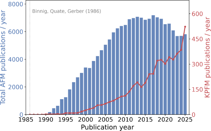
Figure 1: Annual number of publications on atomic force microscopy (blue bars, left axis) and on Kelvin probe force microscopy (red line, right axis), 1986–2025. Source: Web of Science (July 2026).
Figure 1: Annual number of publications on atomic force microscopy (blue bars, left axis) and on Kelvin probe...
Data-driven approaches feature prominently. Gelman et al. demonstrate deep-learning enhancement of low-resolution and noisy scanning probe images, benchmarked against expert human judgment . Barker and Sweetman present an automated pipeline for collecting and categorizing STM images and scanning tunneling spectra, with and without machine learning . Both point toward the increasingly autonomous laboratories of the future. Nanomechanical measurements are represented by Mathias, Bennewitz, and Egberts, who extract mechanical properties from short-term Fourier-transform analysis of AFM thermal deflection noise , and by Çiçek, Kratzer, Teichert, and Mitterer, who quantitatively determine nanoparticle–substrate adhesion by AFM manipulation . Closely related, two contributions from our own collaborations address nanoscale friction on two-dimensional transition metal dichalcogenides. Huang et al. show that exciting the cantilever at its contact resonances in ultrahigh vacuum suppresses the atomic stick–slip friction of a tip sliding on monolayer MoS2/Au(111) by more than an order of magnitude, a reversible and purely mechanical route to dynamic structural superlubricity that persists over the whole load range studied . Complementing these experiments from the theory side, Ravisankar et al. combine density functional theory, machine-learning-based force fields, and molecular dynamics to trace how individual sliding modes of a silicon tip on MoS2, MoSe2, WS2, and WSe2 monolayers supported on Au(111) and Ag(111) give rise to a nonlinear friction–load response with a distinct adhesive contribution . Finally, Lorenzoni contributes a comprehensive review of materials for AFM-based local anodic oxidation, surveying four decades of progress in scanning-probe nanolithography and its emerging applications in two-dimensional materials .
Read together, these fourteen papers are a fair snapshot of a technique that, forty years on, still has no difficulty finding new atoms, new molecules, and new questions to probe. They range from operando reaction chambers to torsional cantilever mechanics, from perovskite photovoltaics to nanoparticle adhesion, from vibration-controlled superlubricity to machine-learning-based friction simulations, and from calibration standards to deep neural networks.
Looking ahead. Binnig, Quate, and Gerber closed their 1986 letter by predicting that microfabrication would soon reduce the mass of the cantilever-stylus unit by orders of magnitude. They also predicted that, cooled below 300 mK, the force sensitivity of their instrument would approach 10−18 N . Both predictions have been realized and surpassed since the proposal of this ambitious objective. What could not have been predicted in 1986 is the sheer diversity of questions AFM would come to answer. It resolves individual chemical bonds and molecular orbital densities. It controls the charge state of a single molecule one electron at a time. It is beginning to probe spin and charge order in quantum materials, and it can image the twisted geometry of a half-Möbius molecule built atom by atom on a surface. As this thematic issue shows, the instrument keeps generating new modes of contrast and new communities of users faster than any single review can capture. On behalf of the editors of the thematic issue “At the cutting edge of atomic force microscopy”, we thank the authors and referees who made this issue possible. We invite the community to keep submitting to the Beilstein Journal of Nanotechnology, fittingly at no cost to authors, readers, and their institutions, as we look forward to the next forty years of atomic force microscopy and the next seventy-five of the Beilstein-Institut.
Thilo Glatzel, Peter Grütter, Shigeki Kawai, and Omur E. Dagdeviren
Basel, Montréal, and Tsukuba, October 2026
Data Availability Statement
The publication data shown in Figure 1 were retrieved from the Web of Science database (July 2026). No other data were generated or analyzed in this study.
References
-
Binnig, G.; Quate, C. F.; Gerber, C. Phys. Rev. Lett. 1986, 56, 930–933. doi:10.1103/physrevlett.56.930
Return to citation in text: [1] [2] -
Ando, T.; Gross, L.; Koehler, M.; Kranz, C.; Mela, I.; Meyer, E.; Pasquina-Lemonche, L. Nat. Rev. Phys. 2026, 8, 405–409. doi:10.1038/s42254-026-00953-6
Return to citation in text: [1] -
Roorda, T.; Achour, H.; van Spronsen, M. A.; Cañas-Ventura, M. E.; Roobol, S. B.; Onderwaater, W.; Bergman, M.; van der Tuijn, P.; van Baarle, G.; Bakker, J. W.; Frenken, J. W. M.; Groot, I. M. N. Beilstein J. Nanotechnol. 2025, 16, 397–406. doi:10.3762/bjnano.16.30
Return to citation in text: [1] -
Khachatryan, K.; Reichling, M. Beilstein J. Nanotechnol. 2025, 16, 2086–2091. doi:10.3762/bjnano.16.143
Return to citation in text: [1] -
Nishida, K.; Yasui, Y.; Sugimoto, Y. Beilstein J. Nanotechnol. 2026, 17, 303–308. doi:10.3762/bjnano.17.21
Return to citation in text: [1] -
Sullivan, N. J.; Valdés, J. J.; Bevan, K. H.; Grutter, P. Beilstein J. Nanotechnol. 2026, 17, 653–667. doi:10.3762/bjnano.17.45
Return to citation in text: [1] -
Rohrbeck, P. N.; Cavar, L. D.; Weber, F.; Reichel, P. G.; Niebling, M.; Weber, S. A. L. Beilstein J. Nanotechnol. 2025, 16, 637–651. doi:10.3762/bjnano.16.49
Return to citation in text: [1] [2] -
Bidinakis, K.; Weber, S. A. L. Beilstein J. Nanotechnol. 2025, 16, 678–689. doi:10.3762/bjnano.16.52
Return to citation in text: [1] [2] -
Kyeyune, B.; Rahe, P.; Reichling, M. Beilstein J. Nanotechnol. 2025, 16, 944–950. doi:10.3762/bjnano.16.72
Return to citation in text: [1] -
Sadewasser, S.; Glatzel, T., Eds. Kelvin Probe Force Microscopy: Measuring and Compensating Electrostatic Forces; Springer Series in Surface Sciences, Vol. 48; Springer: Berlin, Heidelberg, Germany, 2012. doi:10.1007/978-3-642-22566-6
Return to citation in text: [1] -
Sadewasser, S.; Glatzel, T., Eds. Kelvin Probe Force Microscopy: From Single Charge Detection to Device Characterization; Springer Series in Surface Sciences, Vol. 65; Springer International Publishing: Cham, Switzerland, 2018. doi:10.1007/978-3-319-75687-5
Return to citation in text: [1] -
Gelman, S.; Rosenhek-Goldian, I.; Kampf, N.; Patočka, M.; Rios, M.; Penedo, M.; Fantner, G.; Beker, A.; Cohen, S. R.; Azuri, I. Beilstein J. Nanotechnol. 2025, 16, 1129–1140. doi:10.3762/bjnano.16.83
Return to citation in text: [1] -
Barker, D. S.; Sweetman, A. Beilstein J. Nanotechnol. 2025, 16, 1367–1379. doi:10.3762/bjnano.16.99
Return to citation in text: [1] -
Mathias, T.; Bennewitz, R.; Egberts, P. Beilstein J. Nanotechnol. 2025, 16, 1952–1962. doi:10.3762/bjnano.16.136
Return to citation in text: [1] -
Çiçek, A.; Kratzer, M.; Teichert, C.; Mitterer, C. Beilstein J. Nanotechnol. 2026, 17, 1–14. doi:10.3762/bjnano.17.1
Return to citation in text: [1] -
Huang, S.; Song, Y.; Hinaut, A.; Navarro-Marín, G.; Cammarata, A.; Meyer, E.; Glatzel, T. Beilstein J. Nanotechnol. 2026, 17, 1039–1046. doi:10.3762/bjnano.17.71
Return to citation in text: [1] -
Ravisankar, S.; Kumar, R.; Cammarata, A.; Glatzel, T.; Polcar, T. Beilstein J. Nanotechnol. 2026, 17, 1330–1347. doi:10.3762/bjnano.17.92
Return to citation in text: [1] -
Lorenzoni, M. Beilstein J. Nanotechnol. 2026, 17, 275–291. doi:10.3762/bjnano.17.19
Return to citation in text: [1]
© 2026 Glatzel et al.; licensee Beilstein-Institut.
This is an open access article licensed under the terms of the Beilstein-Institut Open Access License Agreement (https://www.beilstein-journals.org/bjnano/terms), which is identical to the Creative Commons Attribution 4.0 International License (https://creativecommons.org/licenses/by/4.0). The reuse of material under this license requires that the author(s), source and license are credited. Third-party material in this article could be subject to other licenses (typically indicated in the credit line), and in this case, users are required to obtain permission from the license holder to reuse the material.