Abstract
This work reports on an experimental investigation of the influence of vertical stacking of quantum dots, the thickness of GaAs potential barriers, and their isovalent doping with bismuth on the photoluminescence properties of InAs/GaAs heterostructures. The experimental samples were grown by ion-beam deposition. We showed that using three vertically stacked layers of InAs quantum dots separated by thin GaAs barrier layers was accompanied by a red-shift of the photoluminescence peak of InAs/GaAs heterostructures. An increase in the thickness of the GaAs barrier layers was accompanied by a blue shift of the photoluminescence peak. The effect of isovalent Bi doping of the GaAs barrier layers on the structural and optical properties of the InAs/GaAs heterostructures was investigated. It was found that the Bi content up to 4.96 atom % in GaAs decreases the density of InAs quantum dots from 1.53 × 10¹⁰ to 0.93 × 1010 cm−2. In addition, the average lateral size of the InAs quantum dots increased from 14 to 20 nm, due to an increase in the surface diffusion of In. It is shown that isovalent doping of GaAs potential barriers by bismuth was accompanied by a red-shift of the photoluminescence peak of InAs quantum dots of 121 meV.
Introduction
The formation of III–V nanoheterostructures with quantum dots (QDs) raises the possibility of developing a new generation of photodetectors in the infrared range [1-3]. The significant problems of existing HgCdTe detectors are low yield and high cost in comparison with quantum-well infrared photodetectors (QWIPs) [4]. The QWIPs, in turn, have a simpler technology but a low quantum efficiency and require cooling. One way to solve these problems is to grow semiconductor heterostructures in which QDs are embedded. The localization of photogenerated charge carriers in a quantum dot along three directions leads to a low thermionic emission and decreases the dark current [5,6]. InAs/GaAs, Ge/Si, and others are examples of such heterostructures [7-14]. The structural and optical properties of InAs QDs rely heavily on their surrounding matrix, which is separated into two parts: the top with a GaAs strain-reducing layer; the bottom with a strained buffer layer (wetting layer) [15-19]. Thus, the simplified energy band diagram of the active region of an InAs/GaAs heterostructure is an InAs quantum dot built into a GaAs matrix in the form of a quantum well. It follows from the above that it is possible to manipulate the photosensitivity spectrum of heterostructures through three methods: 1) the size, shape and density of the quantum dot; 2) the vertical stacking of quantum dot arrays; 3) the material of the barrier layers surrounding the quantum dots. We and other researchers described the first method in [20-23]. Mechanical strains are a technological problem in the development of effective IR devices based on InAs/GaAs nanoheterostructures with vertically stacked QD layers [24,25]. The surface density and geometric sizes of InAs quantum dots varies depending on the mechanism and the sign of the stresses, and consequently, the photoluminescence properties of InAs/GaAs heterostructures will vary too. The distinctive feature of the first and second methods is that the optical transitions are performed inside the potential well formed by the quantum dot. In the case of using the third method, other mechanisms of optical transitions are possible.
The growth of QDs occurs in the Stranski–Krastanov mode, when the substrate wetting layer is elastically stressed. An InAs/GaAs heteropair is ideal for creating conditions for a transition from 2D to 3D growth but has some limitations for a number of functional applications such as manipulating the photosensitivity spectrum in the infrared region. The solution to the problem can be found by the selection of a potential-barrier material for QDs based on III–V multicomponent solid solutions. GaAsSb [26], InGaAs [27], InGaAsN [28,29] and GaAsNSb [30,31] have been used for these purposes. In this work, it is proposed to use GaAsBi. Bismuth is an isovalent doping and forms ternary, quaternary and quinary solid solutions with III–V compounds [18,32-38]. Bi incorporation into GaAs makes it possible to change the lattice mismatches. The GaAs energy band diagram varies greatly due to the large size of Bi atoms. Emerging strains in the GaAsBi layer should have an influence on the surface density and sizes of InAs QDs and also on the photoluminescence properties of InAs/GaAs heterostructures [17,18,20,32,33]. In this regard, a study of the influence of Bi on the optical properties of InAs/GaAs heterostructures is an actual requirement.
In this work, ion-beam deposition (IBD) is used to grow the samples. IBD has significant advantages over many methods of growth from the gas phase, since it allows for a control of the energy of sputtered atomic fluxes and their interaction with the growth surface [39]. IBD plays an important role in the semiconductor materials technology [40-48].
In this work, we study the influence of QD vertical stacking, the thickness of GaAs potential barriers and their doping by bismuth on the structural and photoluminescence properties of InAs/GaAs heterostructures.
Experimental
Heterostructures with vertically stacked InAs QDs
Several samples were grown to investigate the influence of QD vertical stacking on the photoluminescence spectrum of the heterostructures. The heterostructures were synthesized by using an IBD system [21]. The deposition experiments were performed on GaAs(100) substrates. The deposition of the material was carried out by sputtering the target with an Ar+ ion beam. Calibration dependencies of InAs and GaAs sputtering yields on the beam energy, the slope angle and the flux density were obtained earlier [21,49,50]. Two types of samples were grown for the research of vertically stacked QDs. The first type of samples (ST#1) contained one to three QD layers separated by 15 nm GaAs layers. The second type of samples (ST#2) contained three QD layers that were separated by GaAs barriers of 15, 20 and 30 nm.
During the growth process, pressure in the vacuum chamber was 3.7 × 10⁻⁷ Pa. The 500 nm thick n+-GaAs buffer layer at T = 610 °C was grown first. The accelerating voltage of the ion beam was 450 V, and the current density was 3.2 × 10⁻⁴ A/cm2. Furthermore, the i-GaAs barrier layer was deposited under similar conditions, then a 15 second pause was made, and the temperature was reduced to 535 °C. InAs QDs were grown at an accelerating ion voltage of 250 V and an ion current density of 4.5 × 10⁻⁶ A/cm2. The covering of QDs by the i-GaAs layer was accompanied by an increase of the temperature to 550 °C.
InAs/GaAs heterostructures with potential barriers of GaAsBi
Growth of GaAs1−xBix layers was carried out using polycrystalline targets, which were made as follows. The batch composition was calculated in accordance with the solid phase composition of the finished GaAs1−xBix target. The total mass of the melt was calculated taking into account the ingot volume of the polycrystalline target. After chemical treatment, the components of the batch were placed in a graphite container. Heating of the container with the batch was carried out by a resistive heater to a temperature above the melting point of GaAs (1250 °C), which is the highest-melting compound. The melt was kept for 10 min under such conditions, then rapid shutdown of the heating elements was followed by crystallization of the initial melt. The resulting polycrystalline ingot was cut to 2 mm thick plates.
For investigation of the influence of the potential barrier material on the photoluminescence properties of InAs/GaAs heterostructures, experimental samples with GaAs1−xBix potential barriers with bismuth fractions of 1, 3 and 5 atom % were grown. One experimental sample contained GaAs barriers for comparison. The Bi-containing layers were grown at 360 °C. The samples contained one layer of InAs QDs.
Characterization methods
Photoluminescence (PL) measurements of InAs/GaAs heterostructures were carried out at temperatures of 90 and 300 K in the spectral range from 0.9 to 1.4 eV. An injection laser with 402 nm wavelength and power of 12.5 mW was used as a source of excitation optical radiation. The PL signal was recorded by an MDR-23 monochromator with a cooled germanium photodiode PDG-3600. Protection of the monochromator entrance slit from the reflected harmonics of the exciting laser radiation was carried out by means a Y-1.4 optical filter (light yellow color).
A study of the surface morphology of the grown heterostructures was carried out using a Solver HV atomic force microscope. The structural properties of the GaAsBi/GaAs, InAs/GaAs, and InAs/GaAsBi heterointerfaces were investigated by methods of Raman spectroscopy and X-ray diffraction (XRD). X-ray diffraction reflection curves were investigated on a high-resolution TRS-1 X-ray diffractometer with a third-crystal geometry using the Cu Kα emission line (λ = 0.154 nm). A Renishaw InVia Raman spectrometer was used for Raman investigations.
Results and Discussion
Photoluminescence properties of InAs/GaAs heterointerfaces
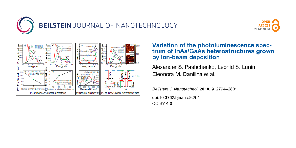
The photoluminescence properties of vertically stacked QD arrays grown by using molecular beam epitaxy (MBE) are well studied in [24,25]. But the IBD method differs from MBE. Therefore, it remains relevant to research the vertical stacking of QDs grown by using IBD. In the beginning, ST#1 samples were studied. The statistical analysis by the threshold tool in the Image Analysis 2.1.2 program showed that the density of QDs in the bottom layer reached 1.42 × 1010 cm−2, the average lateral size of the quantum dots was 17 nm, and their height was 6.3 nm.
During PL excitation, certain steady-state conditions are established in the sample, in which the radiation rate depends on the lifetime of the photogenerated charge carriers in the QD or in the wetting layer. Therefore, the photoluminescence in the grown samples can go through the ground (PLGS) and excited (PLES) states of the electrons at the QDs and also through the energy levels in the wetting layer (PLWL). The measured PL spectra of the vertically stacked QD arrays are shown in Figure 1.
![[2190-4286-9-261-1]](/bjnano/content/figures/2190-4286-9-261-1.png?scale=2.0&max-width=1024&background=FFFFFF)
Figure 1: a) Photoluminescence spectra of samples with vertically stacked InAs QD arrays; b) full width at half maximum of ground state peaks as a function of the number of InAs QD layers.
Figure 1: a) Photoluminescence spectra of samples with vertically stacked InAs QD arrays; b) full width at ha...
The appearance of PLWL peaks indicates the Stranski–Krastanov growth mode of QDs. The photoluminescence of a sample with one QD layer is characterized by the absence of a peak of the excited states, and the PLGS peak has a full width at half maximum (FWHM) of ca. 118 meV with a maximum at an energy of 1.22 eV (Figure 1a). The spectra of samples with two and three QD layers differ by a decrease of FWHM of the PLGS peaks (Figure 1b) and also by an appearance of PLES peaks. In this case, the PLGS peaks in the samples with QD vertical stacking are shifted to lower energies (red-shift). The shift value between PLWL peaks and PLGS peaks in the samples increases by 70 meV. It is seen that the FWHM of PLGS peaks decreases with increasing number of InAs QD layers (Figure 1b). As a result, the luminescence spectrum of vertically stacked QD arrays narrows and becomes similar to the large single-QD spectrum [51-53].
The red-shift is also described in [24] for heterostructures with five QD layers of InAs and in [25] for heterostructures with 1–20 QD layers of In0.5Ga0.5As. In both cases, the heterostructures were grown by using MBE. As a comparison of the possibilities of methods for the formation of heterostructures, we note that the average lateral sizes of quantum dots are the same, and their height, in the case of IBD, is higher. The reasons for this may be related to the difference in the formation of growth fluxes. In IBD, much depends on the calibration of the sputtering yield of the target, which affects the kinetics of the surface processes on the substrate.
The influence of the thickness of the i-GaAs barrier layer (ST#2 samples) on the photoluminescence properties of the InAs/GaAs heterostructures is shown in Figure 2. It is seen that a decrease in the i-GaAs layer thickness from 30 to 15 nm is accompanied by a red-shift of the PL peaks with an increase in the intensity (Figure 2a) and decrease of FWHM (Figure 2b).
![[2190-4286-9-261-2]](/bjnano/content/figures/2190-4286-9-261-2.png?scale=2.0&max-width=1024&background=FFFFFF)
Figure 2: a) Photoluminescence spectra of the InAs/GaAs heterostructures with different thicknesses of the GaAs barrier layer; b) full width at half maximum of ground state photoluminescence peaks as a function of the thickness of the i-GaAs barrier layer.
Figure 2: a) Photoluminescence spectra of the InAs/GaAs heterostructures with different thicknesses of the Ga...
The energy difference between PLGS and PLWL peaks decreased from 205 to 158 meV when the barrier layer thickness was changed from 30 to 15 nm. Obviously, the thicker the i-GaAs layer, the less it is strained. The coating of QDs with a thin i-GaAs layer creates the conditions for the inheritance of properties from the bottom QD layer by a new layer of InAs QDs. Otherwise, if the thickness of the i-GaAs layer is much higher than the height of the QDs, their sizes and density will be determined by the self-organization on the new growth surface, which can cause a fluctuation in QD sizes during vertical stacking and a degradation of the structural and optical properties of the InAs/GaAs heterostructures. The results of the investigations showed that in the case of three QD layers of InAs separated by 15 nm i-GaAs barriers, the density of the array decreased insignificantly from 1.42 × 10¹⁰ to 1.33 × 1010 cm−2. In addition, the average lateral size of the QDs increased by 2 nm. For 30 nm layers of i-GaAs, the average lateral size of the QDs is increased by 2.6 nm, while the density of the top array of quantum dots is decreased to 1.27 × 1010 cm−2. A similar situation was observed in [24,54]. A significant increase in the characteristic QD sizes during vertical stacking was described in [25] only after the deposition of ten QD layers through thin barriers. The observed effect of red-shift and simultaneous increase of PL intensity with increasing number of QD layers was also described in [24] and mainly is explained by a suppression of relative oscillations of QD heights accompanied by high homogeneity of lateral QD sizes. Thus, a decrease in the i-GaAs layer thickness, on the one hand, limits the fluctuation of QD heights in vertically stacked arrays and stabilizes them and, on the other hand, improves the charge transfer due to the resonant tunneling between the vertical InAs QD layers.
Photoluminescence properties of InAs/GaAsBi heterointerfaces
To study the photoluminescence properties of InAs/GaAsBi heterointerfaces, it was necessary to investigate the morphology and structural properties. At first it was necessary to verify the conservation of the stoichiometric composition of GaAs1−xBix during ion-beam deposition. For this purpose, GaAs1−xBix films were grown on GaAs(100) substrates. The thickness of the GaAs1−xBix layers was 20 nm. The results of XRD studies of the epitaxial layer of GaAsBi are shown in Figure 3a.
![[2190-4286-9-261-3]](/bjnano/content/figures/2190-4286-9-261-3.png?scale=2.0&max-width=1024&background=FFFFFF)
Figure 3: a) XRD spectra of GaAs1−xBix films grown on GaAs(100) substrates; b) Raman spectra of InAs/GaAs and InAs/GaAs1−xBix heterostructures.
Figure 3: a) XRD spectra of GaAs1−xBix films grown on GaAs(100) substrates; b) Raman spectra of InAs/GaAs and...
Bi is an isovalent dopant material for III–V compounds and replaces group-V element. Consequently, the GaAs1−xBix crystal lattice should be a zinc blende lattice. Therefore, a diffraction maximum corresponding to the GaAs(004) lattice planes was measured. This is due to its high structure factor, which governs the maximum diffraction intensity. In Figure 3a, two main peaks are identified: a sharp peak of high intensity at 0 radians (θs = 33°) from the GaAs substrate, and low-intensity peaks with negative Δθ due to the strained GaAs1−xBix epitaxial lattice. That kind of peak splitting (with respect to the substrate) is usually observed in epitaxial layers with lattice relaxation. The Bi contents were calculated from obtained X-ray diffraction patterns over the linear dependence x = 6.77Δθ under the condition of a completely strained GaAs1−xBix layer [55]. The calculated values of bismuth contents were xBi = 0.99%, 2.98% and 4.96%. The composition of GaAs1−xBix epitaxial layers differs from the composition of the sputtering targets. In our opinion, this difference is due to the desorption of Bi from the surface, despite the low deposition temperature of 360 °C. The influence of bismuth on the structural properties of InAs/GaAsBi and InAs/GaAs heterointerfaces was investigated by using Raman spectroscopy.
In accordance with the selection rule, modes of longitudinal optical phonons (LO) and longitudinal acoustic phonons (LA) are allowed in the heterointerfaces. The Raman spectroscopy results are shown in Figure 3b. In addition to the allowed modes, forbidden transverse optical (TO) modes for optical phonons of bulk-like GaAs (268 cm−1) and GaBi (182 cm−1) are also observed. This indicates a change in the selection rule under Raman scattering in the InAs/GaAs system caused by lattice strain during the incorporation of Bi into the GaAs matrix. In the spectra we can clearly see the longitudinal modes of optical phonons of bulk GaAs (LO) at 283 cm−1, InAs QDs (LO) at 248 cm−1 and GaBi at 235 cm−1. Effects not detectable in the Raman spectrum of the InAs/GaAs heterostructure appear in the spectra of InAs/GaAs1−xBix samples in the interval of 150–250 cm−1. An increase in xBi is accompanied by the Raman shift of the GaAs-like and GaBi-like peaks to smaller wavenumbers. Due to decrease of lattice mismatch and, thus, less strain in the InAs/GaAs1−xBix heterointerface compared to InAs/GaAs, there is a Raman shift of the peaks of InAs/GaAs1−xBix heterostructures relative to the spectrum of the InAs/GaAs heterostructure. The results of Raman spectroscopy are in good agreement with XRD.
Features of PL in the InAs/GaAs1−xBix heterostructures are shown in Figure 4a.
![[2190-4286-9-261-4]](/bjnano/content/figures/2190-4286-9-261-4.jpg?scale=2.0&max-width=1024&background=FFFFFF)
Figure 4: a) Photoluminescence spectra of InAs/GaAs heterostructures with different Bi content; b) Atomic force microscopy images of the morphology of InAs QDs on GaAs0.95Bi0.05; c) Atomic force microscopy images of the morphology of InAs QDs on GaAs.
Figure 4: a) Photoluminescence spectra of InAs/GaAs heterostructures with different Bi content; b) Atomic for...
The difference between the energies of the PLGS peaks of a sample without Bi and a sample with xBi = 4.96 atom % is 121 meV. This result differs from the red-shift of 23 meV obtained in [56] at room temperature. The reason for this is that InAs QDs in our work have larger sizes and lower density. The results of the investigation of the surface morphology with InAs QDs in samples with GaAs and GaAs1−xBix potential barriers are shown in Figure 4b,c. It is seen that there is a decrease of InAs QD density at growth on the GaAs0.95Bi0.05 surface. The AFM analysis showed that the density of QDs in InAs/GaAs0.95Bi0.05 and InAs/GaAs heterosystems was 0.91 × 1010 cm−2 and 1.53 × 1010 cm−2, respectively. Doping GaAs with bismuth is accompanied by an increase in the average size of QDs from 14 to 20 nm. The results can be explained by an increasing rate of In surface diffusion on GaAs1−xBix during the ion-beam deposition due to the Bi surfactant effect. Note that scientists have different points of view about it. Some argue that doping of Bi during the QD growth process by MBE [17,18,20] and metal-organic vapor-phase epitaxy (MOVPE) [32,33] reduces the In adatom diffusion length and prevents the coalescence of InAs QDs. Then, the QD array density increases. Others argue that the usage of Bi during MOVPE and MBE of InAs QDs [57,58] enhances In adatom diffusion length. In this case, QD sizes increase, and the QD density decreases. Detailed analysis of these works elucidated that in the first case, doping with bismuth was being done during InAs QD growth on GaAs. In the second case [57,58], the QD growth was being done on GaAs1−xBix surfaces. Among other things, temperature conditions also differed from each other. In [20,32,33], doping with Bi on InAs QDs was carried out at 510–530 °C. In our case, as in [52,53], GaAs1−xBix layers were grown at temperatures below 500 °C. We also want to emphasize the fact that the covalent radii [59] of Bi = 148 pm and In = 142 pm atoms are very close. Therefore, in the InAs QD growth process on the GaAs1−xBix surface, In adatom surface diffusion can be realized both through substituting Ga or Bi vacancies. Increase of QD heights in the InAs/GaAs0.95Bi0.05 heterosystem is an indirect demonstration of this. Bi has a large atomic size, so it creates elastic strain in the GaAs lattice and thus influences the QD self-organization. Consequently, using Bi as a surfactant on the GaAs growth surface changes the kinetics of In surface diffusion during ion-beam deposition. Unchanged is the circumstance that with an increase in Bi content in GaAs, an increase in the intensity of photoluminescence is observed in spite of a decrease in InAs QD density (Figure 4b,c). The effect is due to the fact that Bi incorporation leads to valence-band splitting of GaAs, shifting the energy maximum of heavy holes deep into the energy gap. This induces a change in the profiles of the valence and the conduction bands of the InAs/GaAs heterostructure. The InAs/GaAs1−xBix heterointerface forms a type-II misaligned heterojunction [60]. In accordance with the band diagram in Figure 5, the profile of the valence band of the GaAs1−xBix layer shifts in such a way that it forms a well for holes on either side of the InAs QD. In this case, the optical transitions in the direction “quantum dot–barrier layer” begin to appear. The energy of optical transitions is less than the energy of transitions through the ground states of QDs, which explains the red-shift of the PL peaks (Figure 5b). In addition, we suppose that an increase in the lateral QD sizes also contributes to the red-shift of the photoluminescence peak.
![[2190-4286-9-261-5]](/bjnano/content/figures/2190-4286-9-261-5.png?scale=2.0&max-width=1024&background=FFFFFF)
Figure 5: Energy band diagram of heterostructures: a) InAs/GaAs; b) InAs/GaAs1−xBix.
Figure 5: Energy band diagram of heterostructures: a) InAs/GaAs; b) InAs/GaAs1−xBix.
Note that in this case density reduction of the InAs QDs is not a negative factor. QDs being at a more remote distance from each other are able to form a narrower energy sub-band in the conduction band. This effect can be used for the fabrication of multicolor photodetectors.
Conclusion
The properties of InAs/GaAs and InAs/GaAsBi heterointerfaces grown by ion-beam deposition are analyzed. Methods of controlling the photoluminescence spectrum of InAs/GaAs heterostructures are considered. It is demonstrated that the vertical stacking of three QD layers decreases the FWHM of the photoluminescence peaks. According to the studies of photoluminescence, it is established that the variation of the photoluminescence spectrum of InAs/GaAs heterostructures can be carried out by isovalent doping of potential barriers for QDs. It is shown that a content of 4.96 atom % Bi in GaAs leads to a red-shift of the ground-state peaks of the QD array of 121 meV and a decrease of the FWHM. The XRD and Raman spectroscopy methods also show that the InAs/GaAs1−xBix heterosystem has a large lattice relaxation compared to that of InAs/GaAs. It is demonstrated that the surface diffusion of In increases during ion-beam deposition of InAs QDs on the GaAs1−xBix surface.
References
-
Martyniuk, P.; Rogalski, A. Prog. Quantum Electron. 2008, 32, 89–120. doi:10.1016/j.pquantelec.2008.07.001
Return to citation in text: [1] -
Phillips, J.; Kamath, K.; Bhattacharya, P. Appl. Phys. Lett. 1998, 72, 2020–2022. doi:10.1063/1.121252
Return to citation in text: [1] -
Berryman, K. W.; Lyon, S. A.; Segev, M. Appl. Phys. Lett. 1997, 70, 1861–1863. doi:10.1063/1.118714
Return to citation in text: [1] -
Abedin, M. N.; Refaat, T. F.; Zawodny, J. M.; Sandford, S. P.; Singh, U. N.; Bandara, S. V.; Gunapala, S. D.; Bhat, I.; Barnes, N. P. Proc. SPIE 2003, 5152, 10. doi:10.1117/12.505887
Return to citation in text: [1] -
Ryzhii, V. J. Appl. Phys. 2001, 89, 5117. doi:10.1063/1.1359759
Return to citation in text: [1] -
Phillips, J. J. Appl. Phys. 2002, 91, 4590–4594. doi:10.1063/1.1455130
Return to citation in text: [1] -
Kudo, M.; Mishima, T.; Iwamoto, S.; Nakaoka, T.; Arakawa, Y. Phys. E (Amsterdam, Neth.) 2004, 21, 275–278. doi:10.1016/j.physe.2003.11.030
Return to citation in text: [1] -
Shuhui, Z.; Lu, W.; Zhenwu, S.; Yanxiang, C.; Haitao, T.; Huaiju, G.; Haiqiang, J.; Wenxin, W.; Hong, C.; Liancheng, Z. Nanoscale Res. Lett. 2012, 7, 87. doi:10.1186/1556-276x-7-87
Return to citation in text: [1] -
Böhrer, J.; Krost, A.; Bimberg, D. Appl. Phys. Lett. 1994, 64, 1992–1994. doi:10.1063/1.111716
Return to citation in text: [1] -
Hayne, M.; Provoost, R.; Zundel, M. K.; Manz, Y. M.; Eberl, K.; Moshchalkov, V. V. Phys. Rev. B 2000, 62, 10324–10328. doi:10.1103/physrevb.62.10324
Return to citation in text: [1] -
Sugisaki, M.; Ren, H.-W.; Nair, S. V.; Nishi, K.; Masumoto, Y. Phys. Rev. B 2002, 66, 235309. doi:10.1103/physrevb.66.235309
Return to citation in text: [1] -
Ribeiro, E.; Maltez, R. L.; Carvalho, W., Jr.; Ugarte, D.; Medeiros-Ribeiro, G. Appl. Phys. Lett. 2002, 81, 2953–2955. doi:10.1063/1.1513215
Return to citation in text: [1] -
Liu, G.; Chuang, S.-L.; Park, S.-H. J. Appl. Phys. 2000, 88, 5554–5561. doi:10.1063/1.1319328
Return to citation in text: [1] -
Liang, B.; Lin, A.; Pavarelli, N.; Reyner, C.; Tatebayashi, J.; Nunna, K.; He, J.; Ochalski, T. J.; Huyet, G.; Huffaker, D. L. Nanotechnology 2009, 20, 455604. doi:10.1088/0957-4484/20/45/455604
Return to citation in text: [1] -
Souaf, M.; Baira, M.; Nasr, O.; Alouane, M. H. H.; Maaref, H.; Sfaxi, L.; Ilahi, B. Materials 2015, 8, 4699–4709. doi:10.3390/ma8084699
Return to citation in text: [1] -
Nabetani, Y.; Matsumoto, T.; Sasikala, G.; Suemune, I. J. Appl. Phys. 2005, 98, 063502. doi:10.1063/1.2041846
Return to citation in text: [1] -
Wang, L.; Pan, W.; Chen, X.; Wu, X.; Shao, J.; Wang, S. Opt. Mater. Express 2017, 7, 4249–4257. doi:10.1364/ome.7.004249
Return to citation in text: [1] [2] [3] -
Wang, L.; Zhang, L.; Yue, L.; Liang, D.; Chen, X.; Li, Y.; Lu, P.; Shao, J.; Wang, S. Crystals 2017, 7, 63. doi:10.3390/cryst7030063
Return to citation in text: [1] [2] [3] [4] -
Maia, A. D. B.; da Silva, E. C. F.; Quivy, A. A.; Bindilatti, V.; de Aquino, V. M.; Dias, I. F. L. J. Appl. Phys. 2013, 114, 083708. doi:10.1063/1.4818610
Return to citation in text: [1] -
Dasika, V. D.; Krivoy, E. M.; Nair, H. P.; Maddox, S. J.; Park, K. W.; Jung, D.; Lee, M. L.; Yu, E. T.; Bank, S. R. Appl. Phys. Lett. 2014, 105, 253104. doi:10.1063/1.4904825
Return to citation in text: [1] [2] [3] [4] -
Chebotarev, S. N.; Pashchenko, A. S.; Lunin, L. S.; Zhivotova, E. N.; Erimeev, G. A.; Lunina, M. L. Beilstein J. Nanotechnol. 2017, 8, 12–20. doi:10.3762/bjnano.8.2
Return to citation in text: [1] [2] [3] -
Chakrabarti, S.; Stiff-Roberts, A. D.; Su, X. H.; Bhattacharya, P.; Ariyawansa, G.; Perera, A. G. U. J. Phys. D: Appl. Phys. 2005, 38, 2135–2141. doi:10.1088/0022-3727/38/13/009
Return to citation in text: [1] -
Brunkov, P. N.; Kovsh, A. R.; Ustinov, V. M.; Musikhin, Y. G.; Ledentsov, N. N.; Konnikov, S. G.; Polimeni, A.; Patanè, A.; Main, P. C.; Eaves, L.; Kapteyn, C. M. A. J. Electron. Mater. 1999, 28, 486–490. doi:10.1007/s11664-999-0099-6
Return to citation in text: [1] -
Nakata, Y.; Sugiyama, Y.; Futatsugi, T.; Yokoyama, N. J. Cryst. Growth 1997, 175–176, 713–719. doi:10.1016/s0022-0248(96)00814-7
Return to citation in text: [1] [2] [3] [4] [5] -
Tsatsul’nikov, A. F.; Egorov, A. Y.; Zhukov, A. E.; Kovsh, A. R.; Ustinov, V. M.; Ledentsov, N. N.; Maksimov, M. V.; Volovik, B. V.; Suvorova, A. A.; Bert, N. A.; Kop’ev, P. S. Semiconductors 1997, 31, 722–725. doi:10.1134/1.1187076
Return to citation in text: [1] [2] [3] [4] -
Liu, H. Y.; Steer, M. J.; Badcock, T. J.; Mowbray, D. J.; Skolnick, M. S.; Suarez, F.; Ng, J. S.; Hopkinson, M.; David, J. P. R. J. Appl. Phys. 2006, 99, 046104. doi:10.1063/1.2173188
Return to citation in text: [1] -
Popescu, D. P.; Eliseev, P. G.; Stintz, A.; Malloy, K. J. Semicond. Sci. Technol. 2004, 19, 33–38. doi:10.1088/0268-1242/19/1/005
Return to citation in text: [1] -
Mamutin, V. V.; Egorov, A. Y.; Kryzhanovskaya, N. V.; Mikhrin, V. S.; Nadtochy, A. M.; Pirogov, E. V. Semiconductors 2008, 42, 805–812. doi:10.1134/s1063782608070105
Return to citation in text: [1] -
Wu, J.; Shan, W.; Walukiewicz, W. Semicond. Sci. Technol. 2002, 17, 860–869. doi:10.1088/0268-1242/17/8/315
Return to citation in text: [1] -
Keizer, J. G.; Ulloa, J. M.; Utrilla, A. D.; Koenraad, P. M. Appl. Phys. Lett. 2014, 104, 053116. doi:10.1063/1.4864159
Return to citation in text: [1] -
Utrilla, A. D.; Reyes, D. F.; Ulloa, J. M.; González, D.; Ben, T.; Guzman, A.; Hierro, A. Appl. Phys. Lett. 2014, 105, 043105. doi:10.1063/1.4891557
Return to citation in text: [1] -
Zvonkov, B. N.; Karpovich, I. A.; Baidus’, N. V.; Filatov, D. O.; Morozov, S. V. Semiconductors 2001, 35, 93–98. doi:10.1134/1.1340297
Return to citation in text: [1] [2] [3] [4] -
Zvonkov, B. N.; Karpovich, I. A.; Baidus, N. V.; Filatov, D. O.; Morozov, S. V.; Gushina, Y. Y. Nanotechnology 2000, 11, 221–226. doi:10.1088/0957-4484/11/4/306
Return to citation in text: [1] [2] [3] [4] -
Lunin, L. S.; Sysoev, I. A.; Blagina, L. V.; Blagin, A. V.; Barannik, A. A. Inorg. Mater. 2009, 45, 841–845. doi:10.1134/s0020168509080020
Return to citation in text: [1] -
Alfimova, D. L.; Lunin, L. S.; Lunina, M. L. Inorg. Mater. 2014, 50, 113–119. doi:10.1134/s0020168514020010
Return to citation in text: [1] -
Reyes, D. F.; Bastiman, F.; Hunter, C. J.; Sales, D. L.; Sanchez, A. M.; David, J. P. R.; González, D. Nanoscale Res. Lett. 2014, 9, 23. doi:10.1186/1556-276x-9-23
Return to citation in text: [1] -
Ptak, A. J.; France, R.; Beaton, D. A.; Alberi, K.; Simon, J.; Mascarenhas, A.; Jiang, C.-S. J. Cryst. Growth 2012, 338, 107–110. doi:10.1016/j.jcrysgro.2011.10.040
Return to citation in text: [1] -
Janotti, A.; Wei, S.-H.; Zhang, S. B. Phys. Rev. B 2002, 65, 115203. doi:10.1103/physrevb.65.115203
Return to citation in text: [1] -
Greene, J. E. Crit. Rev. Solid State Mater. Sci. 1983, 11, 189–227. doi:10.1080/01611598308244063
Return to citation in text: [1] -
Seshan, K., Ed. Handbook of Thin Film Deposition, 3rd ed.; Elsevier: Amsterdam, Netherlands, 2012. doi:10.1016/c2009-0-64359-2
Return to citation in text: [1] -
Reagor, D. W.; Butko, V. Y. Nat. Mater. 2005, 4, 593–596. doi:10.1038/nmat1402
Return to citation in text: [1] -
Razek, N.; Schindler, A.; Rauschenbach, B. Vacuum 2007, 81, 974–978. doi:10.1016/j.vacuum.2006.12.003
Return to citation in text: [1] -
Kim, W. J.; Koo, W. H.; Jo, S. J.; Kim, C. S.; Baik, H. K.; Lee, J.; Im, S. J. Vac. Sci. Technol., B: Microelectron. Nanometer Struct.–Process., Meas., Phenom. 2005, 23, 2357–2362. doi:10.1116/1.2102969
Return to citation in text: [1] -
Ke, J. J.; Tsai, K. T.; Dai, Y. A.; He, J. H. Appl. Phys. Lett. 2012, 100, 053503. doi:10.1063/1.3680251
Return to citation in text: [1] -
Szczyrbowski, J.; Czapla, A.; Jachimowski, M. Thin Solid Films 1977, 42, 193–200. doi:10.1016/0040-6090(77)90417-5
Return to citation in text: [1] -
Greene, J. E.; Barnett, S. A.; Cadien, K. C.; Ray, M. A. J. Cryst. Growth 1982, 56, 389–401. doi:10.1016/0022-0248(82)90458-4
Return to citation in text: [1] -
Takeuchi, M.; Sakagawa, Y.; Nagasaka, H. Thin Solid Films 1976, 33, 89–98. doi:10.1016/0040-6090(76)90590-3
Return to citation in text: [1] -
Zozime, A.; Cohen-Solal, G.; Bailly, F. Thin Solid Films 1980, 70, 139–152. doi:10.1016/0040-6090(80)90421-6
Return to citation in text: [1] -
Chebotarev, S. N.; Pashchenko, A. S.; Williamson, A.; Lunin, L. S.; Irkha, V. A.; Gamidov, V. A. Tech. Phys. Lett. 2015, 41, 661–664. doi:10.1134/s1063785015070056
Return to citation in text: [1] -
Chebotarev, S. N.; Pashchenko, A. S.; Irkha, V. A.; Lunina, M. L. J. Nanotechnol. 2016, 2016, 5340218. doi:10.1155/2016/5340218
Return to citation in text: [1] -
Dekel, E.; Regelman, D. V.; Gershoni, D.; Ehrenfreund, E.; Schoenfeld, W. V.; Petroff, P. M. Solid State Commun. 2001, 117, 395–400. doi:10.1016/s0038-1098(00)00483-x
Return to citation in text: [1] -
Shtrichman, I.; Metzner, C.; Gerardot, B. D.; Schoenfeld, W. V.; Petroff, P. M. Phys. Rev. B 2002, 65, 081303. doi:10.1103/physrevb.65.081303
Return to citation in text: [1] [2] -
Regelman, D. V.; Dekel, E.; Gershoni, D.; Ehrenfreund, E.; Williamson, A. J.; Shumway, J.; Zunger, A.; Schoenfeld, W. V.; Petroff, P. M. Phys. Rev. B 2001, 64, 165301. doi:10.1103/physrevb.64.165301
Return to citation in text: [1] [2] -
Bimberg, D.; Ledentsov, N. N.; Grundmann, M.; Kirstaedter, N.; Schmidt, O. G.; Mao, M. H.; Ustinov, V. M.; Egorov, A. Y.; Zhukov, A. E.; Kopév, P. S.; Alferov, Zh. I.; Ruvimov, S. S.; Gösele, U.; Heydenreich, J. Jpn. J. Appl. Phys., Part 1 1996, 35, 1311. doi:10.1143/jjap.35.1311
Return to citation in text: [1] -
Steele, J. A.; Lewis, R. A.; Horvat, J.; Nancarrow, M. J. B.; Henini, M.; Fan, D.; Mazur, Y. I.; Schmidbauer, M.; Ware, M. E.; Yu, S.-Q.; Salamo, G. J. Sci. Rep. 2016, 6, 28860. doi:10.1038/srep28860
Return to citation in text: [1] -
Wang, P.; Pan, W.; Wu, X.; Liu, J.; Cao, C.; Wang, S.; Gong, Q. Nanoscale Res. Lett. 2016, 11, 280. doi:10.1186/s11671-016-1470-1
Return to citation in text: [1] -
Okamoto, H.; Tawara, T.; Gotoh, H.; Kamada, H.; Sogawa, T. Jpn. J. Appl. Phys. 2010, 49, 06GJ01. doi:10.1143/jjap.49.06gj01
Return to citation in text: [1] [2] -
Fan, D.; Zeng, Z.; Dorogan, V. G.; Hirono, Y.; Li, C.; Mazur, Y. I.; Yu, S.-Q.; Johnson, S. R.; Wang, Z. M.; Salamo, G. J. J. Mater. Sci.: Mater. Electron. 2013, 24, 1635–1639. doi:10.1007/s10854-012-0987-z
Return to citation in text: [1] [2] -
Kressel, H.; Butler, J. K. Semiconductor lasers and heterojunction LEDs; Academic Press: New York City, NY, U.S.A., 2012. doi:10.1016/b978-0-12-426250-8.x5001-5
Return to citation in text: [1] -
Nag, B. R. Physics of Quantum Well Devices; Kluwer Academic Publishers: Dordrecht, Netherlands, 2000. doi:10.1007/0-306-47127-2
Return to citation in text: [1]
| 32. | Zvonkov, B. N.; Karpovich, I. A.; Baidus’, N. V.; Filatov, D. O.; Morozov, S. V. Semiconductors 2001, 35, 93–98. doi:10.1134/1.1340297 |
| 33. | Zvonkov, B. N.; Karpovich, I. A.; Baidus, N. V.; Filatov, D. O.; Morozov, S. V.; Gushina, Y. Y. Nanotechnology 2000, 11, 221–226. doi:10.1088/0957-4484/11/4/306 |
| 57. | Okamoto, H.; Tawara, T.; Gotoh, H.; Kamada, H.; Sogawa, T. Jpn. J. Appl. Phys. 2010, 49, 06GJ01. doi:10.1143/jjap.49.06gj01 |
| 58. | Fan, D.; Zeng, Z.; Dorogan, V. G.; Hirono, Y.; Li, C.; Mazur, Y. I.; Yu, S.-Q.; Johnson, S. R.; Wang, Z. M.; Salamo, G. J. J. Mater. Sci.: Mater. Electron. 2013, 24, 1635–1639. doi:10.1007/s10854-012-0987-z |
| 1. | Martyniuk, P.; Rogalski, A. Prog. Quantum Electron. 2008, 32, 89–120. doi:10.1016/j.pquantelec.2008.07.001 |
| 2. | Phillips, J.; Kamath, K.; Bhattacharya, P. Appl. Phys. Lett. 1998, 72, 2020–2022. doi:10.1063/1.121252 |
| 3. | Berryman, K. W.; Lyon, S. A.; Segev, M. Appl. Phys. Lett. 1997, 70, 1861–1863. doi:10.1063/1.118714 |
| 15. | Souaf, M.; Baira, M.; Nasr, O.; Alouane, M. H. H.; Maaref, H.; Sfaxi, L.; Ilahi, B. Materials 2015, 8, 4699–4709. doi:10.3390/ma8084699 |
| 16. | Nabetani, Y.; Matsumoto, T.; Sasikala, G.; Suemune, I. J. Appl. Phys. 2005, 98, 063502. doi:10.1063/1.2041846 |
| 17. | Wang, L.; Pan, W.; Chen, X.; Wu, X.; Shao, J.; Wang, S. Opt. Mater. Express 2017, 7, 4249–4257. doi:10.1364/ome.7.004249 |
| 18. | Wang, L.; Zhang, L.; Yue, L.; Liang, D.; Chen, X.; Li, Y.; Lu, P.; Shao, J.; Wang, S. Crystals 2017, 7, 63. doi:10.3390/cryst7030063 |
| 19. | Maia, A. D. B.; da Silva, E. C. F.; Quivy, A. A.; Bindilatti, V.; de Aquino, V. M.; Dias, I. F. L. J. Appl. Phys. 2013, 114, 083708. doi:10.1063/1.4818610 |
| 40. | Seshan, K., Ed. Handbook of Thin Film Deposition, 3rd ed.; Elsevier: Amsterdam, Netherlands, 2012. doi:10.1016/c2009-0-64359-2 |
| 41. | Reagor, D. W.; Butko, V. Y. Nat. Mater. 2005, 4, 593–596. doi:10.1038/nmat1402 |
| 42. | Razek, N.; Schindler, A.; Rauschenbach, B. Vacuum 2007, 81, 974–978. doi:10.1016/j.vacuum.2006.12.003 |
| 43. | Kim, W. J.; Koo, W. H.; Jo, S. J.; Kim, C. S.; Baik, H. K.; Lee, J.; Im, S. J. Vac. Sci. Technol., B: Microelectron. Nanometer Struct.–Process., Meas., Phenom. 2005, 23, 2357–2362. doi:10.1116/1.2102969 |
| 44. | Ke, J. J.; Tsai, K. T.; Dai, Y. A.; He, J. H. Appl. Phys. Lett. 2012, 100, 053503. doi:10.1063/1.3680251 |
| 45. | Szczyrbowski, J.; Czapla, A.; Jachimowski, M. Thin Solid Films 1977, 42, 193–200. doi:10.1016/0040-6090(77)90417-5 |
| 46. | Greene, J. E.; Barnett, S. A.; Cadien, K. C.; Ray, M. A. J. Cryst. Growth 1982, 56, 389–401. doi:10.1016/0022-0248(82)90458-4 |
| 47. | Takeuchi, M.; Sakagawa, Y.; Nagasaka, H. Thin Solid Films 1976, 33, 89–98. doi:10.1016/0040-6090(76)90590-3 |
| 48. | Zozime, A.; Cohen-Solal, G.; Bailly, F. Thin Solid Films 1980, 70, 139–152. doi:10.1016/0040-6090(80)90421-6 |
| 7. | Kudo, M.; Mishima, T.; Iwamoto, S.; Nakaoka, T.; Arakawa, Y. Phys. E (Amsterdam, Neth.) 2004, 21, 275–278. doi:10.1016/j.physe.2003.11.030 |
| 8. | Shuhui, Z.; Lu, W.; Zhenwu, S.; Yanxiang, C.; Haitao, T.; Huaiju, G.; Haiqiang, J.; Wenxin, W.; Hong, C.; Liancheng, Z. Nanoscale Res. Lett. 2012, 7, 87. doi:10.1186/1556-276x-7-87 |
| 9. | Böhrer, J.; Krost, A.; Bimberg, D. Appl. Phys. Lett. 1994, 64, 1992–1994. doi:10.1063/1.111716 |
| 10. | Hayne, M.; Provoost, R.; Zundel, M. K.; Manz, Y. M.; Eberl, K.; Moshchalkov, V. V. Phys. Rev. B 2000, 62, 10324–10328. doi:10.1103/physrevb.62.10324 |
| 11. | Sugisaki, M.; Ren, H.-W.; Nair, S. V.; Nishi, K.; Masumoto, Y. Phys. Rev. B 2002, 66, 235309. doi:10.1103/physrevb.66.235309 |
| 12. | Ribeiro, E.; Maltez, R. L.; Carvalho, W., Jr.; Ugarte, D.; Medeiros-Ribeiro, G. Appl. Phys. Lett. 2002, 81, 2953–2955. doi:10.1063/1.1513215 |
| 13. | Liu, G.; Chuang, S.-L.; Park, S.-H. J. Appl. Phys. 2000, 88, 5554–5561. doi:10.1063/1.1319328 |
| 14. | Liang, B.; Lin, A.; Pavarelli, N.; Reyner, C.; Tatebayashi, J.; Nunna, K.; He, J.; Ochalski, T. J.; Huyet, G.; Huffaker, D. L. Nanotechnology 2009, 20, 455604. doi:10.1088/0957-4484/20/45/455604 |
| 21. | Chebotarev, S. N.; Pashchenko, A. S.; Lunin, L. S.; Zhivotova, E. N.; Erimeev, G. A.; Lunina, M. L. Beilstein J. Nanotechnol. 2017, 8, 12–20. doi:10.3762/bjnano.8.2 |
| 5. | Ryzhii, V. J. Appl. Phys. 2001, 89, 5117. doi:10.1063/1.1359759 |
| 6. | Phillips, J. J. Appl. Phys. 2002, 91, 4590–4594. doi:10.1063/1.1455130 |
| 17. | Wang, L.; Pan, W.; Chen, X.; Wu, X.; Shao, J.; Wang, S. Opt. Mater. Express 2017, 7, 4249–4257. doi:10.1364/ome.7.004249 |
| 18. | Wang, L.; Zhang, L.; Yue, L.; Liang, D.; Chen, X.; Li, Y.; Lu, P.; Shao, J.; Wang, S. Crystals 2017, 7, 63. doi:10.3390/cryst7030063 |
| 20. | Dasika, V. D.; Krivoy, E. M.; Nair, H. P.; Maddox, S. J.; Park, K. W.; Jung, D.; Lee, M. L.; Yu, E. T.; Bank, S. R. Appl. Phys. Lett. 2014, 105, 253104. doi:10.1063/1.4904825 |
| 32. | Zvonkov, B. N.; Karpovich, I. A.; Baidus’, N. V.; Filatov, D. O.; Morozov, S. V. Semiconductors 2001, 35, 93–98. doi:10.1134/1.1340297 |
| 33. | Zvonkov, B. N.; Karpovich, I. A.; Baidus, N. V.; Filatov, D. O.; Morozov, S. V.; Gushina, Y. Y. Nanotechnology 2000, 11, 221–226. doi:10.1088/0957-4484/11/4/306 |
| 4. | Abedin, M. N.; Refaat, T. F.; Zawodny, J. M.; Sandford, S. P.; Singh, U. N.; Bandara, S. V.; Gunapala, S. D.; Bhat, I.; Barnes, N. P. Proc. SPIE 2003, 5152, 10. doi:10.1117/12.505887 |
| 39. | Greene, J. E. Crit. Rev. Solid State Mater. Sci. 1983, 11, 189–227. doi:10.1080/01611598308244063 |
| 27. | Popescu, D. P.; Eliseev, P. G.; Stintz, A.; Malloy, K. J. Semicond. Sci. Technol. 2004, 19, 33–38. doi:10.1088/0268-1242/19/1/005 |
| 30. | Keizer, J. G.; Ulloa, J. M.; Utrilla, A. D.; Koenraad, P. M. Appl. Phys. Lett. 2014, 104, 053116. doi:10.1063/1.4864159 |
| 31. | Utrilla, A. D.; Reyes, D. F.; Ulloa, J. M.; González, D.; Ben, T.; Guzman, A.; Hierro, A. Appl. Phys. Lett. 2014, 105, 043105. doi:10.1063/1.4891557 |
| 59. | Kressel, H.; Butler, J. K. Semiconductor lasers and heterojunction LEDs; Academic Press: New York City, NY, U.S.A., 2012. doi:10.1016/b978-0-12-426250-8.x5001-5 |
| 26. | Liu, H. Y.; Steer, M. J.; Badcock, T. J.; Mowbray, D. J.; Skolnick, M. S.; Suarez, F.; Ng, J. S.; Hopkinson, M.; David, J. P. R. J. Appl. Phys. 2006, 99, 046104. doi:10.1063/1.2173188 |
| 18. | Wang, L.; Zhang, L.; Yue, L.; Liang, D.; Chen, X.; Li, Y.; Lu, P.; Shao, J.; Wang, S. Crystals 2017, 7, 63. doi:10.3390/cryst7030063 |
| 32. | Zvonkov, B. N.; Karpovich, I. A.; Baidus’, N. V.; Filatov, D. O.; Morozov, S. V. Semiconductors 2001, 35, 93–98. doi:10.1134/1.1340297 |
| 33. | Zvonkov, B. N.; Karpovich, I. A.; Baidus, N. V.; Filatov, D. O.; Morozov, S. V.; Gushina, Y. Y. Nanotechnology 2000, 11, 221–226. doi:10.1088/0957-4484/11/4/306 |
| 34. | Lunin, L. S.; Sysoev, I. A.; Blagina, L. V.; Blagin, A. V.; Barannik, A. A. Inorg. Mater. 2009, 45, 841–845. doi:10.1134/s0020168509080020 |
| 35. | Alfimova, D. L.; Lunin, L. S.; Lunina, M. L. Inorg. Mater. 2014, 50, 113–119. doi:10.1134/s0020168514020010 |
| 36. | Reyes, D. F.; Bastiman, F.; Hunter, C. J.; Sales, D. L.; Sanchez, A. M.; David, J. P. R.; González, D. Nanoscale Res. Lett. 2014, 9, 23. doi:10.1186/1556-276x-9-23 |
| 37. | Ptak, A. J.; France, R.; Beaton, D. A.; Alberi, K.; Simon, J.; Mascarenhas, A.; Jiang, C.-S. J. Cryst. Growth 2012, 338, 107–110. doi:10.1016/j.jcrysgro.2011.10.040 |
| 38. | Janotti, A.; Wei, S.-H.; Zhang, S. B. Phys. Rev. B 2002, 65, 115203. doi:10.1103/physrevb.65.115203 |
| 60. | Nag, B. R. Physics of Quantum Well Devices; Kluwer Academic Publishers: Dordrecht, Netherlands, 2000. doi:10.1007/0-306-47127-2 |
| 24. | Nakata, Y.; Sugiyama, Y.; Futatsugi, T.; Yokoyama, N. J. Cryst. Growth 1997, 175–176, 713–719. doi:10.1016/s0022-0248(96)00814-7 |
| 25. | Tsatsul’nikov, A. F.; Egorov, A. Y.; Zhukov, A. E.; Kovsh, A. R.; Ustinov, V. M.; Ledentsov, N. N.; Maksimov, M. V.; Volovik, B. V.; Suvorova, A. A.; Bert, N. A.; Kop’ev, P. S. Semiconductors 1997, 31, 722–725. doi:10.1134/1.1187076 |
| 20. | Dasika, V. D.; Krivoy, E. M.; Nair, H. P.; Maddox, S. J.; Park, K. W.; Jung, D.; Lee, M. L.; Yu, E. T.; Bank, S. R. Appl. Phys. Lett. 2014, 105, 253104. doi:10.1063/1.4904825 |
| 32. | Zvonkov, B. N.; Karpovich, I. A.; Baidus’, N. V.; Filatov, D. O.; Morozov, S. V. Semiconductors 2001, 35, 93–98. doi:10.1134/1.1340297 |
| 33. | Zvonkov, B. N.; Karpovich, I. A.; Baidus, N. V.; Filatov, D. O.; Morozov, S. V.; Gushina, Y. Y. Nanotechnology 2000, 11, 221–226. doi:10.1088/0957-4484/11/4/306 |
| 20. | Dasika, V. D.; Krivoy, E. M.; Nair, H. P.; Maddox, S. J.; Park, K. W.; Jung, D.; Lee, M. L.; Yu, E. T.; Bank, S. R. Appl. Phys. Lett. 2014, 105, 253104. doi:10.1063/1.4904825 |
| 21. | Chebotarev, S. N.; Pashchenko, A. S.; Lunin, L. S.; Zhivotova, E. N.; Erimeev, G. A.; Lunina, M. L. Beilstein J. Nanotechnol. 2017, 8, 12–20. doi:10.3762/bjnano.8.2 |
| 22. | Chakrabarti, S.; Stiff-Roberts, A. D.; Su, X. H.; Bhattacharya, P.; Ariyawansa, G.; Perera, A. G. U. J. Phys. D: Appl. Phys. 2005, 38, 2135–2141. doi:10.1088/0022-3727/38/13/009 |
| 23. | Brunkov, P. N.; Kovsh, A. R.; Ustinov, V. M.; Musikhin, Y. G.; Ledentsov, N. N.; Konnikov, S. G.; Polimeni, A.; Patanè, A.; Main, P. C.; Eaves, L.; Kapteyn, C. M. A. J. Electron. Mater. 1999, 28, 486–490. doi:10.1007/s11664-999-0099-6 |
| 28. | Mamutin, V. V.; Egorov, A. Y.; Kryzhanovskaya, N. V.; Mikhrin, V. S.; Nadtochy, A. M.; Pirogov, E. V. Semiconductors 2008, 42, 805–812. doi:10.1134/s1063782608070105 |
| 29. | Wu, J.; Shan, W.; Walukiewicz, W. Semicond. Sci. Technol. 2002, 17, 860–869. doi:10.1088/0268-1242/17/8/315 |
| 52. | Shtrichman, I.; Metzner, C.; Gerardot, B. D.; Schoenfeld, W. V.; Petroff, P. M. Phys. Rev. B 2002, 65, 081303. doi:10.1103/physrevb.65.081303 |
| 53. | Regelman, D. V.; Dekel, E.; Gershoni, D.; Ehrenfreund, E.; Williamson, A. J.; Shumway, J.; Zunger, A.; Schoenfeld, W. V.; Petroff, P. M. Phys. Rev. B 2001, 64, 165301. doi:10.1103/physrevb.64.165301 |
| 51. | Dekel, E.; Regelman, D. V.; Gershoni, D.; Ehrenfreund, E.; Schoenfeld, W. V.; Petroff, P. M. Solid State Commun. 2001, 117, 395–400. doi:10.1016/s0038-1098(00)00483-x |
| 52. | Shtrichman, I.; Metzner, C.; Gerardot, B. D.; Schoenfeld, W. V.; Petroff, P. M. Phys. Rev. B 2002, 65, 081303. doi:10.1103/physrevb.65.081303 |
| 53. | Regelman, D. V.; Dekel, E.; Gershoni, D.; Ehrenfreund, E.; Williamson, A. J.; Shumway, J.; Zunger, A.; Schoenfeld, W. V.; Petroff, P. M. Phys. Rev. B 2001, 64, 165301. doi:10.1103/physrevb.64.165301 |
| 21. | Chebotarev, S. N.; Pashchenko, A. S.; Lunin, L. S.; Zhivotova, E. N.; Erimeev, G. A.; Lunina, M. L. Beilstein J. Nanotechnol. 2017, 8, 12–20. doi:10.3762/bjnano.8.2 |
| 49. | Chebotarev, S. N.; Pashchenko, A. S.; Williamson, A.; Lunin, L. S.; Irkha, V. A.; Gamidov, V. A. Tech. Phys. Lett. 2015, 41, 661–664. doi:10.1134/s1063785015070056 |
| 50. | Chebotarev, S. N.; Pashchenko, A. S.; Irkha, V. A.; Lunina, M. L. J. Nanotechnol. 2016, 2016, 5340218. doi:10.1155/2016/5340218 |
| 24. | Nakata, Y.; Sugiyama, Y.; Futatsugi, T.; Yokoyama, N. J. Cryst. Growth 1997, 175–176, 713–719. doi:10.1016/s0022-0248(96)00814-7 |
| 25. | Tsatsul’nikov, A. F.; Egorov, A. Y.; Zhukov, A. E.; Kovsh, A. R.; Ustinov, V. M.; Ledentsov, N. N.; Maksimov, M. V.; Volovik, B. V.; Suvorova, A. A.; Bert, N. A.; Kop’ev, P. S. Semiconductors 1997, 31, 722–725. doi:10.1134/1.1187076 |
| 56. | Wang, P.; Pan, W.; Wu, X.; Liu, J.; Cao, C.; Wang, S.; Gong, Q. Nanoscale Res. Lett. 2016, 11, 280. doi:10.1186/s11671-016-1470-1 |
| 17. | Wang, L.; Pan, W.; Chen, X.; Wu, X.; Shao, J.; Wang, S. Opt. Mater. Express 2017, 7, 4249–4257. doi:10.1364/ome.7.004249 |
| 18. | Wang, L.; Zhang, L.; Yue, L.; Liang, D.; Chen, X.; Li, Y.; Lu, P.; Shao, J.; Wang, S. Crystals 2017, 7, 63. doi:10.3390/cryst7030063 |
| 20. | Dasika, V. D.; Krivoy, E. M.; Nair, H. P.; Maddox, S. J.; Park, K. W.; Jung, D.; Lee, M. L.; Yu, E. T.; Bank, S. R. Appl. Phys. Lett. 2014, 105, 253104. doi:10.1063/1.4904825 |
| 24. | Nakata, Y.; Sugiyama, Y.; Futatsugi, T.; Yokoyama, N. J. Cryst. Growth 1997, 175–176, 713–719. doi:10.1016/s0022-0248(96)00814-7 |
| 55. | Steele, J. A.; Lewis, R. A.; Horvat, J.; Nancarrow, M. J. B.; Henini, M.; Fan, D.; Mazur, Y. I.; Schmidbauer, M.; Ware, M. E.; Yu, S.-Q.; Salamo, G. J. Sci. Rep. 2016, 6, 28860. doi:10.1038/srep28860 |
| 24. | Nakata, Y.; Sugiyama, Y.; Futatsugi, T.; Yokoyama, N. J. Cryst. Growth 1997, 175–176, 713–719. doi:10.1016/s0022-0248(96)00814-7 |
| 54. | Bimberg, D.; Ledentsov, N. N.; Grundmann, M.; Kirstaedter, N.; Schmidt, O. G.; Mao, M. H.; Ustinov, V. M.; Egorov, A. Y.; Zhukov, A. E.; Kopév, P. S.; Alferov, Zh. I.; Ruvimov, S. S.; Gösele, U.; Heydenreich, J. Jpn. J. Appl. Phys., Part 1 1996, 35, 1311. doi:10.1143/jjap.35.1311 |
| 25. | Tsatsul’nikov, A. F.; Egorov, A. Y.; Zhukov, A. E.; Kovsh, A. R.; Ustinov, V. M.; Ledentsov, N. N.; Maksimov, M. V.; Volovik, B. V.; Suvorova, A. A.; Bert, N. A.; Kop’ev, P. S. Semiconductors 1997, 31, 722–725. doi:10.1134/1.1187076 |
| 24. | Nakata, Y.; Sugiyama, Y.; Futatsugi, T.; Yokoyama, N. J. Cryst. Growth 1997, 175–176, 713–719. doi:10.1016/s0022-0248(96)00814-7 |
| 25. | Tsatsul’nikov, A. F.; Egorov, A. Y.; Zhukov, A. E.; Kovsh, A. R.; Ustinov, V. M.; Ledentsov, N. N.; Maksimov, M. V.; Volovik, B. V.; Suvorova, A. A.; Bert, N. A.; Kop’ev, P. S. Semiconductors 1997, 31, 722–725. doi:10.1134/1.1187076 |
© 2018 Pashchenko et al.; licensee Beilstein-Institut.
This is an Open Access article under the terms of the Creative Commons Attribution License (http://creativecommons.org/licenses/by/4.0). Please note that the reuse, redistribution and reproduction in particular requires that the authors and source are credited.
The license is subject to the Beilstein Journal of Nanotechnology terms and conditions: (https://www.beilstein-journals.org/bjnano)