Search results
Search for "SEM" in Full Text gives 1106 result(s) in Beilstein Journal of Nanotechnology. Showing first 200.
Observation of multiple bulk bound states in the continuum modes in a photonic crystal cavity
Beilstein J. Nanotechnol. 2023, 14, 544–551, doi:10.3762/bjnano.14.45

- carried out to compensate for deviations between fabricated and designed values. To obtain devices with larger Q factors and higher scattering power, cavities with larger Na = 30 and Nb = 10 were fabricated. Optical and scanning electron microscopy (SEM) images are given in Figure 3a. A microscope was


Carbon nanotube-cellulose ink for rapid solvent identification
Beilstein J. Nanotechnol. 2023, 14, 535–543, doi:10.3762/bjnano.14.44

- length of 5 μm were produced at CTNano/UFMG [59][60][61]. Morphological analysis was carried out by scanning electron microscopy (SEM) in a Quanta 200 FEG, using secondary electrons between 2 and 10 kV. Atomic force microscopy (AFM) was carried out on a Bruker MultiMode8 SPM using the intermittent
- and Centro de Microscopia-UFMG for providing the SEM and TEM images included in Supporting Information File 1. Funding The authors also would like to thank Fapemig (Rede 2D and individual projects), INCT Nanomateriais de Carbono, CNPq/MCT, Petrobras, BNDES and CAPES for the funding support. APMB


Nanoarchitectonics to entrap living cells in silica-based systems: encapsulations with yolk–shell and sepiolite nanomaterials
Beilstein J. Nanotechnol. 2023, 14, 522–534, doi:10.3762/bjnano.14.43

- sol–gel methods, as well as pre-synthesised yolk–shell bionanohybrids have been studied subsequently. Optical microscopy and SEM confirm that the silica shell microstructures provide a reduced contact between cells. The inorganic matrix increases the survival of the cells and maintains their
- systems were studied by means of optical and electron microscopy (SEM and FE-SEM). Both techniques allowed us to study in detail the cellular arrangement of the microorganisms and their interaction with the inorganic matrix system. FE-SEM microscopy images of the different gel encapsulation systems are
- improved transport of nutrients and metabolites across the material. The FE-SEM images in Figure 2C and Figure 2D show the same microorganism cells but previously encapsulated in yolk–shell microstructures. They are arranged differently from those immobilized freely in the silica gel substrate. In the


On the use of Raman spectroscopy to characterize mass-produced graphene nanoplatelets
Beilstein J. Nanotechnol. 2023, 14, 509–521, doi:10.3762/bjnano.14.42

- widely used characterization tool for GR2Ms [8]. A search of Web of Science showed that of 97,532 articles published in the last five years with “Graphene” in the abstract, 9.3% also mentioned “Raman”. This is compared with atomic force microscopy (AFM) (2.4%), scanning electron microscopy (SEM) (11.4
- production of materials. (A) Example AFM image of flakes from the GNPref sample; the scale bar is 2 μm. (B) Example SEM image of flakes from the GNPref sample; the scale bar is 200 nm. (C) Flake sizes of GNPref sample measured by AFM with histograms of the lateral size and thickness distributions. (A


The origin of black and white coloration of the Asian tiger mosquito Aedes albopictus (Diptera: Culicidae)
Beilstein J. Nanotechnol. 2023, 14, 496–508, doi:10.3762/bjnano.14.41

- analysed using scanning electron microscopy, transmission electron microscopy, and fluorescence microscopy. Reflectance spectra of the white areas are measured. No clear difference is present in the morphology of micro- and nanostructures of black and white scales in SEM and TEM, but black scales contain a
- SEM reveal that the body of Ae. albopictus is covered with scales and microtrichia (Figure 2). Scales of different shape are present on different body parts. Spatulate scales are the most common kind of scales. They are found on the thorax (Figure 2a,b), wings (Figure 2c), halters (Figure 2d), head
- in the morphology of black and white scales in SEM and TEM, except for the small difference in the distance between the longitudinal ridges. The scales appear black and white only under reflected light at low magnification. In our observation of the tarsal scales of Ae. aegypti in a light microscope


Mixed oxides with corundum-type structure obtained from recycling can seals as paint pigments: color stability
Beilstein J. Nanotechnol. 2023, 14, 467–477, doi:10.3762/bjnano.14.37

- results. The Raman modes are A1g (ca. 149 cm−1 and ca. 501 cm−1) and E1g (ca. 222 cm−1, ca. 290 cm−1, ca. 298 cm−1, ca. 402 cm−1, and ca. 615 cm−1, where 290 cm−1 and 298 cm−1 usually are a doublet with E1g symmetry and cannot be easily resolved [20]. Scanning electron microscopy (SEM) The morphology of
- together with recycled boehmite. The absorbance spectra indicate the presence of 3+ ions in the samples, which are responsible for the colors. The spectra also confirm the type of structure found via XRD and the oxidation state determined from XPS. SEM images show the characteristic morphology of this type
- -performance detector, with a power of 300 W. A scanning electron microscope Hitachi SU8020 SEM (Tokyo, Japan) was used to obtain morphology information. The oxidation state and composition of the chemical elements at the surface were evaluated by X-ray photoelectron spectroscopy (XPS) (Versaprobe PHI 5000


Conjugated photothermal materials and structure design for solar steam generation
Beilstein J. Nanotechnol. 2023, 14, 454–466, doi:10.3762/bjnano.14.36

- hydrophobicity allows the material to float on water. Microporous structures were obtained by NaCl particulate leaching. The pore sizes could be flexibly controlled by the size of the NaCl template. This method is simple, low-cost, and easy to scale up. The surface structures of the foam were visualized by SEM
- B. Shao et al., J. Mater. Chem. A, vol. 8, issue 23, © 2020); permission conveyed through Copyright Clearance Center, Inc. This content is not subject to CC BY 4.0. (a) SEM images of the poly(1,3,5-hexahydro-1,3,5-triazine)-based foams at low (up) and high (down) magnifications. (b) An optical image


Evaluation of electrosynthesized reduced graphene oxide–Ni/Fe/Co-based (oxy)hydroxide catalysts towards the oxygen evolution reaction
Beilstein J. Nanotechnol. 2023, 14, 420–433, doi:10.3762/bjnano.14.34
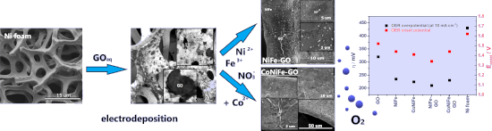
- the electrodeposition of conductive films on active metals [26]. The morphology of the deposits was analyzed by scanning electron microscopy (SEM) and is presented in Figure 1b–f. Typical GO flakes regularly distributed over the surface of the nickel foam were successfully obtained after the one-step
- interconnected nanoflakes, which formed a porous 3D structure uniformly distributed over the entire surface of the nickel foam (Figure 1d). The morphology of the catalysts changed after the combination of GO with NiFe and CoNiFe (Figure 1e,f). In each case, the SEM images clearly show the complete coverage of
- . Deposition of CoNiFe on the GO/Ni foam changed the shape of the GO flakes, with some visible agglomerations (Figure 1f). In contrast, the presence of GO resulted in the formation of a CoNiFe layer, which only remained an interconnected 3D porous material in some areas. Additional SEM images with different


Plasmonic nanotechnology for photothermal applications – an evaluation
Beilstein J. Nanotechnol. 2023, 14, 380–419, doi:10.3762/bjnano.14.33


The steep road to nonviral nanomedicines: Frequent challenges and culprits in designing nanoparticles for gene therapy
Beilstein J. Nanotechnol. 2023, 14, 351–361, doi:10.3762/bjnano.14.30

- offer some standards on the most commonly found techniques with an intent to provide a starting point for the development of repeatable protocols. Imaging (individual analysis) For particles that are readily measured in the solid state, we recommend the ISO 19749:221 standard for SEM analysis, ASTM


Polymer nanoparticles from low-energy nanoemulsions for biomedical applications
Beilstein J. Nanotechnol. 2023, 14, 339–350, doi:10.3762/bjnano.14.29

- 80/20 and water contents above 87 wt %, with droplet sizes in the 180–190 nm range. The derived ethyl cellulose nanoparticles had a size between 107 and 161 nm as estimated by TEM and SEM (Figure 2). Dexamethasone (DXM), a steroid with potent anti-inflammatory and immunosuppressant activity, was
- concentration of nanoparticles in the as-prepared samples, improving surface functionalization, and expanding the range of polymers that can be processed by this approach. Schematics of the structural and curvature changes during (a) PIT and (b) PIC nanoemulsification. (A) TEM and (B) SEM images of ethyl


Biocatalytic synthesis and ordered self-assembly of silica nanoparticles via a silica-binding peptide
Beilstein J. Nanotechnol. 2023, 14, 280–290, doi:10.3762/bjnano.14.25

- scattering (DLS). The efficiency of the self-assembly was evaluated with scanning electron microscopy (SEM), UV–vis spectroscopy, and qualitative visual demonstration. Results and Discussion SiBP alone as catalyst Reaction kinetics were studied via OD measurements of the particles and GC analysis of
- of the particles into interconnected particle strands [26]. Our findings indicate that, when used alone, the positively charged SiBP can also act as a cationic emulsifier resulting in the branched fibrillar networks observed by SEM. In this aspect, when used alone, the SiBP mimicked the in vitro
- . To further investigate this interesting profile, samples were collected from the NH3 + 1 mM SiBP reaction and the NH3 only reaction in the middle of the steep increase (20 min), at the peak point (45 min), and in the middle of the steep decrease (55 min) (Figure 4a). SEM analysis showed that very


A novel approach to pulsed laser deposition of platinum catalyst on carbon particles for use in polymer electrolyte membrane fuel cells
Beilstein J. Nanotechnol. 2023, 14, 190–204, doi:10.3762/bjnano.14.19

- ; ORR; PEMFCs; PLD deposition; Pt catalyst; rotating ring-disk electrode (RRDE); SEM; TEM; XPS; Introduction Fuel cells, which cleanly and efficiently convert the chemical energy of hydrogen or other fuels to electrical energy, are a good alternative to dirty and wasteful combustion engines for
- , morphology, and chemical composition of the fabricated catalysts were investigated using TEM, SEM, EDX, XPS, and Raman spectroscopy. Electrochemical measurements determined the performance of the fabricated catalysts. Results and Discussion Synthesis of a highly graphitized carbon material The synthesis of
- morphology of the carbon material was examined using a scanning electron microscope (SEM) equipped with energy-dispersive spectroscopy (EDS). SEM measurements of the samples were carried out using a Quanta 3D FEG microscope (FEI, United States). The surface imaging and EDS measurements were done at an


Batch preparation of nanofibers containing nanoparticles by an electrospinning device with multiple air inlets
Beilstein J. Nanotechnol. 2023, 14, 141–150, doi:10.3762/bjnano.14.15

- using an electronic balance (XJ120A, Precisa LTD.). The nanofiber morphology was investigated by a scanning electron microscopy (SEM, Hitachi S4800, Hitachi LTD.), and Image J software (National Institute of Mental Health) was used to characterize the fiber diameter distribution by random selection of
- 100 nanofibers from ten SEM images of each sample. Simultaneously, the element distribution on the sample surface was characterized by a desktop SEM (TM3030, Hitachi LTD.). Electric field simulation Maxwell 3D was used to simulate the electric field distribution of EMAI under different voltages (40


Formation of nanoflowers: Au and Ni silicide cores surrounded by SiOx branches
Beilstein J. Nanotechnol. 2023, 14, 133–140, doi:10.3762/bjnano.14.14

- annealed at 1050 °C was named 15Au5Ni. The morphology was imaged by optical microscopy (OM, Zeiss Axiotech) and high-resolution scanning electron microscopy (HR-SEM, Hitachi S-4800) equipped with energy-dispersive X-ray spectroscopy (EDS, Thermo Scientific). The SEM images were recorded by using mixed
- epitaxial line structures inside the decomposition cavity. Supporting Information Supporting Information File 36: Additional OM, LSM, SEM, EDS and XRD measurements. Acknowledgements Joachim Döll from the Center of Micro- and Nanotechnology (ZMN), a DFG-funded core facility at TU Ilmenau, is acknowledged


Antimicrobial and mechanical properties of functionalized textile by nanoarchitectured photoinduced Ag@polymer coating
Beilstein J. Nanotechnol. 2023, 14, 95–109, doi:10.3762/bjnano.14.11

- electron microscopy (SEM), transmission electron microscopy (TEM), and reflectance measurements to assess the optical properties and the durability of the functionalized textiles. Results and Discussion Photoinduced synthesis of the Ag@polymer coating Specific monomers poly(ethylene glycol) 600 diacrylate
- , as well as the final thickness of the metallic layer, account for this difference in reflectivity. Scanning electron microscopy (SEM) carried out on the surface of functionalized textiles revealed the homogenous distribution of AgNPs, with average sizes of 62 ± 2 nm and 58 ± 1 nm for the Ag@PEG600DA
- surface roughness, as can be seen in the SEM images (Figure 7c). Consequently, the diffuse reflectivity drops in favor of the specular reflectance. The particles are no longer simply juxtaposed but form a continuous silver layer, especially after 1000 friction cycles. The characteristic silver plasmon


Combining physical vapor deposition structuration with dealloying for the creation of a highly efficient SERS platform
Beilstein J. Nanotechnol. 2023, 14, 83–94, doi:10.3762/bjnano.14.10

- SERS properties of the nanoporous structure. Using scanning electron microscopy (SEM) and X-ray photoelectron spectroscopy (XPS) the morphology and surface composition of each nanoporous structure were respectively evaluated and used to describe the SERS properties of the samples. Results and
- to the alloy thin film. Three different silver compositions were selected, namely 18, 30, and 36 atom %, and characterized by SEM/energy-dispersive X-ray spectroscopy (EDX). Figure 1 displays the SEM micrograph of the as-deposited thin films. The thin films exhibit a columnar morphology (see the
- cross-section SEM images in Figure 1d–f). The top view images (Figure 1a–c) reveal the presence of dispersed hexagonal columns. A possible explanation for the formation of the hexagonal structure is due to the Guinier–Preston (GP) zone of the silver–aluminum alloy system [31]. The GP zone induces the


Gap-directed chemical lift-off lithographic nanoarchitectonics for arbitrary sub-micrometer patterning
Beilstein J. Nanotechnol. 2023, 14, 34–44, doi:10.3762/bjnano.14.4

- to etch the exposed underlying Au film. After 30 min of etching, the substrate was rinsed with deionized water and blown dry with nitrogen gas. The transferred metal structures were then characterized by optical microscopy, scanning election microscopy (SEM, JEOL JSM-7600F, Tokyo, Japan) and atomic
- stamp line features. These gaps therefore result in parallel linear SAM strips ranging from 150 nm to sub-10 nm wide after the CLL operation, and a new resolution limit of 5 nm in the CLL process is achieved when the used stamp height is 700 nm (Figure 4B–F). For 150 nm features SEM (JEOL JSM-7600F
- characterized by SEM for feature line width of (B) 150 nm and by AFM for feature line widths (C) 80 nm (D) 50 nm (E) 35 nm, and (F) 5 nm. Scale bar is 2 μm (B) and (C), and 100 nm (D–F). Illustration of the gap-directed chemical lift-off lithography process. (A) The selective removal of alkanethiols from an Au


Observation of collective excitation of surface plasmon resonances in large Josephson junction arrays
Beilstein J. Nanotechnol. 2022, 13, 1578–1588, doi:10.3762/bjnano.13.132

- characteristics from (a). The peak represents the resonant step. It reduces with decreasing N and is not visible for N = 106 (lower panel) below the threshold number of JJs. (a) Optical image of a superconducting detector with a log-periodic microwave antenna. (b) SEM image of a nanoscale sensor junction (false


Induced electric conductivity in organic polymers
Beilstein J. Nanotechnol. 2022, 13, 1551–1557, doi:10.3762/bjnano.13.128

- Center FSRC “Crystallography and Photonics” RAS, and were partly supported by the Ministry of Science and Higher Education of the Russian Federation within the State assignment FSRC “Crystallography and Photonics” RAS in the part of SEM and TEM measurements. Funding The work was supported by the Mirror


Photoelectrochemical water oxidation over TiO2 nanotubes modified with MoS2 and g-C3N4
Beilstein J. Nanotechnol. 2022, 13, 1541–1550, doi:10.3762/bjnano.13.127

- materials (Figure 1b). This agrees with the results of previous publications in which hydrothermal methods were applied [24][25][26]. The SEM image of the g-C3N4 material shows the uniform nanosheets that were fabricated by the melamine pyrolysis method (Figure 1c). After the deposition of 2D materials MoS2
- and g-C3N4 onto the TNAs substrate, we examined the morphology of these heterostructures by using SEM (Figure 2). There are some small pieces that are randomly distributed on the surface of TNAs in Figure 2a, which were attributed to be MoS2. There is a similar result in the SEM image of g-C3N4/TNAs
- ultrasonic treatment peeled the MoS2 material into thinner layered structures. This is in agreement with the SEM images, in which material with rather small and thinner structures scattered on the surface of TNAs was observed. The functional groups and chemical bonds of the as-prepared materials were


Non-stoichiometric magnetite as catalyst for the photocatalytic degradation of phenol and 2,6-dibromo-4-methylphenol – a new approach in water treatment
Beilstein J. Nanotechnol. 2022, 13, 1531–1540, doi:10.3762/bjnano.13.126

- SEM, X-ray diffraction, and ultraviolet–visible (UV–vis) analysis. The XRD and UV–vis results were published in our previous article [17]. We present this data again in this article as it is necessary for the discussion of the results. Zeta potential measurements were also presented in another
- forms a porous structure. There is a disparity in the particle sizes measured with SEM and XRD, because the particle sizes detected using these two techniques are not in the same orientation, and the terms "particle size" and "crystal size" refer to different concepts (a particle may contain several or
- SEM reveals the maximum size of the particles. The catalytic activity of commercially available M1 and M2 was evaluated through the photocatalytic degradation of phenol and DBMP. The photocatalytic activity was compared with the efficiency of ozonolysis. The photocatalytic efficiency is improved by


A TiO2@MWCNTs nanocomposite photoanode for solar-driven water splitting
Beilstein J. Nanotechnol. 2022, 13, 1520–1530, doi:10.3762/bjnano.13.125

- nanocomposite characterizations The surface morphology of MWCNTs and the TiO2@MWCNTs nanocomposite is characterized by using field-emission scanning electron microscopy (FE-SEM, S4800) and transmission electron microscopy (TEM, JEOL-1400). The crystallization behavior of the catalysts is analyzed by X-ray
- surface with a size of 1.5 × 2.0 cm2 is irradiated. A schematic of the experimental apparatus is described in Figure 1. Results and Discussion Characterization of the TiO2@MWCNTs nanocomposite catalyst FE-SEM images of the morphology of the MWCNTs, TiO2 powder, and the TiO2@MWCNTs nanocomposite are shown
- performance of the TiO2@MWCNTs electrode for water splitting. Schematic of the photoelectrochemical water splitting experimental apparatus. SEM images of (a) MWCNTs, (b) TiO2, and (c) the TiO2@MWCNTs nanocomposite. TEM images of (a) MWCNTs, (b) TiO2, and (c) the TiO2@MWCNTs nanocomposite. EDX spectra of


In search of cytotoxic selectivity on cancer cells with biogenically synthesized Ag/AgCl nanoparticles
Beilstein J. Nanotechnol. 2022, 13, 1505–1519, doi:10.3762/bjnano.13.124

- ° in 2θ. Quantitative chemical analysis was performed on a JEOL JSM-7401F field-emission scanning electron microscope (FE-SEM) using EDX. An acceleration voltage of 15 kV and a working distance of 8 mm were used. The samples were precoated with Au/Pd for 10 seconds. Ultraviolet–visible spectroscopy was


Hydroxyapatite–bioglass nanocomposites: Structural, mechanical, and biological aspects
Beilstein J. Nanotechnol. 2022, 13, 1490–1504, doi:10.3762/bjnano.13.123

- microstructural characteristics, such as micromorphology of the obtained materials and the qualitative and quantitative distribution of the granular phase and pores, SEM analysis of the sintered materials was performed using a Zeiss Auriga FESEM-FIB electron microscope. For SEM analysis, the samples underwent a
- special cleaning process for 6 min in a nitrogen plasma jet in a FISCHONE Plasma Cleaner. A FEI QUANTA INSPECT S SEM was used to observe structure and morphology of the samples after their treatment in simulated body fluid, as well as to acquire the energy-dispersive X-ray (EDX) spectra and the elemental
- (mm2) [40]. The surface of the samples was then investigated by SEM to estimate their apatite-forming ability. The pH value of SBF was measured before and after the insertion of the sintered sample using a Multi-parameter analyzer Consort C1010, version 2.0, which has the possibility to measure pH