Search results
Search for "atomic resolution" in Full Text gives 90 result(s) in Beilstein Journal of Nanotechnology.
Comparing a porphyrin- and a coumarin-based dye adsorbed on NiO(001)
Beilstein J. Nanotechnol. 2019, 10, 874–881, doi:10.3762/bjnano.10.88

- study and do not influence the reported results. Figure 2b shows the frequency-shift signal acquired using the multipass imaging technique [14][15][35][36] clearly showing atomic resolution of the NiO(001) surface. Employing this method, the crystallographic directions of the substrate are resolved with
- ). (b) Frequency-shift (Δf1) signal of the same surface at atomic resolution, recorded in the second line scan of the multipass technique with following scan parameters: = 4 nm, Δf1 = −42 Hz) and zoffset = −700 pm. (a) Large-scale topographic image showing that Cu-TCPP molecules form islands on the
Polymorphic self-assembly of pyrazine-based tectons at the solution–solid interface
Beilstein J. Nanotechnol. 2019, 10, 494–499, doi:10.3762/bjnano.10.50

- means of split-image technique [21] in which both the adsorbate layer and the substrate are recorded with molecular and atomic resolution, respectively, in a single frame. The details of calibration and correction of STM images is given in the section 5 of Supporting Information File 1. A 5 μL droplet
Intuitive human interface to a scanning tunnelling microscope: observation of parity oscillations for a single atomic chain
Beilstein J. Nanotechnol. 2019, 10, 337–348, doi:10.3762/bjnano.10.33

- detailed discussion about this is given in Supporting Information File 1. This elegant and accurate approach allows us to determine the background lattice without the need to work towards atomic resolution of the Au(111) surface each time. The method is not limited to the Au(111) surface. A similar
Investigation of CVD graphene as-grown on Cu foil using simultaneous scanning tunneling/atomic force microscopy
Beilstein J. Nanotechnol. 2018, 9, 2953–2959, doi:10.3762/bjnano.9.274

- (graphene–3d metals) or lattice-mismatched configuration (graphene–4d/5d metals) [10]. Material and termination of the apex of the tip play an important role in STM and AFM results as well. Due to the strong interaction between metal tip and carbon atoms, atomic resolution can be obtained in both attractive
Charged particle single nanometre manufacturing
Beilstein J. Nanotechnol. 2018, 9, 2855–2882, doi:10.3762/bjnano.9.266

- capable of writing with 8 nm feature resolution over a 200 mm wafer substrate [8]. The state of the art for lithography using scanning proximity probes is the Zyvector system from Zyvex Labs [9]. It provides atomic resolution – the removal of a single atom – using scanning tunneling lithography, but the
- in principle achieve atomic resolution [124]. Writing speeds for this process are up to 100 nm·s−1, which is similar to values achieved for scanning electron beam systems as described in Section 2.2 [9]. 3.1 Field-emission electron scanning probe lithography In this review, we will focus on field
Quantitative comparison of wideband low-latency phase-locked loop circuit designs for high-speed frequency modulation atomic force microscopy
Beilstein J. Nanotechnol. 2018, 9, 1844–1855, doi:10.3762/bjnano.9.176

- imaging of calcite dissolution in water at 0.5 s/frame with true atomic resolution. The high-speed and high-resolution imaging capabilities of the proposed design will enable a wide range of studies to be conducted on various atomic-scale dynamic phenomena at solid–liquid interfaces. Keywords: calcite
- dissolution process; frequency modulation atomic force microscopy; high-speed atomic-resolution imaging; phase-locked loop; Introduction Frequency modulation atomic force microscopy (FM-AFM) is a powerful tool for investigating atomic- and molecular-scale structures of sample surfaces in various environments
- ][7], as well as atomic-scale imaging of intramolecular structures at low temperatures [8]. The liquid-environment applications of FM-AFM have also been intensively explored. So far, this method has been utilized for atomic-resolution imaging of inorganic crystals [9][10][11][12][13][14] and
Combined pulsed laser deposition and non-contact atomic force microscopy system for studies of insulator metal oxide thin films
Beilstein J. Nanotechnol. 2018, 9, 686–692, doi:10.3762/bjnano.9.63

- not required. The performance of the combined system is demonstrated for the preparation and high-resolution NC-AFM imaging of atomically flat thin films of anatase TiO2(001) and LaAlO3(100). Keywords: atomic resolution; frequency modulation atomic force microscopy; insulator thin film; pulsed laser
- transmission electron microscopy [5][8][9][10][11][12][13]. As atomic resolution methods, scanning probe microscopy including scanning tunneling microscopy (STM) [13][14][15][16][17][18][19][20][21][22][23][24][25][26][27][28][29][30] and non-contact atomic force microscopy (NC-AFM) [19][23][29][31][32][33][34
- systems allow for in situ observations, from sample preparation to measurements. In order to image surface atoms of insulator metal oxides with atomic resolution, in this study, we have developed a combined system consisting of an NC-AFM and PLD operated in ultra-high vacuum (UHV) at room temperature. In
Anchoring of a dye precursor on NiO(001) studied by non-contact atomic force microscopy
Beilstein J. Nanotechnol. 2018, 9, 242–249, doi:10.3762/bjnano.9.26

- 1), adsorbed on an atomically clean NiO(001) crystal surface. It adsorbs either as single molecule or forms specific assemblies increasing in size from small clusters up to complete islands inducing a clear change of the surface potential. Results and Discussion Atomic resolution of NiO(001) Figure
- ) was resolved with atomic resolution using the first resonance and the torsional resonance. Depending on the deposition rate, single molecules, molecular clusters, and molecular islands have been imaged. Through the so-called multipass technique, submolecular resolution could be achieved and direct
- ) changes to a cis-conformation upon coordination of a metal ion Mn+ (right). From large-scale imaging to atomic resolution on NiO(001). (a) Large scale topographic image of the NiO(001) crystal presenting clean terraces (scan parameters: = 4 nm, Δf1 = −30 Hz). The high-resolution topographic image (b
A robust AFM-based method for locally measuring the elasticity of samples
Beilstein J. Nanotechnol. 2018, 9, 1–10, doi:10.3762/bjnano.9.1

- soft FDTS-coating of the tip is sensed (see also Figure 8). The effect of collecting Teflon-like molecules with AFM tips has been known for a long time. It has been successfully used for the topographic imaging of the Si(111) surface with atomic resolution. Howald et al. [27] studied the Si(111) 7 × 7
Robust procedure for creating and characterizing the atomic structure of scanning tunneling microscope tips
Beilstein J. Nanotechnol. 2017, 8, 2389–2395, doi:10.3762/bjnano.8.238

- [1][2], it became possible to image conducting surfaces with atomic resolution. STM operates by bringing the apex of a fine metallic wire into tunneling distance from a surface of interest. By providing feedback in the tunnel current and scanning the tip over the surface one can make topographic maps
- of the surface with atomic resolution. STM has found its applications in many fields of science. Apart from studying surface topography, STM has been used for, e.g., manipulating single atoms [3][4][5], for doing spectroscopy [6], for fabricating nano-structures with novel engineered electronic
Stable Au–C bonds to the substrate for fullerene-based nanostructures
Beilstein J. Nanotechnol. 2017, 8, 1073–1079, doi:10.3762/bjnano.8.109

- annealing (600 °C, 5 min) were required to obtain samples with overall cleanliness suitable for achieving the atomic resolution by means of STM. For deposition, we employed a custom-made thermal evaporation source, which contained a pocket made of tantalum, suitable for the evaporation of molecules such as
Self-assembly of silicon nanowires studied by advanced transmission electron microscopy
Beilstein J. Nanotechnol. 2017, 8, 440–445, doi:10.3762/bjnano.8.47

- technique to study a larger range of volumes, while still offering reasonable spatial resolution from ≈1 nm3 [6] down to atomic resolution in very recently developed microscopes [7][8]. Electron tomography is accomplished through the reconstruction of a sequence of projection images acquired by tilting the
Noise in NC-AFM measurements with significant tip–sample interaction
Beilstein J. Nanotechnol. 2016, 7, 1885–1904, doi:10.3762/bjnano.7.181

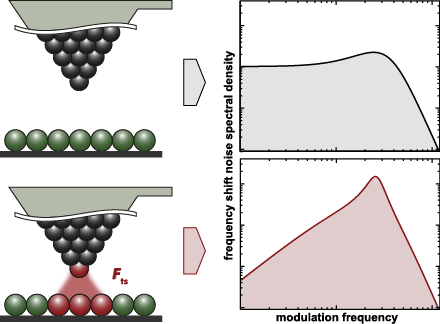
- always the experimental task defining the desired spatial resolution λ that is, for instance, a fraction of the atomic periodicity in atomic resolution imaging, and the available time for the measurement expressed by the scan speed vscan. Assuming perfectly band-limited output signals, the sampling
Nanostructured germanium deposited on heated substrates with enhanced photoelectric properties
Beilstein J. Nanotechnol. 2016, 7, 1492–1500, doi:10.3762/bjnano.7.142

- masking technique were deposited by magnetron sputtering (Varian ER3119) and e-beam assisted thermal evaporation (Bestec), respectively. Ge:SiO2 films were characterized using an X-ray diffractometer (BRUKER-AXS with Cu Kα1 radiation of λ = 0.15406 nm) and an advanced analytical atomic resolution electron
Noncontact atomic force microscopy III
Beilstein J. Nanotechnol. 2016, 7, 946–947, doi:10.3762/bjnano.7.86

- tunneling microscopy (STM) relies on quantum mechanical tunneling of electrons to enable the atomic-resolution imaging of (semi-)conducting sample surfaces, it was the atomic force microscope (AFM) that eventually allowed for nanometer-scale imaging of sample surfaces with no limitations on electrical
Optical absorption signature of a self-assembled dye monolayer on graphene
Beilstein J. Nanotechnol. 2016, 7, 862–868, doi:10.3762/bjnano.7.78

- means of atomic resolution obtained on HOPG images in XY-directions and with flame-annealed gold through the height of steps in the Z-direction. All the images were obtained at a quasi-constant current, i.e., in the variable-height mode. The images in Figure 1a,b were corrected for the thermal drift by
Coupled molecular and cantilever dynamics model for frequency-modulated atomic force microscopy
Beilstein J. Nanotechnol. 2016, 7, 708–720, doi:10.3762/bjnano.7.63

- different forces on approach and retraction of the tip [16][17]. The second type of simulation models focuses on the atomic dissipation mechanisms of the interaction between tip and sample. Small regions of tip and sample, where the interaction takes place, are simulated with atomic resolution. Some of
- frequency shift and the dissipation (with atomic resolution) are compared for three such scans with different nominal distances d. For the largest distance, d = 1.30, the hysteresis is not present. Hence the damping is only minute (note the different ΔE-scale compared to the panels for the other two d
Length-extension resonator as a force sensor for high-resolution frequency-modulation atomic force microscopy in air
Beilstein J. Nanotechnol. 2016, 7, 432–438, doi:10.3762/bjnano.7.38

- Hannes Beyer Tino Wagner Andreas Stemmer Nanotechnology Group, ETH Zürich, Säumerstrasse 4, 8803 Rüschlikon, Switzerland 10.3762/bjnano.7.38 Abstract Frequency-modulation atomic force microscopy has turned into a well-established method to obtain atomic resolution on flat surfaces, but is often
- ; frequency-modulation atomic force microscopy; high-resolution; length-extension resonator; Introduction Frequency-modulated atomic force microscopy (FM-AFM) is the method of choice to image nanoscale structures on surfaces down to the atomic level. Whereas atomic resolution is routinely achieved in ultra
- forces. A viable strategy to circumvent meniscus forces and to achieve atomic resolution is to measure in liquid [1]. Operation with small amplitudes can further help to stay within a single water layer, minimising disturbances which may arise by penetrating several water layers per oscillation [2]. To
Efficiency improvement in the cantilever photothermal excitation method using a photothermal conversion layer
Beilstein J. Nanotechnol. 2016, 7, 409–417, doi:10.3762/bjnano.7.36

- of AC55 with a spring constant of ≈140 N/m were improved by 6.1 times and 2.5 times, respectively, by coating with a PTC layer. We experimentally demonstrate high stability of the PTC layer in liquid by AFM imaging of a mica surface with atomic resolution in phosphate buffer saline solution for more
- due to its great potential for many applications. For example, recent advancements in instrumentation of dynamic-mode AFM have enabled atomic-resolution imaging not only in vacuum [2][3][4] but also in liquid [5][6]. In addition, other advanced AFM techniques such as high-speed AFM [7][8][9] and
- nominal spring constants of 42 and 85 N/m (PPP-NCHAuD and AC55). In addition, we demonstrate high stability of the PTC layer in liquid by long-term FM-AFM imaging of mica with atomic resolution in phosphate buffer saline (PBS) solution. Results and Discussion Preparation of PTC layers Figure 1 shows a
Synthesis and applications of carbon nanomaterials for energy generation and storage
Beilstein J. Nanotechnol. 2016, 7, 149–196, doi:10.3762/bjnano.7.17

Large area scanning probe microscope in ultra-high vacuum demonstrated for electrostatic force measurements on high-voltage devices
Beilstein J. Nanotechnol. 2015, 6, 2485–2497, doi:10.3762/bjnano.6.258

- of the scanner is in the sub-nanometre regime for both lateral and vertical directions according to the specifications provided by the manufacturer. Atomic resolution in vertical direction is demonstrated later in our article (see Figure 6). The scan unit is placed on two custom designed
Sub-monolayer film growth of a volatile lanthanide complex on metallic surfaces
Beilstein J. Nanotechnol. 2015, 6, 2412–2416, doi:10.3762/bjnano.6.248

- metal surfaces without decomposition. Results and Discussion Figure 2a–f shows STM topographies of Tb(thd)3 deposited on Cu(111), Ag(111) and Au(111), respectively. The imaging parameters are given in the caption. The white arrows indicate the direction, which was determined from images with atomic
- resolution of each surface. We observed large molecular islands of Tb(thd)3 growing homogeneously on Cu(111) and Ag(111) and very few isolated molecules on these substrates (see Figure 2a,c). Thus, the deposited molecules diffuse at room temperature on the substrate, and eventually, nucleation of islands
A simple method for the determination of qPlus sensor spring constants
Beilstein J. Nanotechnol. 2015, 6, 1733–1742, doi:10.3762/bjnano.6.177

- technique opens up a broad materials spectrum to the possibility of atomic-scale analysis. This new capability has led to imaging with sub-atomic resolution [1], and chemical identification of surface atoms [2] and molecules [3], as well as dynamic force spectroscopy in a wet chemical environment [4
Possibilities and limitations of advanced transmission electron microscopy for carbon-based nanomaterials
Beilstein J. Nanotechnol. 2015, 6, 1541–1557, doi:10.3762/bjnano.6.158

- , can now be carried out in unprecedented detail by aberration-corrected transmission electron microscopy (AC-TEM). This review discusses new possibilities and limits of AC-TEM at low voltage, including the structural imaging at atomic resolution, in three dimensions and spectroscopic investigation of
- performance of electron microscopes. Therefore, the advances of the instruments have led to a dramatic improvement in imaging at low voltage. Atomic resolution has been achieved at low voltages of 60–80 kV or even as low as 20 kV [13], and energy resolution has been increased up to 0.1 eV [12]. These recent
- -based nanomaterials are then reviewed, including structural imaging at atomic resolution and in three-dimensional (3D) reconstruction, spectroscopy of the chemistry at defects and interfaces, and in situ TEM under external stimuli along with dynamic TEM. 2 Basics of TEM: Lower the voltage 2.1 Aberration
Heterometal nanoparticles from Ru-based molecular clusters covalently anchored onto functionalized carbon nanotubes and nanofibers
Beilstein J. Nanotechnol. 2015, 6, 1287–1297, doi:10.3762/bjnano.6.133

- agglomeration, and in particular, their characterization at atomic resolution to prove their bimetal nature within individual nanoparticles. In order to test their potential application in catalysis, the carbon-supported nanoparticles are evaluated in ammonia synthesis, as a reference reaction with mature
- contrast features. As shown in Figure 5, metal nanoparticles appear as a white contrast at atomic resolution. In addition, free standing individual atoms are visible on the CNT surface as well, which is likely to be caused by cluster collapse. This finding is important as it is generally believed that


![[Graphic 32]](/bjnano/content/inline/2190-4286-7-181-i73.png?max-width=637&scale=1.18182) wit...
wit...

![[Graphic 34]](/bjnano/content/inline/2190-4286-7-181-i75.png?max-width=637&scale=1.18182) wit...
wit...


![[Graphic 9]](/bjnano/content/inline/2190-4286-6-177-i28.png?max-width=637&scale=1.18182) vs b is plotted where kI is the spring constan...
vs b is plotted where kI is the spring constan...


