Search results
Search for "electron microscopy" in Full Text gives 1097 result(s) in Beilstein Journal of Nanotechnology. Showing first 200.
Titania nanoparticles for photocatalytic degradation of ethanol under simulated solar light
Beilstein J. Nanotechnol. 2023, 14, 616–630, doi:10.3762/bjnano.14.51

- transmission electron microscopy. Also, specific surface area and photoluminescence with optical absorbance were evaluated. By varying the synthesis parameters (especially the working pressure), different TiO2 nanopowders were obtained, whose photodegradation properties were tested compared to a commercial
- diffraction (XRD) patterns, measured by an X-ray diffractometer Panalytical X’Pert MPD theta–theta, and the morphological properties were determined by transmission electron microscopy (TEM), high-resolution transmission electron microscopy (HRTEM), and selected-area electron diffraction (SAED) measurements


SERS performance of GaN/Ag substrates fabricated by Ag coating of GaN platforms
Beilstein J. Nanotechnol. 2023, 14, 552–564, doi:10.3762/bjnano.14.46

- substrates were examined regarding their optical properties using UV–vis spectroscopy and regarding their morphology using scanning electron microscopy. SERS properties of the fabricated GaN/Ag substrates were evaluated by measuring SERS spectra of 4-mercaptobenzoic acid molecules adsorbed on them. For all
- morphology of fabricated Ag layers examined by scanning electron microscopy (SEM). Then, we present the results of their optical properties determined using UV–vis spectroscopy. Finally, we compare the SERS performance of the GaN/Ag substrates toward 4-mercaptobenzoic acid (pMBA) molecules adsorbed on them


Observation of multiple bulk bound states in the continuum modes in a photonic crystal cavity
Beilstein J. Nanotechnol. 2023, 14, 544–551, doi:10.3762/bjnano.14.45

- carried out to compensate for deviations between fabricated and designed values. To obtain devices with larger Q factors and higher scattering power, cavities with larger Na = 30 and Nb = 10 were fabricated. Optical and scanning electron microscopy (SEM) images are given in Figure 3a. A microscope was
- all bulk modes. (d) The near-field H field intensity distribution of modes M11 to M44. The yellow dashed squares indicate the boundary of region A. Fabricated samples and optical setup. (a) Optical and scanning electron microscopy images of fabricated samples, scale bar: 10 μm. (b) Diagram of the


Carbon nanotube-cellulose ink for rapid solvent identification
Beilstein J. Nanotechnol. 2023, 14, 535–543, doi:10.3762/bjnano.14.44

- length of 5 μm were produced at CTNano/UFMG [59][60][61]. Morphological analysis was carried out by scanning electron microscopy (SEM) in a Quanta 200 FEG, using secondary electrons between 2 and 10 kV. Atomic force microscopy (AFM) was carried out on a Bruker MultiMode8 SPM using the intermittent


Nanoarchitectonics to entrap living cells in silica-based systems: encapsulations with yolk–shell and sepiolite nanomaterials
Beilstein J. Nanotechnol. 2023, 14, 522–534, doi:10.3762/bjnano.14.43

- systems were studied by means of optical and electron microscopy (SEM and FE-SEM). Both techniques allowed us to study in detail the cellular arrangement of the microorganisms and their interaction with the inorganic matrix system. FE-SEM microscopy images of the different gel encapsulation systems are
- without staining. Electron microscopy imaging was conducted using a field-emission scanning electron microscope FEI-NOVA NanoSEM 230 equipped with an Apollo XL silicon drift detector from EDAX-Ametek or using a high-resolution JEOL IT500HR/LA microscope equipped with an energy dispersive X-ray


On the use of Raman spectroscopy to characterize mass-produced graphene nanoplatelets
Beilstein J. Nanotechnol. 2023, 14, 509–521, doi:10.3762/bjnano.14.42

- widely used characterization tool for GR2Ms [8]. A search of Web of Science showed that of 97,532 articles published in the last five years with “Graphene” in the abstract, 9.3% also mentioned “Raman”. This is compared with atomic force microscopy (AFM) (2.4%), scanning electron microscopy (SEM) (11.4
- %), transmission electron microscopy (TEM) (7.2%) or X-ray photoelectron spectroscopy (XPS) (5.6%). It has the advantages of relatively low cost, simple sample preparation, quick measurements, and automated analysis, offering clear benefits for quality control applications. It has been demonstrated in several


The origin of black and white coloration of the Asian tiger mosquito Aedes albopictus (Diptera: Culicidae)
Beilstein J. Nanotechnol. 2023, 14, 496–508, doi:10.3762/bjnano.14.41

- analysed using scanning electron microscopy, transmission electron microscopy, and fluorescence microscopy. Reflectance spectra of the white areas are measured. No clear difference is present in the morphology of micro- and nanostructures of black and white scales in SEM and TEM, but black scales contain a
- . The ultrastructure of the white and black scales on the hindlegs of Ae. albopictus is analysed using scanning electron microscopy, transmission electron microscopy, and fluorescence microscopy. Moreover, reflectance spectra of the white areas are measured. The scales are present also on other body
- , and with an excitation filter 546 nm, chromatic beam splitter FT 580 nm, emission 590 nm. Insect legs were removed from the insects and placed on a microscope slide for observations. Scanning electron microscopy and cryo-SEM Dry insects were observed in a scanning electron microscope Hitachi S-4800


Mixed oxides with corundum-type structure obtained from recycling can seals as paint pigments: color stability
Beilstein J. Nanotechnol. 2023, 14, 467–477, doi:10.3762/bjnano.14.37

- results. The Raman modes are A1g (ca. 149 cm−1 and ca. 501 cm−1) and E1g (ca. 222 cm−1, ca. 290 cm−1, ca. 298 cm−1, ca. 402 cm−1, and ca. 615 cm−1, where 290 cm−1 and 298 cm−1 usually are a doublet with E1g symmetry and cannot be easily resolved [20]. Scanning electron microscopy (SEM) The morphology of
- the four vibrational modes E1g characteristic of Cr2O3, and (b) sample 2, which features the seven characteristic vibrational modes (two A1g modes and five E1g modes) of Fe2O3. Scanning electron microscopy images of (a) sample 1 (low magnification, SE detector), (b) sample 1 (high magnification, SE


Evaluation of electrosynthesized reduced graphene oxide–Ni/Fe/Co-based (oxy)hydroxide catalysts towards the oxygen evolution reaction
Beilstein J. Nanotechnol. 2023, 14, 420–433, doi:10.3762/bjnano.14.34
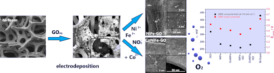
- the electrodeposition of conductive films on active metals [26]. The morphology of the deposits was analyzed by scanning electron microscopy (SEM) and is presented in Figure 1b–f. Typical GO flakes regularly distributed over the surface of the nickel foam were successfully obtained after the one-step


Quercetin- and caffeic acid-functionalized chitosan-capped colloidal silver nanoparticles: one-pot synthesis, characterization, and anticancer and antibacterial activities
Beilstein J. Nanotechnol. 2023, 14, 362–376, doi:10.3762/bjnano.14.31

- infrared (FTIR) spectroscopy, and transmission electron microscopy (TEM). The characteristic surface plasmon resonance (SPR) absorption band has been found at 417 and 424 nm for Ch/Q- and Ch/CA-Ag NPs, respectively. The formation of a chitosan shell comprising quercetin and caffeic acid, which surround the
- of quercetin, caffeic acid, chitosan, Ch/Q-, and Ch/CA-Ag NPs were monitored using a Perkin Elmer Spectrum Two FTIR-ATR spectrophotometer in the range of 4000–400 cm−1. Transmission electron microscopy (TEM) measurements were performed with a JEOL JEM-2100 electron microscope operating at 200 kV


The steep road to nonviral nanomedicines: Frequent challenges and culprits in designing nanoparticles for gene therapy
Beilstein J. Nanotechnol. 2023, 14, 351–361, doi:10.3762/bjnano.14.30

- cells. (f) Annual prevalence of reporting imaging and (or) flow cytometry during the last five years. Of note, “imaging” refers to images captured by widefield fluorescence microscopy or confocal microscopy (electron microscopy excluded). See details about experimental approach and scope in Supporting


Biocatalytic synthesis and ordered self-assembly of silica nanoparticles via a silica-binding peptide
Beilstein J. Nanotechnol. 2023, 14, 280–290, doi:10.3762/bjnano.14.25

- scattering (DLS). The efficiency of the self-assembly was evaluated with scanning electron microscopy (SEM), UV–vis spectroscopy, and qualitative visual demonstration. Results and Discussion SiBP alone as catalyst Reaction kinetics were studied via OD measurements of the particles and GC analysis of
- covers were moved completely out of the colloidal solution. Scanning electron microscopy To analyze as-synthesized particles, 50 µL aliquots of the reaction solutions were placed on standard microscope cover slides. The excess liquid was removed by absorbing on a clean absorbent paper. To analyze self


A novel approach to pulsed laser deposition of platinum catalyst on carbon particles for use in polymer electrolyte membrane fuel cells
Beilstein J. Nanotechnol. 2023, 14, 190–204, doi:10.3762/bjnano.14.19

- at the Biological and Chemical Research Centre, University of Warsaw, Poland, on the TALOS F200X (Thermo Fisher Scientific, United States) equipped with a four-detector windowless Super X-EDS system. The EDX measurements were performed in STEM (scanning transmission electron microscopy) mode with a


Structural, optical, and bioimaging characterization of carbon quantum dots solvothermally synthesized from o-phenylenediamine
Beilstein J. Nanotechnol. 2023, 14, 165–174, doi:10.3762/bjnano.14.17

- 2.2 mg/mL. These specimens were designated as CQDs/PU. For bioimaging studies, toluene was evaporated, and a thin film of CQDs was redissolved in water and filtered. The prepared QCD samples were characterized by transmission electron microscopy (TEM), atomic force microscopy (AFM), Fourier


Batch preparation of nanofibers containing nanoparticles by an electrospinning device with multiple air inlets
Beilstein J. Nanotechnol. 2023, 14, 141–150, doi:10.3762/bjnano.14.15

- using an electronic balance (XJ120A, Precisa LTD.). The nanofiber morphology was investigated by a scanning electron microscopy (SEM, Hitachi S4800, Hitachi LTD.), and Image J software (National Institute of Mental Health) was used to characterize the fiber diameter distribution by random selection of


Formation of nanoflowers: Au and Ni silicide cores surrounded by SiOx branches
Beilstein J. Nanotechnol. 2023, 14, 133–140, doi:10.3762/bjnano.14.14

- annealed at 1050 °C was named 15Au5Ni. The morphology was imaged by optical microscopy (OM, Zeiss Axiotech) and high-resolution scanning electron microscopy (HR-SEM, Hitachi S-4800) equipped with energy-dispersive X-ray spectroscopy (EDS, Thermo Scientific). The SEM images were recorded by using mixed


Antimicrobial and mechanical properties of functionalized textile by nanoarchitectured photoinduced Ag@polymer coating
Beilstein J. Nanotechnol. 2023, 14, 95–109, doi:10.3762/bjnano.14.11

- electron microscopy (SEM), transmission electron microscopy (TEM), and reflectance measurements to assess the optical properties and the durability of the functionalized textiles. Results and Discussion Photoinduced synthesis of the Ag@polymer coating Specific monomers poly(ethylene glycol) 600 diacrylate
- , as well as the final thickness of the metallic layer, account for this difference in reflectivity. Scanning electron microscopy (SEM) carried out on the surface of functionalized textiles revealed the homogenous distribution of AgNPs, with average sizes of 62 ± 2 nm and 58 ± 1 nm for the Ag@PEG600DA
- (Figure 4a) and Ag@PEG600DA/PETIA (Figure 4b) coatings, respectively. The AgNP size dispersion is also slightly higher in the case of Ag@PEG600DA. Both results are coherent with the peak and FMWH values calculated from the absorbance spectra (Figure 2c). Transmission electron microscopy (TEM) cross


Combining physical vapor deposition structuration with dealloying for the creation of a highly efficient SERS platform
Beilstein J. Nanotechnol. 2023, 14, 83–94, doi:10.3762/bjnano.14.10

- SERS properties of the nanoporous structure. Using scanning electron microscopy (SEM) and X-ray photoelectron spectroscopy (XPS) the morphology and surface composition of each nanoporous structure were respectively evaluated and used to describe the SERS properties of the samples. Results and
- 20 min to stop the dealloying process and to ensure a good cleaning of the samples. Characterization Scanning electron microscopy micrographs were recorded using a HITACHI STEM-FEG with an acceleration voltage of 5 kV. The images were treated with the ImageJ software [58] to assess the size of the


Solvent-induced assembly of mono- and divalent silica nanoparticles
Beilstein J. Nanotechnol. 2023, 14, 52–60, doi:10.3762/bjnano.14.6

- shell of approx. 15 nm (as estimated by transmission electron microscopy, TEM). The latter is made up of covalently grafted PS macromolecules resulting from the copolymerization of styrene with methacryloxymethyl groups. As previously demonstrated [25], these PS chains can serve as sticky patches when
- . Characterization methods Transmission electron microscopy experiments were performed using a Hitachi H600 microscope operating at an acceleration voltage of 75 kV. The samples were prepared by depositing one drop of the colloidal dispersion on conventional carbon-coated copper grids. The liquid evaporated in the


Two-step single-reactor synthesis of oleic acid- or undecylenic acid-stabilized magnetic nanoparticles by thermal decomposition
Beilstein J. Nanotechnol. 2023, 14, 11–22, doi:10.3762/bjnano.14.2

- , and composition via several techniques, such as transmission electron microscopy, dynamic light scattering, thermogravimetric analysis, Fourier-transform infrared spectroscopy/attenuated total reflectance, 57Fe Mössbauer spectroscopy, and X-ray diffraction. The effect of unsaturated oleic (OA) and
- be adsorbed on the surface of the particles, ensures nanodispersion stability (Figure 1) [31]. Transmission electron microscopy (TEM) micrographs of the prepared nanoparticles confirmed their size between 8 and 16 nm (Figure 2 and Figure 3). The diameter and shape of the nanoparticles depend on the
- diameter, X-ray diffraction, transmission electron microscopy, and dynamic light scattering (Malvern Zetasizer Nano S, Palaiseau, France) were used. Transmission electron microscopy observations were conducted using a JEOL JEM 2100 HR microscope (Croissy Sur Seine, France) equipped with a LaB6 source, and


Induced electric conductivity in organic polymers
Beilstein J. Nanotechnol. 2022, 13, 1551–1557, doi:10.3762/bjnano.13.128

- polymer film thickness, which is just 3 nm. After electric measurements, a number of heterostructures was sent for analysis by high resolution transmission electron microscopy and/or scanning electron microscopy. None of the studied samples showed a systematic ‘sticking’ of lead electrodes through the
- the preparation of a sample for electron microscopy. Results and Discussion The experiment was carried out in a four-contact configuration at direct or alternating currents. Both R(T) and V(I) dependences of the Pb–PDP–Pb sandwich could be measured, as shown in Figure 2a, and the transport
- : either macroscopic ‘pinholes’ or formation of multiple thin dendrites. The second possibility seems unlikely: neither the previous studies, nor the selective microscopic analysis of several samples by scanning and transmission electron microscopy revealed signs of the presence of dendrites. While the


Photoelectrochemical water oxidation over TiO2 nanotubes modified with MoS2 and g-C3N4
Beilstein J. Nanotechnol. 2022, 13, 1541–1550, doi:10.3762/bjnano.13.127

- of materials The morphology, the phase, and the vibrational characteristics of the surface functional groups of the materials were observed by field-emission scanning electron microscopy (FESEM), X-ray diffraction (XRD), and Fourier-transform infrared spectroscopy (FTIR). Diffuse reflectance


A TiO2@MWCNTs nanocomposite photoanode for solar-driven water splitting
Beilstein J. Nanotechnol. 2022, 13, 1520–1530, doi:10.3762/bjnano.13.125

- -scanning electron microscopy, transmission electron microscopy, X-ray diffraction, and linear sweep voltammetry. The results show that the TiO2@MWCNTs nanocomposite has an optical bandgap of 2.5 eV, which is a significant improvement in visible-light absorption capability compared to TiO2 (3.14 eV). The
- nanocomposite characterizations The surface morphology of MWCNTs and the TiO2@MWCNTs nanocomposite is characterized by using field-emission scanning electron microscopy (FE-SEM, S4800) and transmission electron microscopy (TEM, JEOL-1400). The crystallization behavior of the catalysts is analyzed by X-ray


In search of cytotoxic selectivity on cancer cells with biogenically synthesized Ag/AgCl nanoparticles
Beilstein J. Nanotechnol. 2022, 13, 1505–1519, doi:10.3762/bjnano.13.124

- -dispersive X-ray spectroscopy (EDX), and transmission electron microscopy (TEM) techniques were used to characterize nanoparticle development. The breast cancer cell line MCF-7 was used as a test model to study the cytotoxic behavior of Ag/AgCl nanoparticles and, as a counterpart, the nanoparticles were also
- . Transmission electron microscopy images were acquired on a JEOL 1010 microscope, with an accelerating voltage of 80 kV. For that, samples were pre-prepared in acetone and sonicated for 20 min, then dried at room temperature. Thermogravimetric analysis was performed on a Perkin Elmer STA 6000 simultaneous


Hydroxyapatite–bioglass nanocomposites: Structural, mechanical, and biological aspects
Beilstein J. Nanotechnol. 2022, 13, 1490–1504, doi:10.3762/bjnano.13.123

- intensity ratio between the peaks associated with the crystallographic planes (112) and (300) expressed in counts per second, and I300 represents the intensity of the characteristic peak of the crystallographic plane (300) expressed in counts per second. Scanning electron microscopy To determine the