Search results
Search for "scanning electron microscopy (SEM)" in Full Text gives 465 result(s) in Beilstein Journal of Nanotechnology. Showing first 200.
A comparison of tarsal morphology and traction force in the two burying beetles Nicrophorus nepalensis and Nicrophorus vespilloides (Coleoptera, Silphidae)
Beilstein J. Nanotechnol. 2019, 10, 47–61, doi:10.3762/bjnano.10.5

- traction force measurements of entire animals, whereas the performance of single fore tarsi has been measured with a nanotribometer. Both the number and the special morphology of the tarsal tenent hairs of the fore, middle and hind tarsi have been investigated by scanning electron microscopy (SEM). The
- masses amounted to 204 ± 44 mg in N. vespilloides (males: 212 ± 46 mg, females: 196 ± 41 mg) and 279 ± 41 mg in N. nepalensis (males: 276 ± 35 mg, females: 281 ± 48 mg; arithmetic means ± s.d. n = 10 for both males and females). Morphology Scanning electron microscopy (SEM) was used to characterize the


Zn/F-doped tin oxide nanoparticles synthesized by laser pyrolysis: structural and optical properties
Beilstein J. Nanotechnol. 2019, 10, 9–21, doi:10.3762/bjnano.10.2

- the crystalline domains by selected area electron diffraction (SAED) analysis. Energy dispersion X-ray spectroscopy (EDX) was performed using a scanning electron microscopy (SEM-Inspect TM S50) with an acceleration voltage of 15 kV, using a SiLi detector cooled with liquid nitrogen. This method can


A novel polyhedral oligomeric silsesquioxane-modified layered double hydroxide: preparation, characterization and properties
Beilstein J. Nanotechnol. 2018, 9, 3053–3068, doi:10.3762/bjnano.9.284

- hood before characterization. Scanning electron microscopy (SEM) images were recorded on an SU-70 or Utral 55 to study the surface morphologies. The samples were placed on a conducting carbon cement holder and were then coated with a thin layer of platinum using a sputter coater. Thermogravimetric


Femtosecond laser-assisted fabrication of chalcopyrite micro-concentrator photovoltaics
Beilstein J. Nanotechnol. 2018, 9, 3025–3038, doi:10.3762/bjnano.9.281
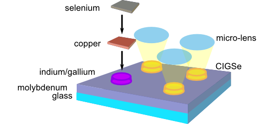
- array. The series of spots at the surface illustrates, that a stronger surface modification or even the formation of a crater can be achieved by increasing the number of laser pulses per spot as well as by increasing the laser fluence (energy density). Selected scanning electron microscopy (SEM) images


Colloidal chemistry with patchy silica nanoparticles
Beilstein J. Nanotechnol. 2018, 9, 2989–2998, doi:10.3762/bjnano.9.278

- CMs can be clearly seen on the scanning electron microscopy (SEM) images shown as inserts of Figure 3e,f. We also aimed to mimic at the colloidal scale the possible bonding of atoms of different natures to a same central atom, which is the source of the richness of the organic molecules. We focused on
- operating at 120 kV. We prepared the samples by depositing one drop of the colloidal dispersion on conventional carbon-coated copper grids. We let the liquid evaporate in the open air at room temperature and placed the grids in a box away from dust. Scanning electron microscopy (SEM) We performed high


Co-intercalated layered double hydroxides as thermal and photo-oxidation stabilizers for polypropylene
Beilstein J. Nanotechnol. 2018, 9, 2980–2988, doi:10.3762/bjnano.9.277

- analysis (TG-DTA) was performed on a PCT-IA instrument in the range of 25 to 700 °C at 5 °C·min−1 under flowing air. Scanning electron microscopy (SEM) images were taken with a Zeiss scanning electron microscope by dropping dilute ethanol suspension at room temperature. Elemental analysis for metal


Investigation of CVD graphene as-grown on Cu foil using simultaneous scanning tunneling/atomic force microscopy
Beilstein J. Nanotechnol. 2018, 9, 2953–2959, doi:10.3762/bjnano.9.274

- temperature, the Ar flow was stopped and the H2 flow was reduced while CH4 was let in to the quartz tube as the carbon source. As-grown samples, as well as graphene crsytals transferred on to dielectric substrates were investigated by optical microscopy and scanning electron microscopy (SEM). Raman spectra


Time-resolved universal temperature measurements using NaYF4:Er3+,Yb3+ upconverting nanoparticles in an electrospray jet
Beilstein J. Nanotechnol. 2018, 9, 2916–2924, doi:10.3762/bjnano.9.270

- scanning electron microscopy (SEM) image of a thin film of NaYF4:Er3+,Yb3+ UCNPs drop-cast on a glass coverslip is shown in Figure 1. The UCNPs are relatively uniform in size and shape with an average diameter around 300 nm and a height of around 80 nm. The height (thickness) of the UCNPs is determined by


Layered calcium phenylphosphonate: a hybrid material for a new generation of nanofillers
Beilstein J. Nanotechnol. 2018, 9, 2906–2915, doi:10.3762/bjnano.9.269

- (AFM) analysis. The samples prepared by the “drop by drop” and “several portions” methods contained thinner particles in comparison those where the portion was all added at once. Nevertheless, as it can be seen in scanning electron microscopy (SEM) images (Figure 2) and as was also verified by AFM


Nanostructure-induced performance degradation of WO3·nH2O for energy conversion and storage devices
Beilstein J. Nanotechnol. 2018, 9, 2845–2854, doi:10.3762/bjnano.9.265

- by a facile hydrothermal method for the first time at the different termperatures to investigate the disadvantages of 2D structures. The growth mechanism analyzed by X-ray diffraction (XRD) and scanning electron microscopy (SEM) suggested a 2D to 3D structural transition upon temperature increment


Controlling surface morphology and sensitivity of granular and porous silver films for surface-enhanced Raman scattering, SERS
Beilstein J. Nanotechnol. 2018, 9, 2813–2831, doi:10.3762/bjnano.9.263

- surfaces act as efficient SERS substrates showing greater enhancement factors compared to as prepared, sputtered, but untreated silver films when using rhodamine B as Raman probe molecule. The obtained roughened silver films were fully characterized by scanning electron microscopy (SEM), atomic force
- silver films were characterized using atomic force microscopy (AFM) in contact mode on a CP-II AFM (Bruker-Veeco) with SiC cantilevers to determine the topography and surface roughness (root mean square roughness, Rq). Scanning electron microscopy (SEM) of the silver films was performed on a Philips XL


Low cost tips for tip-enhanced Raman spectroscopy fabricated by two-step electrochemical etching of 125 µm diameter gold wires
Beilstein J. Nanotechnol. 2018, 9, 2718–2729, doi:10.3762/bjnano.9.254

- on the applications of TERS can be found in [20][21][22][23][24][25]. TERS features unique advantages as compared with scanning electron microscopy (SEM), scanning near-field Raman microscopy (Raman-SNOM) [26] and far-field nanoscopy [27][28]: (i) it is a label-free technique, i.e., it does not
- drops of HCl and rinsing in ethanol and water. This eliminates residual impurities from the surface. Scanning electron microscopy (SEM) inspection of the produced tips is carried out to characterize the tip apex using a Zeiss Merlin field emission electron microscope, equipped with a Gemini II column


Optimization of Mo/Cr bilayer back contacts for thin-film solar cells
Beilstein J. Nanotechnol. 2018, 9, 2700–2707, doi:10.3762/bjnano.9.252

- scanning electron microscopy (SEM), atomic force microscopy (AFM), UV–vis–NIR spectroscopy and X-ray photoelectron spectroscopy. A careful analysis of the resulting Mo/Cr thin film across all the sputtering parameters led us to the best combination, optimizing both the electro-optical response of the Mo/Cr


Nanoantenna structures for the detection of phonons in nanocrystals
Beilstein J. Nanotechnol. 2018, 9, 2646–2656, doi:10.3762/bjnano.9.246

- well as the structural parameters of nanoantennas (length, width, and lateral periodicity) were measured by scanning electron microscopy (SEM) using the same Raith-150 system at 10 kV acceleration voltage, 30 µm aperture, and 6 mm working distance. The structural parameters of the microantennas were


Impact of the anodization time on the photocatalytic activity of TiO2 nanotubes
Beilstein J. Nanotechnol. 2018, 9, 2628–2643, doi:10.3762/bjnano.9.244

- anodization time, ta, was investigated via scanning electron microscopy (SEM), as presented in Figure 1. Well-defined, regular, nanotubular arrays are observed at the surface of these materials. It has been previously reported that ta determines some of the geometrical characteristics of such nanostructures


Characterization of the microscopic tribological properties of sandfish (Scincus scincus) scales by atomic force microscopy
Beilstein J. Nanotechnol. 2018, 9, 2618–2627, doi:10.3762/bjnano.9.243

- gives the averaged frictional force
. The corresponding frictional coefficient μ was obtained by subsequently fitting the data with Ffric = Fad + µ·Fload. Cantilevers and the cross section of a sandfish dorsal scale were imaged by scanning electron microscopy (SEM, SUPRA 60 VP, Zeiss, Germany


Friction reduction through biologically inspired scale-like laser surface textures
Beilstein J. Nanotechnol. 2018, 9, 2561–2572, doi:10.3762/bjnano.9.238

- surface (dimple diameter ≈30 µm, height ≈5 µm). Figure 1 presents scanning electron microscopy (SEM) and white light profilometry results for these textures (Figure 1a and 1b), demonstrating that such samples were successfully fabricated with high reproducibility for the scale-like texturing elements. For
- pearlitic steel with round dimples, no microstructural changes within the 100Cr6 samples in the heat affected zone were expected [15]. Figure S2, Supporting Information File 1 shows a scanning electron microscopy (SEM) image of a cross-section through a laser-textured sample prepared by focused ion beam


Hydrothermal-derived carbon as a stabilizing matrix for improved cycling performance of silicon-based anodes for lithium-ion full cells
Beilstein J. Nanotechnol. 2018, 9, 2381–2395, doi:10.3762/bjnano.9.223

- range between 25 °C and 800 °C on a TGA Q500 (TA Instruments) in an oxygen/nitrogen atmosphere (nitrogen flow: 10 mL min−1, oxygen flow: 25 mL min−1) in order to determine the Si content. A heating ramp of 20 °C min−1 was applied. Scanning electron microscopy (SEM) with a field emission gun (Schottky


High-throughput micro-nanostructuring by microdroplet inkjet printing
Beilstein J. Nanotechnol. 2018, 9, 2372–2380, doi:10.3762/bjnano.9.222

- nm thickness (prepared by Fraunhofer ISIT, Itzehoe), 200 nm thick nickel–titanium thin film sputtered on a 4 µm thick copper layer and glass substrate (prepared by Acquandas, Kiel) and free-standing nickel–titanium foil of 50 µm thickness (prepared by Acquandas, Kiel). Scanning electron microscopy
- (SEM) and image analysis SEM (Supra 55VP, Zeiss, Germany) imaging was carried out at 5 kV using the in-lens detector at a working distance of 5 mm. The SEM images were processed via ImageJ. The nanoparticles were then segmented in the images and indicated as maxima. For analysis of the coordinates of


Nanoscale characterization of the temporary adhesive of the sea urchin Paracentrotus lividus
Beilstein J. Nanotechnol. 2018, 9, 2277–2286, doi:10.3762/bjnano.9.212

- around 20 nm could clearly be distinguished (Figure 4a,c). These results are consistent with previous reports, describing the sea urchin footprint microstructure, observed by scanning electron microscopy (SEM), as a dense meshwork with smaller mesh-like areas (<1 μm) delimited by very fine fibrils (about


The role of adatoms in chloride-activated colloidal silver nanoparticles for surface-enhanced Raman scattering enhancement
Beilstein J. Nanotechnol. 2018, 9, 2236–2247, doi:10.3762/bjnano.9.208

- exposure is shown in Figure S2B (using crystal violet as an analyte). The formation of AgNPs from AgCl precursor microparticles can be also followed in the scanning electron microscopy (SEM) micrographs depicted in Figure 3. Figure 3a shows 1–2 μm AgCl particles formed after mixing silver nitrate and


Filling nanopipettes with apertures smaller than 50 nm: dynamic microdistillation
Beilstein J. Nanotechnol. 2018, 9, 2181–2187, doi:10.3762/bjnano.9.204

- different methods: first, by using the velocity of the water meniscus and an optical microscopy view of the tip [14]; second, by using scanning electron microscopy (SEM) after capillary use (this method is destructive); third, via electrical characterization of the nanopipette. Figure 1 shows the SEM view


Localized photodeposition of catalysts using nanophotonic resonances in silicon photocathodes
Beilstein J. Nanotechnol. 2018, 9, 2097–2105, doi:10.3762/bjnano.9.198

- precision and implementing the approach in a state-of-the art photo-electrode/catalyst system in order to demonstrate the potential for solar fuel production enhancement. Experimental General Chemicals were purchased from major chemical suppliers and used as received. Scanning electron microscopy (SEM) was


The structural and chemical basis of temporary adhesion in the sea star Asterina gibbosa
Beilstein J. Nanotechnol. 2018, 9, 2071–2086, doi:10.3762/bjnano.9.196

- electron microscope. Scanning electron microscopy (SEM) Footprints were collected on clean glass coverslips and tube feet were cut off while being unattached. All samples were chemically fixed at room temperature in Bouin’s fluid for at least 24 h. All samples were then dehydrated in graded ethanol and


Biomimetic and biodegradable cellulose acetate scaffolds loaded with dexamethasone for bone implants
Beilstein J. Nanotechnol. 2018, 9, 1986–1994, doi:10.3762/bjnano.9.189

- work, a drug-delivery nanoplatform system consisting of polymeric celluloce acetate (CA) scaffolds loaded with dexamethasone was fabricated through electrospinning. Atomic force microscopy (AFM) and scanning electron microscopy (SEM) indicated the successful fabrication of these structures


