Search results
Search for "surface structures" in Full Text gives 86 result(s) in Beilstein Journal of Nanotechnology.
A comparison of tarsal morphology and traction force in the two burying beetles Nicrophorus nepalensis and Nicrophorus vespilloides (Coleoptera, Silphidae)
Beilstein J. Nanotechnol. 2019, 10, 47–61, doi:10.3762/bjnano.10.5

- ]. For theoretical access to the problem of frictional anisotropy considering factors such as the slope of the surface structures, the rigidity of the joints, and sliding speed, the reader is referred to [44]. Our results are in full accordance with previous measurements of the frictional directionality


Apparent tunneling barrier height and local work function of atomic arrays
Beilstein J. Nanotechnol. 2018, 9, 3048–3052, doi:10.3762/bjnano.9.283

- have been used to estimate variations of the local work function Φ of surface structures. We experimentally show that Φapp can fail as a measure of Φ. The discrepancies are attributed to a kinetic-energy contribution to Φapp. This contribution depends on the lateral extent of the tunneling current


Femtosecond laser-assisted fabrication of chalcopyrite micro-concentrator photovoltaics
Beilstein J. Nanotechnol. 2018, 9, 3025–3038, doi:10.3762/bjnano.9.281
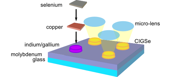
- -induced periodic surface structures (LIPSS [19]) and round melting features form on the glass surface (Figure 5b). The LIPSS with periods in the sub-micrometer range are generated via intra-pulse scattering and interference of the fs-laser radiation at the roughened glass surface, leading to the spatially
- fluences F (from left to right: F = 1.63 J/cm2, N = 100; F = 1.83 J/cm2, N = 30; F = 1.83 J/cm2, N = 100; F = 2.04 J/cm2, N = 100). For all depicted laser spots, the laser-generated surface structures constitute a diffusion trap for evaporated indium during the PVD process. The fact that the strongest


Controlling surface morphology and sensitivity of granular and porous silver films for surface-enhanced Raman scattering, SERS
Beilstein J. Nanotechnol. 2018, 9, 2813–2831, doi:10.3762/bjnano.9.263

- plasmas on the surface morphology of thin silver films. It was found that different surface structures and different degrees of surface roughness could be obtained by a systematic variation of the plasma type and condition as well as plasma power and treatment time. The differently roughened silver
- mixture as oxidizing plasma (Figure 8b). Even the silver film oxidized by air plasma for 15 min and 30 min as well as the silver film oxidized by the argon/oxygen plasma mixture show different surface structures compared to the silver films oxidized by pure oxygen plasma (Figure S16, Supporting


Biomimetic surface structures in steel fabricated with femtosecond laser pulses: influence of laser rescanning on morphology and wettability
Beilstein J. Nanotechnol. 2018, 9, 2802–2812, doi:10.3762/bjnano.9.262

- surface topography needs to be mimicked, but often also a specific function of the structure. An alternative approach to laser direct writing of complex structures is the generation of laser-induced periodic surface structures (LIPSS), which is based on directed self-organization of the material and
- surface structures; laser rescanning; steel; wettability; Introduction Complex structures found in nature often present properties that are attractive for applications in science and technology. The hydrophobicity found at the lotus leaf surface [1], the exceptional adhesion capability of gecko feet [2
- [12][13][14]. Such structures are commonly referred to as laser-induced periodic surface structures (LIPSSs). Two main mechanisms have been proposed to explain the origin of these structures. One of them takes into account laser light scattered at a rough surface, which interferes with the incident


Friction reduction through biologically inspired scale-like laser surface textures
Beilstein J. Nanotechnol. 2018, 9, 2561–2572, doi:10.3762/bjnano.9.238

- maximum friction reduction [15]. A similar effect may exist for the optimum diameter of the scale-like surface morphologies as a function of oil viscosity, something to be tested in future experiments. Among our ongoing work, we have started to test these surface structures in reciprocating contacts, in


Quantitative comparison of wideband low-latency phase-locked loop circuit designs for high-speed frequency modulation atomic force microscopy
Beilstein J. Nanotechnol. 2018, 9, 1844–1855, doi:10.3762/bjnano.9.176

- subnanometer-scale imaging of biomolecules [15][16][17]. Moreover, three-dimensional (3D) imaging techniques have been developed based on FM-AFM and used to visualize 3D distributions of hydration structures as well as flexible surface structures at solid–liquid interfaces [18][19][20][21][22]. Although FM-AFM


Direct AFM-based nanoscale mapping and tomography of open-circuit voltages for photovoltaics
Beilstein J. Nanotechnol. 2018, 9, 1802–1808, doi:10.3762/bjnano.9.171

- ability to serially mill a surface, in order to reveal underlying surface structures and uniquely develop three-dimensional (3D) nanoscale property maps. The most notable examples are based on pure current detection with the AFM to resolve conduction pathways in filamentary semiconducting devices and


Heterostuctures of 4-(chloromethyl)phenyltrichlorosilane and 5,10,15,20-tetra(4-pyridyl)-21H,23H-porphine prepared on Si(111) using particle lithography: Nanoscale characterization of the main steps of nanopatterning
Beilstein J. Nanotechnol. 2018, 9, 1211–1219, doi:10.3762/bjnano.9.112

- triple-decker sandwich complex of phthalocyanine compounds prepared on graphite was studied using STM by Lei et al. [17]. A method of photocatalytic lithography was reported for making porphyrin surface structures that were applied for preparing protein arrays [18][19]. The assembly of porphyrins at


Periodic structures on liquid-phase smectic A, nematic and isotropic free surfaces
Beilstein J. Nanotechnol. 2018, 9, 342–352, doi:10.3762/bjnano.9.34

- surface and in the bulk. Comparison of our images with FCD images [15] shows great similarity with FCD domains observed earlier. Figure 2 shows three-dimensional images of SmA surface structures obtained by the interferometric surface structure analyzer (ISSA). We see a system of craters and hills similar
- region corresponded to a 10 nm distance between the tip and the surface but can be shifted to other values. The interaction between the tip and surface begins at a distance of 30 nm. Image of the 8CB surface structures on the SmA free surface (a) and in bulk (b), as measured with an optical polarized


Combined scanning probe electronic and thermal characterization of an indium arsenide nanowire
Beilstein J. Nanotechnol. 2018, 9, 129–136, doi:10.3762/bjnano.9.15

- long-range electrostatic force between surface structures and the tip, care has to be taken to minimize long-range averaging effects on device structures with exposed electrodes [9]. Highest resolution and accuracy is enabled by detecting the force gradient [17][18][19]. On typical devices with large


Nanoprofilometry study of focal conic domain structures in a liquid crystalline free surface
Beilstein J. Nanotechnol. 2017, 8, 2544–2551, doi:10.3762/bjnano.8.254

- following liquid crystalline phase sequence: isotropic (41 °C), nematic (32 °C), smectic-A (22.2 °C). The profilometer is an interferometric surface structure analyzer (ISSA) that allows three dimensional images of the surface structures to be obtained. Its vertical scan range is 150 μm, the vertical
- and the LC surface. Experimental As was previously mentioned, the free surface structure of the liquid crystal compound 8CB has been studied using a profilometer. Scanning interferometry of white light was used in ISSA to obtain images and to calculate and analyze surface structures of the test parts
- substrate without electrodes. On the glass substrate the same surface structures were observed. It is important to note that the FCDs are not observed on the smectic-free surface. The conditions for FCD formation are given in [4]. The nanoprofilometer presented in this work is a noncontact method which can


Surfactant-induced enhancement of droplet adhesion in superhydrophobic soybean (Glycine max L.) leaves
Beilstein J. Nanotechnol. 2017, 8, 2345–2356, doi:10.3762/bjnano.8.234

- and five variations of nonionic surfactants) have been investigated. The leaf surface structures show a hierarchical organization, built up by convex epidermal cells (microstructure) and superimposed epicuticular platelet-shaped wax crystals (micro- to nanostructure). Chemical analysis of the
- applied liquid also depends on the wetting mode. In the Wenzel mode [26] an applied water droplet penetrates into cavities formed by the surface structures, increasing the contact area, and resulting in high hysteresis of the applied liquid. In Cassie–Baxter mode [27] air remains in the surface cavities
- distortion from desiccation, without changing the leaf surface structures [36][51]. The leaves were cut into 0.5 cm2 pieces and placed on a wet paper towel inside a petri dish. Glycerol (90%) was added drop-wise over a period of 20 h. The process ensures that glycerol infiltrates the tissue through the cut


(Metallo)porphyrins for potential materials science applications
Beilstein J. Nanotechnol. 2017, 8, 1786–1800, doi:10.3762/bjnano.8.180

- determine surface structures of deposited CuTPP(Br)8 on a Au(111) surface. After deposition of about one monolayer only small periodically structured arrangements were observed. Annealing significantly enlarged the size of ordered areas, which is attributed to a remarkable surface mobility [71] of the


Collembola cuticles and the three-phase line tension
Beilstein J. Nanotechnol. 2017, 8, 1714–1722, doi:10.3762/bjnano.8.172

- springtails (Collembola) are superhydrophobic, but the mechanism has not been described in detail. Previous studies have suggested that overhanging surface structures play an important role, but such structures are not a universal trait among springtails with superhydrophobic cuticles. A novel wetting
- experiment with a fluorescent dye revealed the extent of wetting on exposed surface structures. Using simple wetting models to describe the composite wetting of the cuticular surface structures results in underestimating the contact angles of water. Including the three-phase line tension allows for a
- . clavatus does not have overhanging surface structures. This large change in observed contact angles can be explained with a modest change of the three-phase line tension. Keywords: springtails (Collembola); superhydrophobicity; three-phase line tension; Introduction Collembola, a group of small


![[Graphic 16]](/bjnano/content/inline/2190-4286-8-172-i22.png?max-width=637&scale=1.18182) . A system with θ0 = 105°, S = 0.1 μm a...
. A system with θ0 = 105°, S = 0.1 μm a...


Air–water interface of submerged superhydrophobic surfaces imaged by atomic force microscopy
Beilstein J. Nanotechnol. 2017, 8, 1671–1679, doi:10.3762/bjnano.8.167

- sensory systems. Biological surfaces are the basis of the discovery and are models for the development of biomimetic surfaces. The conquest of land some 450 million years ago led to the evolution of an almost endless variety of surface structures and functionalities in plants and animals [3]. One of the
- sputter-coated onto the surface to enhance their conductivity. Biological role models of air-retaining Salvinia effect surfaces. a) The floating fern Salvinia molesta has one of the most complex surface structures in plants. Reproduced with permission from [5], copyright 2010 Wiley-VCH Verlag GmbH & Co


Micro- and nano-surface structures based on vapor-deposited polymers
Beilstein J. Nanotechnol. 2017, 8, 1366–1374, doi:10.3762/bjnano.8.138

- materials (with the exception for the case of selective deposition on transition metals and charged surfaces). Because of the well-established and available photolithography and soft lithography techniques, promising patterned surface structures have been created. Attempts were conducted to produce


Calculating free energies of organic molecules on insulating substrates
Beilstein J. Nanotechnol. 2017, 8, 667–674, doi:10.3762/bjnano.8.71

- of attention due to their versatility, functionality, and technological potential. Understanding the behaviour of self-assembling molecules is important for catalysis [1], coatings [2], sensors [3][4] and molecular electronics [5][6][7]. To design and fabricate surface structures relevant for these


Analysis and modification of defective surface aggregates on PCDTBT:PCBM solar cell blends using combined Kelvin probe, conductive and bimodal atomic force microscopy
Beilstein J. Nanotechnol. 2017, 8, 579–589, doi:10.3762/bjnano.8.62

- transport characteristics and the distribution of carriers and materials, which are relevant to device performance [16]. In this work, we use a series of AFM techniques to characterize electrically defective surface structures aggregated on test PSC specimens. The active layer of the test PSCs comprises the


Biological and biomimetic materials and surfaces
Beilstein J. Nanotechnol. 2017, 8, 403–407, doi:10.3762/bjnano.8.42

- , which later proved to be wax crystals that are formed by self-organisation processes and show specific shapes characteristic for different plant groups. To study these surface structures in the SEM, the leaves have to be prepared and cleaned, and Wilhelm Barthlott soon realized that the leaves of some
Nano- and microstructured materials for in vitro studies of the physiology of vascular cells
Beilstein J. Nanotechnol. 2016, 7, 1620–1641, doi:10.3762/bjnano.7.155

- /nanotopographies. We define micro/nanostructured substrates as materials having fabricated surface structures in all three dimensions and, consequently, not having a planar surface. The number of possible architectures of micro/nanostructured substrates is huge and it is often difficult to keep track with all
- desired surface feature size. In particular, some methods are not suited for structuring surface topographies in the nanometer range. 1.2 Microfabrication techniques Microfabrication techniques are mainly used to generate surface structures in the micrometer range, which is the size scale of cells. In
- using metals [58]. The surface structures made by photolithography are typically further used as a master structure for further processing. Hot embossing also replicates micro- and nanofeatures of master substrates. In that case, a thermoplastic material is pressed on the mold at a high temperature to


Surface roughness rather than surface chemistry essentially affects insect adhesion
Beilstein J. Nanotechnol. 2016, 7, 1471–1479, doi:10.3762/bjnano.7.139

- attachment of the beetles. Surface roughness was found to be the dominant factor, strongly affecting the attachment ability of the beetles. Keywords: insect attachment; superhydrophilicity; superhydrophobicity; superoleophobicity; surface structures; Introduction The development of functional coatings that
- also produce capillary forces. Inspired by this idea, artificial silicone polymer structures with underwater adhesive properties were fabricated [34]. Thus, the relationship between surface structures and the attachment of insects, in combination with their particular chemical/physical properties, has
- flows on textured substrates with different roughness parameters and surface energies [39]. These numerical studies demonstrated that a higher density of geometrical surface structures of the rigid substrate results in a greater loss of fluid from the pad. The draining rate of the pad fluid is more


Three-gradient regular solution model for simple liquids wetting complex surface topologies
Beilstein J. Nanotechnol. 2016, 7, 1377–1396, doi:10.3762/bjnano.7.129

- approach, without a discontinuity in the water front shape or in the water advancing contact angle θ. Therefore, air entrapment cannot be the main reason why the contact angle θ for an advancing water front varies. Rather, the contact line is pinned and curved due to the surface structures, inducing
- surface is hydrophobic (apparent contact angle θ > 90°) [1]. Recently, different surface structures have been designed and fabricated from hydrophilic materials that show hydrophobic contact angles [2][3][4][5][6][7][8][9][10]. An example is an inverse opal as schematically shown in Figure 1. Our group
- scale, and does not entail details about the droplet shape close to the surface structures on a microscopic level. Another explanation of the difference in θ for a structured and unstructured surface of the same material is contact line pinning [17][18][19][20]. The three-phase contact line is hereby


![[Graphic 10]](/bjnano/content/inline/2190-4286-7-129-i31.png?max-width=637&scale=1.18182) and standard deviation as a function of the cavity diameter d (in...
and standard deviation as a function of the cavity diameter d (in...

![[Graphic 20]](/bjnano/content/inline/2190-4286-7-129-i41.png?max-width=637&scale=1.18182) and the corresponding fluctuations (measured along the x-direction) indica...
and the corresponding fluctuations (measured along the x-direction) indica...

![[Graphic 28]](/bjnano/content/inline/2190-4286-7-129-i49.png?max-width=637&scale=1.18182) and the corresponding standard deviation as measure of variation in the ...
and the corresponding standard deviation as measure of variation in the ...

![[Graphic 39]](/bjnano/content/inline/2190-4286-7-129-i60.png?max-width=637&scale=1.18182) and standard deviation as function of cut-off fraction c for fixed ...
and standard deviation as function of cut-off fraction c for fixed ...
Functional diversity of resilin in Arthropoda
Beilstein J. Nanotechnol. 2016, 7, 1241–1259, doi:10.3762/bjnano.7.115

- is transported to the mouth region of the beetle where it can be seized with the mandibles [116][117][118]. The sticky pads feature a surface that is subdivided into numerous terminally branched outgrowths. During the prey capture, these surface structures are completely covered by an adhesive


Frog tongue surface microstructures: functional and evolutionary patterns
Beilstein J. Nanotechnol. 2016, 7, 893–903, doi:10.3762/bjnano.7.81

- been shown before, that the anatomy of frog tongues can be very diverse in different anuran taxa [14], little is known about the diversity of tongue surface structures in frogs. Besides a study on the ornamentation of the tongue in the dicroglossid frog Fejervarya cancrivora [15] (the frog is referred
- and tongue adhesion in frogs of the genus Ceratophrys [13][31]. The Litoria caerulea specimen studied herein was previously used for a study on toe-pad anatomy in tree frogs [32]. We examined the surface structures of frog tongues by using scanning electron microscopy (SEM). For SEM we prepared pieces
- package Amira 6.0 (FEI SAS, Mérignac Cedex, France). The micro-CT data of the Ceratophrys ornata specimen was already used in a previous study [13] and is accessible at http://dx.doi.org/10.5061/dryad.066mr. Results Tongue surface structures: Two types of papillae cover the dorsal surface of frog tongues


