Search results
Search for "absorption spectroscopy" in Full Text gives 142 result(s) in Beilstein Journal of Nanotechnology.
Properties of tin oxide films grown by atomic layer deposition from tin tetraiodide and ozone
Beilstein J. Nanotechnol. 2023, 14, 1085–1092, doi:10.3762/bjnano.14.89

- grazing incidence X-ray diffractometry (GIXRD), using an X-ray diffractometer SmartLab Rigaku with Cu Kα radiation, which corresponds to an X-ray wavelength of 0.15406 nm. X-ray photoelectron emission and X-ray absorption spectroscopy (XPS and XAS, respectively) measurements were made at the FinEstBeAMS


A visible-light photodetector based on heterojunctions between CuO nanoparticles and ZnO nanorods
Beilstein J. Nanotechnol. 2023, 14, 1018–1027, doi:10.3762/bjnano.14.84

- . The energy-dispersive X-ray spectroscopy (EDS) results in Figure 1c show Zn, Cu, and O, which indicates the presence of ZnO and CuO. No further impurities were found. The results from X-ray diffractometry (XRD) and UV–vis absorption spectroscopy confirm that the nanoparticles covering the surfaces and
- composition of the materials. The morphology of pure CuO NPs and CuO NPs/ZnO NRs was examined by field-emission scanning electron microscopy. UV–vis absorption spectroscopy showed the optical properties of the materials. The photodetector performance was studied through the current–voltage (I–V


Upscaling the urea method synthesis of CoAl layered double hydroxides
Beilstein J. Nanotechnol. 2023, 14, 927–938, doi:10.3762/bjnano.14.76

- process and aiming to quantify the amount of this Co-based α-LH impurity, X-ray absorption spectroscopy (XAS) measurements were performed at the CLÆSS BL22 beamline at the ALBA synchrotron. Figure 2A depicts the X-ray absorption near-edge structure (XANES) spectra for the Co K edge. In all samples, the
- h at 523 K and 5 × 10⁻⁵ bar. The desorption branch of the N2 isotherm was used to determine the pore size distribution using the BJH method. The surface area was determined using the BET method. The micropores volumes were determined by applying t-plot and DR methods. X-ray absorption spectroscopy X
- -ray absorption spectroscopy (XAS) measurements were performed at the BL-22 (CLÆSS) beamline of the ALBA synchrotron (Barcelona, Spain), proposal: 2022097096. XANES and EXAFS Co K edge spectra were measured at room temperature in transmission mode. Absorbents of as-synthesized fresh samples were


Evaluation of electrosynthesized reduced graphene oxide–Ni/Fe/Co-based (oxy)hydroxide catalysts towards the oxygen evolution reaction
Beilstein J. Nanotechnol. 2023, 14, 420–433, doi:10.3762/bjnano.14.34
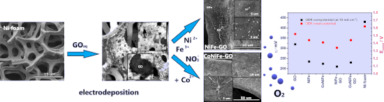
- probably inhibited the electrodeposition process of NiFe and CoNiFe on its surface. This may be the reason for the slower stabilization of the synthesis current density observed in the chronoamperograms (Figure 1a). X-ray diffraction, X-ray photoemission spectroscopy and X-ray absorption spectroscopy
- ). Characterizations The morphology and structure of the catalysts were characterized using a scanning electron microscope (FEI QUANTA FEG 250) with an energy-dispersive X-ray (EDX) sensor. X-ray absorption spectroscopy (XAS) was performed at the 04BM beamline at the National Synchrotron Radiation Centre SOLARIS [41


Quercetin- and caffeic acid-functionalized chitosan-capped colloidal silver nanoparticles: one-pot synthesis, characterization, and anticancer and antibacterial activities
Beilstein J. Nanotechnol. 2023, 14, 362–376, doi:10.3762/bjnano.14.31

- was calculated from the obtained results. Results and Discussion The characterization of the synthesized silver nanoparticles (Ag NPs) was carried out by using UV–vis absorption spectroscopy at ambient temperature. Figure 2 shows the UV–vis absorption measurement results of different Ag NPs, which are


A nonenzymatic reduced graphene oxide-based nanosensor for parathion
Beilstein J. Nanotechnol. 2022, 13, 730–744, doi:10.3762/bjnano.13.65

- performance of the nanosensor was validated by adding PT to natural samples and comparing the data via absorption spectroscopy. PT detection results encourage the design of easy-to-use nanosensor-based analytical tools for rapidly monitoring other environmental samples. Keywords: electrochemical nanosensor


Impact of device design on the electronic and optoelectronic properties of integrated Ru-terpyridine complexes
Beilstein J. Nanotechnol. 2022, 13, 219–229, doi:10.3762/bjnano.13.16

- was verified by infrared reflection absorption spectroscopy (IRRAS) and surface-enhanced Raman spectroscopy in combination with density functional theory calculations, as well as variable angle spectroscopic ellipsometry. Based on this wire formation protocol the on-chip preparation of Ru(TP)2-complex


Fate and transformation of silver nanoparticles in different biological conditions
Beilstein J. Nanotechnol. 2021, 12, 665–679, doi:10.3762/bjnano.12.53

- absorption spectroscopy (GF-AAS), nuclear magnetic resonance (NMR) spectroscopy, and transmission electron microscopy (TEM) experiments. Physicochemical characteristics of freshly prepared AgNPs Freshly prepared AgNPs coated with PVP, sodium bis(2-ethylhexyl)sulfosuccinate (AOT), and poly(ʟ-lysine) (PLL


High-yield synthesis of silver nanowires for transparent conducting PET films
Beilstein J. Nanotechnol. 2021, 12, 624–632, doi:10.3762/bjnano.12.51

- circuit including a white LED was utilized. Results and Discussion To determine the morphology, the as-prepared silver nanostructures were first characterized by UV–vis absorption spectroscopy. The absorption spectrum of AgNWs is a function of the dielectric material, the chemicals used, and the particle


A review on nanostructured silver as a basic ingredient in medicine: physicochemical parameters and characterization
Beilstein J. Nanotechnol. 2021, 12, 440–461, doi:10.3762/bjnano.12.36

- –NIR absorption spectroscopy: Metallic nanoparticles are known to emit characteristic colors in the visible region of the electromagnetic spectrum due to a phenomenon known as surface plasmon resonance. The color of a colloidal nanoparticle solution is mainly dependent on the size and shape of the


Nickel nanoparticle-decorated reduced graphene oxide/WO3 nanocomposite – a promising candidate for gas sensing
Beilstein J. Nanotechnol. 2021, 12, 343–353, doi:10.3762/bjnano.12.28

- comparison to pure nickel nanoparticles from [BMIm][NTf2] (size pure nickel nanoparticles 11 ± 2 nm) [46]. Nickel nanoparticles supported on pristine graphene sheets were synthesized with a size 35 ± 5 nm [51]. The nickel content was measured using atomic absorption spectroscopy (AAS). Ni@rGO contained 8
- precipitated with acetonitrile from the nanoparticle/ionic liquid dispersion and washed several times with acetonitrile. P-XRD patterns were recorded for 1 h (2θ = 5–100°). Atomic absorption spectroscopy, AAS for metal analysis was performed on a PerkinElmer PinAAcle 900T, equipped with a flame furnace. Flame


Differences in surface chemistry of iron oxide nanoparticles result in different routes of internalization
Beilstein J. Nanotechnol. 2021, 12, 270–281, doi:10.3762/bjnano.12.22

- or cytoskeleton dynamics was quantified by atomic absorption spectroscopy (AAS) and the uptake was verified by fluorescent microscopy. The uptake route of the tested MNPs differed depending on the surface coating. While BSA-SO-MNPs were internalized via CME, PEG-SO-MNPs were preferentially taken up


A review on the green and sustainable synthesis of silver nanoparticles and one-dimensional silver nanostructures
Beilstein J. Nanotechnol. 2021, 12, 102–136, doi:10.3762/bjnano.12.9


Fusion of purple membranes triggered by immobilization on carbon nanomembranes
Beilstein J. Nanotechnol. 2021, 12, 93–101, doi:10.3762/bjnano.12.8

- reflection absorption spectroscopy (IRRAS). Experimental Preparation of SAMs and CNMs NBPT was purchased from Taros Chemicals (Dortmund, Germany). Thermally evaporated Au films (300 nm) on mica supports (Georg Albert physical vapor deposition coatings) were used as substrates for the SAM preparation


Cu2O nanoparticles for the degradation of methyl parathion
Beilstein J. Nanotechnol. 2020, 11, 1546–1555, doi:10.3762/bjnano.11.137

- 4-NPh makes quantification of the degradation much easier because 4-NPh absorbs light in the UV–vis range. Hence absorption spectroscopy was used along with the Beer–Lambert law [44]. The molar absorptivity coefficients were determined to be 10080 M−1·cm−1 (λ = 320 nm) for 4-nitrophenol and 17632 M


A few-layer graphene/chlorin e6 hybrid nanomaterial and its application in photodynamic therapy against Candida albicans
Beilstein J. Nanotechnol. 2020, 11, 1054–1061, doi:10.3762/bjnano.11.90

- concentration of Ce6 in the FLG-Ce6 hybrid was calculated using UV–vis absorption spectroscopy. Ce6 presents an intense absorption band at 407 nm, corresponding to the Soret band. A Ce6 calibration curve at this wavelength is then produced. Then, in order to obtain a correct approximation of the real


Nickel nanoparticles supported on a covalent triazine framework as electrocatalyst for oxygen evolution reaction and oxygen reduction reactions
Beilstein J. Nanotechnol. 2020, 11, 770–781, doi:10.3762/bjnano.11.62

- immobilized on the CTFs via the decomposition of the metal precursor in the IL (Scheme 1). The composites were designated Ni/CTF-1-400-X and Ni/CTF-1-600-X, where X represents the weight percentage of nickel in the composite material based on flame atomic absorption spectroscopy (AAS). Nickel loadings of 20
- atomic absorption spectroscopy (AAS) for the determination of the metal content was conducted with a Vario 6 from Analytic Jena. For AAS the sample was treated with aqua regia. Ion chromatography (IC) measurements were performed with a Dionex ICS 1100 instrument with an IonPac AS 22column, combined with


Interfacial charge transfer processes in 2D and 3D semiconducting hybrid perovskites: azobenzene as photoswitchable ligand
Beilstein J. Nanotechnol. 2020, 11, 466–479, doi:10.3762/bjnano.11.38

- conformational change and the associated symmetry breaking makes the transition S0→S1 more likely for the cis-isomer. By irradiating a solution of the ligands in an appropriate solvent at 313 nm (3.96 eV) the photoswitching can be observed via UV–vis absorption spectroscopy (see Figure 1). In the following, we
- presence of the azo compounds was verified using a combination of Fourier-transform infrared (FTIR) and UV–vis absorption spectroscopy. A characteristic vibration at 2991 cm−1, which can be associated to the N–H stretching vibrations of the ammonium headgroup (Figure 4E), vanishes completely for 3D-AzoC2
- photoisomerisation through a direct connection of the azobenzene to the perovskite. Finally, transient absorption spectroscopy (TAS) was applied. TAS is a versatile technique to observe rapid charge-carrier mechanisms in semiconductor–chromophore systems [55][56]. The observation of the excited bandgap of the


Rational design of block copolymer self-assemblies in photodynamic therapy
Beilstein J. Nanotechnol. 2020, 11, 180–212, doi:10.3762/bjnano.11.15

- the integrity of the self-assembled objects, since it is often coupled to orthogonal detection techniques, such as refractive index (RI) or light scattering measurements and absorption spectroscopy. Even if the application is PDT, this step is general and is performed without the PS inside the vector


Mobility of charge carriers in self-assembled monolayers
Beilstein J. Nanotechnol. 2019, 10, 2449–2458, doi:10.3762/bjnano.10.235

- °) to determine the molecular orientation. Infrared reflection-absorption spectroscopy (IRRAS) was performed on a Bruker VERTEX80 spectrometer (Bruker Optics GmbH) by investigating the PAT-SAM on Au/silicon wafers. As a background sample we used a SAM of deuterated octadecanethiol on an Au/silicon wafer
- , Figure S1). Further characterization methods such as infrared reflection absorption spectroscopy (IRRAS) and attenuated transmission reflection (ATR) spectroscopy were also applied. The assignment of the molecular vibrations was aided by quantum-chemical calculations, from which the transition dipole


Use of data processing for rapid detection of the prostate-specific antigen biomarker using immunomagnetic sandwich-type sensors
Beilstein J. Nanotechnol. 2019, 10, 2171–2181, doi:10.3762/bjnano.10.210

- PBS and incubated with 5 μL of bovine serum albumin (BSA) (2% w/w) diluted in PBS to avoid non-specific binding (NBS). Each step of the modification was monitored with polarization-modulated infrared reflection absorption spectroscopy (PM-IRRAS, see Figure S1F in Supporting Information File 1). The
- spectroscopy (FTIR), field-emission scanning electron microscopy (FE-SEM), energy-dispersive X-ray spectroscopy (EDX), and polarization-modulated infrared reflection absorption spectroscopy (PM-IRRAS, Figure S1) is described. The Silhouette coefficients calculated for IDMAP, Sammon’s mapping (SM), principal


Green and scalable synthesis of nanocrystalline kuramite
Beilstein J. Nanotechnol. 2019, 10, 2073–2083, doi:10.3762/bjnano.10.202

- microscopy (SEM), principal component analysis (PCA) of the wavelength dispersion spectroscopy (WDS) data, X-ray absorption spectroscopy (XAS) and Raman spectroscopy. Materials and Methods Synthesis The reactants necessary for the three syntheses are: CuCl2·2H2O (Merck), ZnCl2 (Merck), SnCl2·2H2O (Riedel-de
- and confirmed by the Raman spectra of sphalerite showing only one sharp and very intense peak (RUFF ID: R040136; [80][81]). X-ray absorption spectroscopy EXAFS (at Cu and Sn K-edge) of samples S1, S2 and S3, along with the respective Fourier transforms, are shown in Figure 5 together with the


Pulsed laser synthesis of highly active Ag–Rh and Ag–Pt antenna–reactor-type plasmonic catalysts
Beilstein J. Nanotechnol. 2019, 10, 1958–1963, doi:10.3762/bjnano.10.192

- of the reduction can be directly observed via UV–vis absorption spectroscopy. Ag–Rh/Pt heterostructures exhibited a significantly increased catalytic activity compared to the constituents. The observed increase is attributed to the heterostructures forming antenna–reactor-type plasmonic catalysts
- , eliminating any potential changes in catalytic activity due to alloying in the Ag–Pt and Ag–Rh bimetallic systems. UV–vis absorption spectroscopy measurements were made using an OceanOptics USB200 UV–vis spectrometer. Bright-field images of NPs were obtained with a Zeiss Libra transmission electron microscope


Microfluidic manufacturing of different niosomes nanoparticles for curcumin encapsulation: Physical characteristics, encapsulation efficacy, and drug release
Beilstein J. Nanotechnol. 2019, 10, 1826–1832, doi:10.3762/bjnano.10.177

- mL was taken from the dialysis media and the amount of curcumin was measured using UV absorption spectroscopy at 421 nm using a HELIOS ALPHA ThermoSpectronic spectrophotometer (Thermo Fisher Scientific, UK). The curcumin concentration was determined using a calibration curve of the pure drug in


Biocatalytic oligomerization-induced self-assembly of crystalline cellulose oligomers into nanoribbon networks assisted by organic solvents
Beilstein J. Nanotechnol. 2019, 10, 1778–1788, doi:10.3762/bjnano.10.173

- discussed further below. The crystal structure of the representative products was analyzed by X-ray diffraction (XRD) measurements and attenuated total reflection Fourier-transform infrared (ATR-FTIR) absorption spectroscopy. The XRD profiles showed three peaks at 2θ (θ is the Bragg angle) of 12.2, 19.9
- dispersions was dried at 105 °C for 24 h, followed by weighing. For 1H NMR spectroscopy, ATR-FTIR absorption spectroscopy, and XRD measurements, as much as possible of the supernatant after the final centrifugation was removed by pipette, followed by adding water to the products. The resultant product aqueous
- from 15–20° for each profile. The χc was estimated according to the following equation: where Ic(2θ) is the diffraction intensity from the crystalline phase, and I(2θ) is the intensity from both the crystalline and amorphous phases. For ATR-FTIR absorption spectroscopy, the lyophilized products in a


