Search results
Search for "plasmon" in Full Text gives 316 result(s) in Beilstein Journal of Nanotechnology. Showing first 200.
The role of Ag+, Ca2+, Pb2+ and Al3+ adions in the SERS turn-on effect of anionic analytes
Beilstein J. Nanotechnol. 2019, 10, 2338–2345, doi:10.3762/bjnano.10.224

- surface of the AgNPs. Therefore, the observed blue shift and damping of the surface plasmon resonance (SPR) peak, which is observed only after the formation of Ag+ adions, indicates an electronic contact between the AgNPs and citrate (Figure 1B) [28][29][30]. No SERS spectra of citrate were obtained at pH


Nonlinear absorption and scattering of a single plasmonic nanostructure characterized by x-scan technique
Beilstein J. Nanotechnol. 2019, 10, 2182–2191, doi:10.3762/bjnano.10.211

- properties of plasmonic nanostructures differ significantly from those of the corresponding bulk materials, mainly because of two reasons, i.e., the enhancement in the surface-to-volume ratio and the appearance of resonance effects such as surface plasmon resonance (SPR). For example, the color, or more
- plasmonic nanostructures [4][5][6]. The potential applications of nonlinear nanoplasmonics include nanolasers [7], nanoantennas [8], surface plasmon polariton (SPP)-based waveguides [9], nanostructure-based optical limiters [10], nanoscopy instruments [11][12], and nanoelectronics as integrated optical


Gold-coated plant virus as computed tomography imaging contrast agent
Beilstein J. Nanotechnol. 2019, 10, 1983–1993, doi:10.3762/bjnano.10.195

- confirms the formation of spherical particles for all three sizes. The surface plasmon resonance depends on the shape and the size of the NPs. For instance, ellipsoid shapes with three different axes have three different dipole modes. When the size of the spherical AuNPs increases, their SPR does not red
- functionalization of Au-CPMV. The localized surface plasmon resonance (LSPR) spectrum shifted by almost 4 nm (Figure 3A). This shift of the extinction maximum from 534 nm to 538 nm is a result of an increase in the local refractive index at the Au-CPMV surface as reported in the literature following surface


Porous silver-coated pNIPAM-co-AAc hydrogel nanocapsules
Beilstein J. Nanotechnol. 2019, 10, 1973–1982, doi:10.3762/bjnano.10.194

- -isopropylacrylamide (NIPAM) and acrylic acid (AAc). The hydrogel cores were then encased within either a porous or complete silver shell for which the localized surface plasmon resonance (LSPR) extends from visible to near-infrared (NIR) wavelengths (i.e., λmax varies from 550 to 1050 nm, depending on the porosity
- increased, water was expelled from the particle, causing the core to collapse and leading to a smaller size. Thermochromic effects in the particles can be detected by examining the extinction maximum shift from 550 to ≈600 nm, corresponding to the plasmon band of the THPC gold seeds. The red-shift observed
- broadening of the spectra is likely the result of several contributing factors, such as polydispersity of the hydrogel core particles, capsule roughness, variable capsule thicknesses, and/or overlap of multipole surface plasmon resonances. The contribution from overlapping multipole surface plasmon


Pulsed laser synthesis of highly active Ag–Rh and Ag–Pt antenna–reactor-type plasmonic catalysts
Beilstein J. Nanotechnol. 2019, 10, 1958–1963, doi:10.3762/bjnano.10.192

- –reactor; catalysis; heterostructures; laser ablation; multicomponent; nanoparticles; 4-nitrophenol; plasmonic; Pt; Rh; Introduction Metal nanoparticles can interact with visible light through an excitation of the localized surface plasmon resonance (LSPR). The LSPR is a resonant, collective oscillation
- of the free electrons of the metal that occurs when the dielectric constants of the metal and the medium (through which the free electrons oscillate) are appropriately matched and the wavelength of the incident light is longer than the size of the nanoparticle (NP). A consequence of plasmon
- plasmon decay for monometallic Ag. However, after Pt deposition the primary pathway for plasmon decay changed to absorption, indicating the thin Pt layer provided an alternate pathway for the dissipation of energy. Combined with electrodynamic simulations of spatial distributions of LSPR energy


The influence of porosity on nanoparticle formation in hierarchical aluminophosphates
Beilstein J. Nanotechnol. 2019, 10, 1952–1957, doi:10.3762/bjnano.10.191

- techniques were used to explore the nature of the Au species. UV–vis measurements show signals attributed to localised surface plasmon resonance for both Au/MP-SAPO-5 (Figure S8, Supporting Information File 1) and Au/HP-SAPO-5 (Figure S9, Supporting Information File 1) systems suggesting that nanoparticles


Growth dynamics and light scattering of gold nanoparticles in situ synthesized at high concentration in thin polymer films
Beilstein J. Nanotechnol. 2019, 10, 1768–1777, doi:10.3762/bjnano.10.172

- mass percentages of gold were also tested (1–3 wt %). At the end of the annealing, the films became reddish. The optical transmission was measured showing a plasmon resonance near 530 nm (data not shown). On the basis of these experiments, in order to achieve AuNP synthesis within reasonable time, 2
- angle θi was set to −20°. The wavelength was set to λ = 570 ± 10 nm, slightly off-resonance with the plasmon excitation at ca. 530 nm. The results are presented in Figure 3 on a logarithmic scale. Forward scattering and backscattering correspond, respectively, to the left and right parts of the circular
- the refractive index. As the operating wavelength of the ellipsometer is off-resonance with respect to the localized plasmon wavelength (λSPR ≃ 530 nm) and the volume fraction of gold in the sample is low (fAu < 0.20%), the extinction coefficient of the film can be assumed to be small and therefore


Remarkable electronic and optical anisotropy of layered 1T’-WTe2 2D materials
Beilstein J. Nanotechnol. 2019, 10, 1745–1753, doi:10.3762/bjnano.10.170

- its semi-metal bandgap structure and high anisotropy. In addition to angle-dependent photodetectors, its angle-resolved photoelectric properties may permit the development of plasmonic devices in which the surface plasmon polariton frequency has a highly directional dependence on the wave vector


Highly ordered mesoporous silica film nanocomposites containing gold nanoparticles for the catalytic reduction of 4-nitrophenol
Beilstein J. Nanotechnol. 2019, 10, 1368–1379, doi:10.3762/bjnano.10.135

- diffraction peak at 2θ = 38.2°, a surface plasmon resonance peak between 500–580 nm, and the spherical shape observed in the transmission electron microscope images, as well as the visual change in color from pink to purple. Interestingly, by simply dipping the material into a reaction solution of 4
- TEM 3D tomography at low accelerating voltage with topography-based reconstruction to show the pore orientation at the various angles with the presence of AuNPs (see Supporting Information File 1 for the movie). Optical properties of AuNPs Surface plasmon resonance (SPR) peaks in the UV–vis spectrum
- 140 minutes. Summary of the d100 XRD peaks, d-spacing of mesoporous silica, the crystallite size based on calculations using Scherrer’s equation, and the surface plasmon resonance (SPR) peak maxima of the AuNPs after both types of heat treatments. Supporting Information Supporting Information File


Construction of a 0D/1D composite based on Au nanoparticles/CuBi2O4 microrods for efficient visible-light-driven photocatalytic activity
Beilstein J. Nanotechnol. 2019, 10, 1360–1367, doi:10.3762/bjnano.10.134

- the carriers migrating to the surface of the semiconductor to participate in the photoreactions [15]. Decorating semiconductors with noble metals, such as Ag, Au, and Pt, is a strategy to enhance the photocatalytic performance. Certain noble metals exhibiting surface plasmon resonance (SPR) can
- photogenerated electron–hole pairs, thus greatly improving the photocatalytic activity of the semiconductor photocatalyst. Conclusion 0D/1D heterostructure Au/CBO composite photocatalysts were synthesized by a simple in situ thermal reduction–precipitation method. Due to the plasmon resonance effect of the Au


Janus-micromotor-based on–off luminescence sensor for active TNT detection
Beilstein J. Nanotechnol. 2019, 10, 1324–1331, doi:10.3762/bjnano.10.131

- ], surface plasmon resonance [10], molecularly imprinted polymers [6], and fluorescence polarization [11] have been proposed to detect TNT. However, most of these techniques have major limitations such as cumbersome pretreatment, complicated operation, long detection time and high cost. In recent years


A silver-nanoparticle/cellulose-nanofiber composite as a highly effective substrate for surface-enhanced Raman spectroscopy
Beilstein J. Nanotechnol. 2019, 10, 1270–1279, doi:10.3762/bjnano.10.126

- electromagnetic field caused by localized surface plasmon resonance [46]. In order to create more nanogaps and to generate more hot spots to improve the SERS effect, a number of nanostructures based on metal particles were prepared by different methods, such as thermal evaporation [47], electrospray [48], inject
- results. The reflectance UV–vis spectra of the samples are presented in Figure 3b. No obvious absorption band was observed for the bare cellulose filter paper (Supporting Information File 1, Figure S4). For sample Ag-NP/cellulose-NF–A, the strong surface plasmon resonance absorption band of silver
- nanoparticles was observed at around 400 nm. With increasing size of the silver nanoparticles, this band gradually broadened and red-shifted to 450 nm for sample Ag-NP/cellulose-NF–E. It is known that, along with the increment of the silver nanoparticle sizes, the corresponding surface plasmon resonance band


Revisiting semicontinuous silver films as surface-enhanced Raman spectroscopy substrates
Beilstein J. Nanotechnol. 2019, 10, 1048–1055, doi:10.3762/bjnano.10.105

- percolation threshold has the SERS signal about four times lower than the highest signal sample. Keywords: metal island film; plasmon resonance; semicontinuous silver film; SERS; surface-enhanced Raman spectroscopy; Introduction Noble metal nanostructures exhibit exceptional optical properties. They can
- efficiently absorb and/or scatter visible and near infrared electromagnetic radiation [1]. The origin of the above phenomena lies in localized surface plasmon resonances (LSPR). LSPRs are light induced oscillations of free electrons in metallic nanostructures. The spectral position of an LSPR depends on the


Enhanced inhibition of influenza virus infection by peptide–noble-metal nanoparticle conjugates
Beilstein J. Nanotechnol. 2019, 10, 1038–1047, doi:10.3762/bjnano.10.104

- exert its antiviral activity from the outside of the cell. Thus, the addition of FluPep to cells in culture prevents infection by influenza viruses, as does intranasal delivery of the peptide in a murine model of human influenza [15]. Noble-metal nanoparticles possess a strong plasmon absorbance, which
- electrolyte-induced aggregation of the nanoparticles, demonstrated by a decrease in the plasmon absorption at 520 nm. Gold nanoparticles with a ligand shell incorporating 5% (mol/mol) FluPep ligand had a very similar resistance to ligand exchange with DTT as the control mixed-matrix-protected gold
- nanoparticles were then applied to the column, the unbound fraction was recovered. Columns were washed with PBS and eluted with 1 M NaCl and then 2 M NaCl in 8 mM Na2HPO4, 15 mM KH2PO4, pH 7.4. Calculation of the aggregation parameter (AP) The surface plasmon absorption peak of 8.8 nm diameter gold


Correlation of surface-enhanced Raman scattering (SERS) with the surface density of gold nanoparticles: evaluation of the critical number of SERS tags for a detectable signal
Beilstein J. Nanotechnol. 2019, 10, 1016–1023, doi:10.3762/bjnano.10.102

- suitability of plasmonic SERS labels for ultrasensitive analytical and biomedical applications is evident. Keywords: discrete dipole approximation (DDA); gold nanoparticles (AuNPs); nanotags; surface-enhanced Raman scattering (SERS); surface plasmon resonance (SPR); Introduction In surface-enhanced Raman
- phenomena, the local electric field enhancement due to the surface plasmon resonance of the metal nanostructure (electromagnetic enhancement) and the charge transfer between the molecule and the metal substrate (chemical enhancement) [6][7][8]. In addition, given the generally low Raman scattering cross


Structural and optical properties of penicillamine-protected gold nanocluster fractions separated by sequential size-selective fractionation
Beilstein J. Nanotechnol. 2019, 10, 955–966, doi:10.3762/bjnano.10.96

- is smaller than 3 nm, the surface plasmon resonance band broadens into the baseline and the absorption spectra show only the characteristic exponential decay curve [40]. For even smaller AuNCs, some molecular features may begin to appear because of the presence of HOMO–LUMO band gaps [41]. The inset
- transition of the HOMO–LUMO bandgap of the subnanometer-sized NCs [42]. For the final precipitated fraction (F90%, Au11 clusters), its UV absorption decays to visible light in an approximately exponential manner with no detectable surface plasmon spectral bands. All the normalized PL spectra for the crude


Fabrication of silver nanoisland films by pulsed laser deposition for surface-enhanced Raman spectroscopy
Beilstein J. Nanotechnol. 2019, 10, 882–893, doi:10.3762/bjnano.10.89

- cheap, reliable, reproducible and efficient SERS substrates. The SERS effect is generally assumed to mainly originate in the electromagnetic field enhancement caused by a localized surface plasmon excitation in nanostructures through the incident laser light. With respect to the substrate. It depends on
- ][8][9], photovoltaics [10] or optical sensing through localized surface plasmon resonance (LSPR) [11]. It is therefore not surprising that quite a number of studies have been initiated and performed in order to design and fabricate highly active SERS substrates based on metallic nanoparticles and
- ,respectively. The samples with the smallest dimensions of silver nanoislands (samples A, B, F, G, H, and I) have completely different shapes of spectra. These samples have a much lower reflectance in the range of 350 to 850 nm with one characteristic minimum between 400 and 430 nm, which corresponds to plasmon


Polydopamine-coated Au nanorods for targeted fluorescent cell imaging and photothermal therapy
Beilstein J. Nanotechnol. 2019, 10, 794–803, doi:10.3762/bjnano.10.79

- having a thickness of 10 ± 3 nm. No uncoated AuNRs and free PDA particles were observed on the TEM images of the sample (Figure 1B). From an optical point of view the PDA coating leads to a red-shift of plasmon bands by 5–7 nm and sligth decrease in extinction. At the second stage, PDA-coated nanorods
- tetrachloroaurate trihydrate (HAuCl4·3H2O) and silver nitrate (AgNO3, >99%) were purchased from Alfa Aesar. Ultrapure water obtained from a Milli-Q Integral 5 system was used in all experiments. Synthesis of AuNRs AuNRs with a plasmon peak at around 800 nm were obtained by the seed-mediated growth method [41


Features and advantages of flexible silicon nanowires for SERS applications
Beilstein J. Nanotechnol. 2019, 10, 725–734, doi:10.3762/bjnano.10.72

- important for SERS enhancement, but also the quality of the hot spots. We can see that when Ag sputtering freezes the SiNW structure, SiNWs cannot aggregate to bundles and consequently the SERS intensity decreases. A possible shift of the localized surface plasmon absorption band is out of the scope of this


Biomimetic synthesis of Ag-coated glasswing butterfly arrays as ultra-sensitive SERS substrates for efficient trace detection of pesticides
Beilstein J. Nanotechnol. 2019, 10, 578–588, doi:10.3762/bjnano.10.59

- signal intensity [3]. When incident light interacts with the free conduction electrons near the metallic plasmonic nanostructures, the collective oscillation of these electrons is significantly enhanced at metal–dielectric interfaces, which is known as localized surface plasmon resonance (LSPR). Namely


Quantification and coupling of the electromagnetic and chemical contributions in surface-enhanced Raman scattering
Beilstein J. Nanotechnol. 2019, 10, 549–556, doi:10.3762/bjnano.10.56

- plasmon resonances of the nanostructured metal surface when excited by incident light. The generally weaker chemical enhancement mechanism (CE) is thought to be associated with electronic interactions such as charge redistribution, hybridization, or other interactions between molecular adsorbate and the
- Raman active modes of benzenethiol on different substrates and when limited to within a fraction of the localized surface plasmon bandwidth. Representative Raman spectra of self-assembled monolayers of benzenethiol acquired on four different metal substrates, in comparison to neat benzenethiol are


Gold nanoparticles embedded in a polymer as a 3D-printable dichroic nanocomposite material
Beilstein J. Nanotechnol. 2019, 10, 442–447, doi:10.3762/bjnano.10.43

- , where craftsmen, unaware of the existence of surface plasmon resonance [3], used metallic nanoparticles for coloring mosaic tiles, pottery and glass [4][5]. Metallic nanoparticles were also used for staining glass during medieval times, examples of which can still be found in many churches and
- S1). In addition to the surface plasmon resonance color [15], the large size of the nanoparticles increases the Mie scattering [16], giving rise to the opaque reflection. However, the elongated shape of the nanoparticles may also contribute to the dichroism, as nanoparticles with an aspect ratio
- transparent color to the PVA, here named “ruby plastic” as reference to the first reproducible nanoparticle embedded glass “ruby glass” (Supporting Information File 1, Figure S8). The surface plasmon resonance band of the gold nanoparticles in the PVA film shows a redshift of 20 nm with respect to the


Sub-wavelength waveguide properties of 1D and surface-functionalized SnO2 nanostructures of various morphologies
Beilstein J. Nanotechnol. 2019, 10, 379–388, doi:10.3762/bjnano.10.37

- tapered Ag NW waveguides showed that plasmon polaritons are slowed near the tip and subsequent accumulation of energy and giant local fields appear at the tip [9][10]. A NW waveguide was reported for use as a single photon emitter [4][6][7][11]. In particular, InAsP quantum dots embedded on the axis of an


Electromagnetic analysis of the lasing thresholds of hybrid plasmon modes of a silver tube nanolaser with active core and active shell
Beilstein J. Nanotechnol. 2019, 10, 294–304, doi:10.3762/bjnano.10.28

- 2RD, UK 10.3762/bjnano.10.28 Abstract Results from the electromagnetic modeling of the threshold conditions of hybrid plasmon modes of a laser based on a silver nanotube with an active core and covered with an active shell are presented. We study the modes of such a nanolaser that have their emission
- surface plasmon (HLSP) modes of the metal tube, the core modes, and the shell modes. The latter two types can be kept off the visible range in thin enough configurations. Keeping this in mind, we focus on the HLSP modes and study how their threshold gain values change with variations in the geometrical
- 3 can be several times lower, with emission in the violet or blue parts of the spectrum. Keywords: hybrid localized plasmon mode; nanolaser; nanotube; threshold; Introduction The promise of greatly enhanced light–matter interaction in nanostructured metal configurations, combined with controlled


![[Graphic 29]](/bjnano/content/inline/2190-4286-10-28-i46.svg?max-width=637&scale=1.18182) λ = 565.03...
λ = 565.03...

![[Graphic 36]](/bjnano/content/inline/2190-4286-10-28-i53.svg?max-width=637&scale=1.18182) ...
...


Magnetic-field sensor with self-reference characteristic based on a magnetic fluid and independent plasmonic dual resonances
Beilstein J. Nanotechnol. 2019, 10, 247–255, doi:10.3762/bjnano.10.23
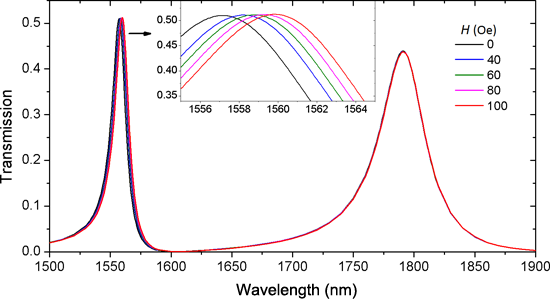
- compactness of the MDM waveguide structure. This research may open new opportunities to design nanoscale magnetic sensors with good performance. Keywords: dual resonance; magnetic fluid; magnetic sensor; plasmonic waveguide; self-reference; surface plasmon polaritons; Introduction Sensors that can detect
- fabrication and compactness. In recent years, compact optical devices based on surface plasmon polaritons (SPPs) have been reported. SPPs propagate along the dielectric–metal interface with the amplitudes decaying exponentially into both sides [16]. The deep subwavelength confinement of SPPs leads to the


