Search results
Search for "cytotoxicity" in Full Text gives 227 result(s) in Beilstein Journal of Nanotechnology. Showing first 200.
Involvement of two uptake mechanisms of gold and iron oxide nanoparticles in a co-exposure scenario using mouse macrophages
Beilstein J. Nanotechnol. 2017, 8, 2396–2409, doi:10.3762/bjnano.8.239
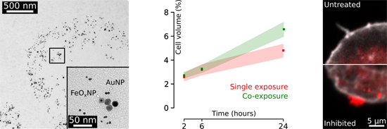
- Morada 11 megapixel camera (Münster, Germany). Lactate dehydrogenase assay (LDH) One millilitre of the supernatant of each experiment was collected and stored at 4 °C to determine cytotoxicity. Triton X (0.2% in unsupplemented RPMI) was used for cell lysis as a positive control. The supernatant of
- untreated cells was used as negative control. The LDH assay was performed with the Cytotoxicity Detection Kit (Roche Applied Science, Mannheim, Germany) according to the supplier's manual. Trypan blue exclusion assay The assay was carried out according to the manufacturer’s manual (Sigma Aldrich, Steinheim



Evaluating the toxicity of TiO2-based nanoparticles to Chinese hamster ovary cells and Escherichia coli: a complementary experimental and computational approach
Beilstein J. Nanotechnol. 2017, 8, 2171–2180, doi:10.3762/bjnano.8.216
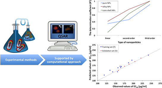
- obtained cytotoxicity data were analyzed by means of computational methods (quantitative structure–activity relationships, QSAR approach). Based on a combined experimental and computational approach, predictive models were developed, and relationships between cytotoxicity, size, and specific surface area
- bactericidal activity (towards Gram-positive B. subtilis and Gram-negative P. putida) than NPs activated by UV [4]. At the same time, no significant cytotoxicity has been detected for TiO2 doped with nitrogen (N), gold (Au) or selenium (Sn) [20][21]. Whereas, copper oxide-doped TiO2 and iron/nitrogen co-doped
- approach is based on defining mathematical dependencies between the variance in molecular structures, encoded by so-called molecular descriptors, and the variance in a given physicochemical property or biological (e.g., cytotoxicity) property in a set of compounds (“endpoints”) [5][25][26][27][28][29][30


Carbon nano-onions as fluorescent on/off modulated nanoprobes for diagnostics
Beilstein J. Nanotechnol. 2017, 8, 1878–1888, doi:10.3762/bjnano.8.188
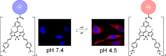
- recent reports have shown that CNOs exhibit weak inflammatory potential and low cytotoxicity [16], and they are readily internalized by cancer cells and localize in the lysosomes [18][19]. Moreover, our in vivo studies performed on zebrafish (Danio Rerio) during the development stage demonstrated their
- dye molecules (Table 2). Cytotoxicity studies The possible adverse effects of fluo-CNOs on HeLa cells were tested by using a colorimetric assay (WST1). Cells were exposed to different concentrations of fluo-CNOs (1, 2, 5, 10 and 20 μg mL−1) for different time periods (12, 24, 48 and 72 h). Cells
- objective. In order to switch the fluorescence on and off, the cells were incubated respectively with an acidic PBS solution (pH 4.5) and a basic PBS solution (pH 8.5) for 1 h before imaging. Viability assay For the cytotoxicity assays, the cells were seeded at 5 × 10⁴ cells in in 96-well plates and




Synthesis and functionalization of NaGdF4:Yb,Er@NaGdF4 core–shell nanoparticles for possible application as multimodal contrast agents
Beilstein J. Nanotechnol. 2017, 8, 1815–1824, doi:10.3762/bjnano.8.183
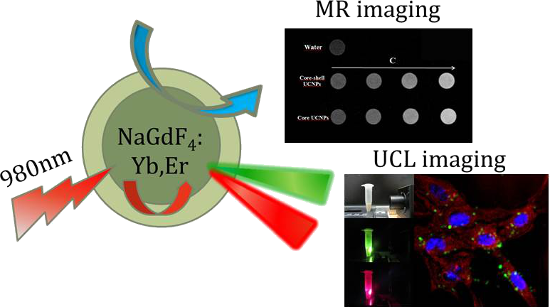
- , Tween 80-coated core–shell nanoparticles presented enhanced optical and MR signal intensity, good colloidal stability, low cytotoxicity and nonspecific internalization into two different breast cancer cell lines, which indicates that these nanoparticles could be applied as an efficient, dual-modal
- . Our results showed that Tween 80-coated UCNPs exhibited low cytotoxicity even at a high-dose concentration. Results and Discussion The SEM images of the NaGdF4:Yb,Er core and NaGdF4:Yb,Er@NaGdF4 core–shell nanoparticles are shown in Figure 1. Core nanoparticles are monodisperse, and have a spherical
- to 100 μg/mL, the viability of human breast cancer MCF-7 cells remained over 92–100% and the viability of MDA-MB-231 cells remained 85–93%. These results clearly express that core–shell gadolinium-based UCNPs have low cytotoxicity and are in good agreement with previous studies [35][36]. Conclusion


Uptake and intracellular accumulation of diamond nanoparticles – a metabolic and cytotoxic study
Beilstein J. Nanotechnol. 2017, 8, 1649–1657, doi:10.3762/bjnano.8.165
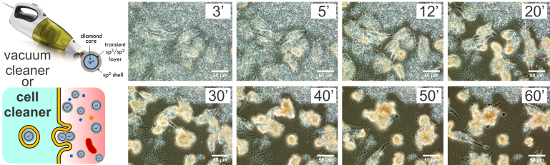
- ][27], particle size reduction [22] and surface oxidation (i.e., the surface is covered by defined starting functional groups). The cytotoxicity of NDs depends on their origin (i.e., DNDs or HPHT NDs), their size (distribution), their tendency to form aggregates (surface charge), the presence of
- impurities, and surface functionalization groups. Adverse effects on cell viability have been reported when using DNDs [24][28][29][30][31][32], while HPHT NDs often appear to be nontoxic [33][34]. Factors influencing the cytotoxicity of nanoparticles are their size [24][35][36] and surface functionalization
- [37]. In this work, we focus on cytotoxicity studies of NDs as a function of their synthesis route (DNDs versus HPHT NDs), their concentration in the medium (from 10 to 1000 mg/mL, 3 to 300 µg/cm2), their size (5 nm DND, 18–210 nm HPHT NDs) and their surface potential/termination (as-received and


Luminescent supramolecular hydrogels from a tripeptide and nitrogen-doped carbon nanodots
Beilstein J. Nanotechnol. 2017, 8, 1553–1562, doi:10.3762/bjnano.8.157
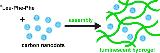
- by rheometry, fluorescence, circular dichroism (CD), FTIR spectroscopy, transmission electron microscopy (TEM), and differential scanning calorimetry (DSC). Given that this tripeptide is capable of forming a hydrogel with mild antimicrobial activity and a lack of cytotoxicity in vitro [26], this new


A nanocomplex of C60 fullerene with cisplatin: design, characterization and toxicity
Beilstein J. Nanotechnol. 2017, 8, 1494–1501, doi:10.3762/bjnano.8.149
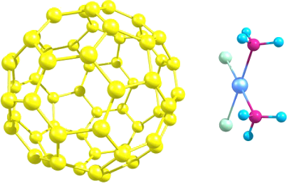
- compounds. The genotoxicity of С60 fullerene, Cis and their complex was evaluated in vitro with the comet assay using human resting lymphocytes and lymphocytes after blast transformation. The cytotoxicity of the mentioned compounds was estimated by Annexin V/PI double staining followed by flow cytometry



Calcium fluoride based multifunctional nanoparticles for multimodal imaging
Beilstein J. Nanotechnol. 2017, 8, 1484–1493, doi:10.3762/bjnano.8.148
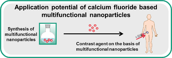
- synthesis and determine the long-term stability of the CAs. No cytotoxicity of NP concentrations between 0.5 and 1 mg·mL−1 was observed after exposure to human dermal fibroblasts over 24 h. Overall this study shows, that the CaF2:(Tb3+,Gd3+) NPs are suitable for medical imaging. Keywords: calcium fluoride
- cytotoxicity of the NPs was tested by a cell culture based viability assay. Results and Discussion Synthesis and characterization of the multifunctional nanoparticles The synthesis of the CaF2:(Tb3+,Gd3+) NPs was carried out in analogy to the reported wet-chemical procedure that is based on a co-precipitation
- cells with the NP dispersions (cf. Figure 7b). NP concentrations of 0.5, 0.75 and 1.0 mg·mL−1 yield cell viabilities of more than 80% with respect to the positive control. Thus in this concentration range no cytotoxicity of CaF2:(Tb3+,Gd3+) NPs is observed on hdF. Conclusion In summary, we have


Development of polycationic amphiphilic cyclodextrin nanoparticles for anticancer drug delivery
Beilstein J. Nanotechnol. 2017, 8, 1457–1468, doi:10.3762/bjnano.8.145
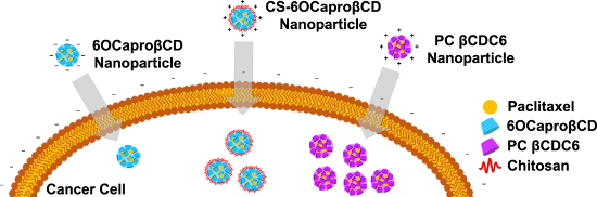
- capacity, in vitro release profiles and cytotoxicity studies. Results: Paclitaxel-loaded cyclodextrin nanoparticles were obtained in the diameter range of 80−125 nm (depending on the nature of the cyclodextrin derivative) where the smallest diameter nanoparticles were obtained with polycationic (PC) βCDC6
- presence of PF68 in CD nanoparticle formulations on cytotoxicity on L929, a healthy mouse fibroblast cell line. According to these results, it was suggested that PF68 has no significant effect on size and drug loading capacity of nanoparticles but dose-dependent toxicity could occur on L929 fibroblast
- and MCF-7 human breast cancer cell lines were used, respectively. Both cell lines were grown and incubated in appropriate conditions (see Experimental section for full experimental details). The cytotoxicity of blank amphiphilic CD nanoparticles was determined on L929 mouse fibroblast cells with MTT


Cationic PEGylated polycaprolactone nanoparticles carrying post-operation docetaxel for glioma treatment
Beilstein J. Nanotechnol. 2017, 8, 1446–1456, doi:10.3762/bjnano.8.144
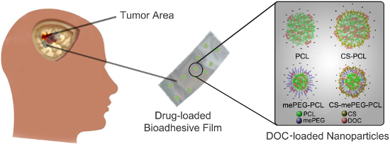
- particles, and the drug release rate from the nanoparticles was slowed down to 48 h by dispersing the nanoparticles in a hydroxypropyl cellulose film. Cell culture studies revealed that docetaxel-loaded nanoparticles cause higher cytotoxicity compared to the free docetaxel solution in DMSO. Conclusion
- recurrence during the first 2 days. Cell culture studies Cytotoxicity assay for blank nanoparticles Mouse fibroblast cell lines L929 (recommended by the USP for the cytotoxicity evaluation of polymeric systems) were used to determine the cytotoxicity of blank nanoparticles with MTT assay. According to MTT
- after surgical operation of glioma treatment. All formulations were characterized in terms of mean particle size, polydispersity index, zeta potential, drug loading capacity, drug release profile and cytotoxicity. When nanoparticle formulations are compared with each other, mePEG-PCL nanoparticles have




Nano-engineered skin mesenchymal stem cells: potential vehicles for tumour-targeted quantum-dot delivery
Beilstein J. Nanotechnol. 2017, 8, 1218–1230, doi:10.3762/bjnano.8.123
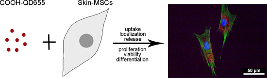
- MSCs to assess their potential use as vectors for the targeting of SC or other tumours. Results Optimal QD labelling conditions for MSCs The concentration-dependent cytotoxicity of QDs was analysed in MSC cultures after 24 and 48 h using a colorimetric CCK-8 assay, which measures intracellular





Silicon microgrooves for contact guidance of human aortic endothelial cells
Beilstein J. Nanotechnol. 2017, 8, 675–681, doi:10.3762/bjnano.8.72
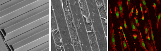
- cell adhesion and surface stability following the 3-amimoptopyl triethoxylane (APTES)–glutaraldehyde (GTA)–collagen sequence as described in Experimental section. Cytotoxicity of silicon substrates Cytotoxicity was assessed by measuring LDH activity 24 h, 2 days, 3 days, 6 days and 7 days (D1–D7) after
- incubating the silicon substrates with human aortic endothelial cells (HAECs). Blank control values (cells seeded in the absence of any silicon substrates) were set at 100% and the other conditions were calculated in relation to this reference value. As shown in Figure 3, no cytotoxicity was observed as no
- , Spain) with an atmosphere containing 5% CO2. Cell viability and cytotoxicity Cell viability was assessed by morphology using phase-contrast microscopy and by trypan blue dye exclusion test (Merck). Viability 97% was required for the thawed HAECs in order to guarantee the viability of the cells before


Dispersion of single-wall carbon nanotubes with supramolecular Congo red – properties of the complexes and mechanism of the interaction
Beilstein J. Nanotechnol. 2017, 8, 636–648, doi:10.3762/bjnano.8.68
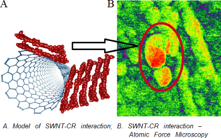
- amphiphilic molecules that cover the hydrophobic CNT surface with hydrophilic groups [15]. Functionalization leads not only to the increased water dispersibility of CNTs but also improves their biocompatibility due to enhanced penetration through biological membranes and reduced cytotoxicity [16][17


Facile fabrication of luminescent organic dots by thermolysis of citric acid in urea melt, and their use for cell staining and polyelectrolyte microcapsule labelling
Beilstein J. Nanotechnol. 2016, 7, 1905–1917, doi:10.3762/bjnano.7.182
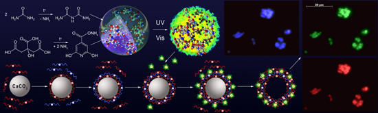
- details, see Supporting Information File 1, Table S1 and Table S2). It is demonstrated that a mixture of citric acid and urea, without heating (“0 min” sample), possessed some cytotoxicity. At concentrations greater than ca. 300 μg/mL, this sample decreased the number of living ST-cells. The enzymatic
- 100 μg/mL [50]. “Green” carbon dots from coriander leaves extract became toxic for normal lung cells (L-132) or cancer cell line (A549) at concentrations higher than 500 μg/mL [51], the cancer cells being somewhat more sensitive. Microcapsules decorated by O-dots had a very low cytotoxicity
- gentamicin (Arterium, Ukraine), at a concentration of 40 μg/mL. Cultures formed a uniform monolayer of cells. Oxidative stress was induced by introducing into the cellular medium 3% hydrogen peroxide solution, at a final concentration of 8 μg/mL, in a well. The cytotoxicity of microcapsules was studied using




Low temperature co-fired ceramic packaging of CMOS capacitive sensor chip towards cell viability monitoring
Beilstein J. Nanotechnol. 2016, 7, 1871–1877, doi:10.3762/bjnano.7.179
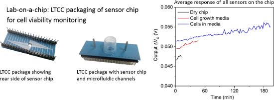
- effective methods for evaluating cytotoxicity, both short and long term. Traditional in vitro cytotoxicity evaluation methods include cell cultivation and label-based assay kits, which are often expensive and time-consuming end-point measurements. Furthermore, the labelling techniques used for cell
- spectroscopy [1], electrochemical quartz crystal microbalance measurements [2], optical sensing [3], impedimetric sensing [4][5][6], and capacitive sensing [7][8][9][10][11]. The lab-on-a-chip (LoC) concept is an excellent way to implement label-free, noninvasive, cost-effective cytotoxicity assessment. LoCs
- capacitance sensing and the intention is to develop a method for nanoparticle exposure of cells to establish cytotoxicity assessment of nanomaterials. Capacitance measurements reflect the surface attachment of adherent cells. While healthy cells attach to the cultivation surface and spread out, dying cells


Chitosan-based nanoparticles for improved anticancer efficacy and bioavailability of mifepristone
Beilstein J. Nanotechnol. 2016, 7, 1861–1870, doi:10.3762/bjnano.7.178
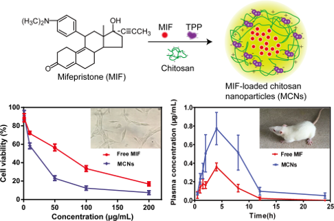
- crosslinking agent for controlled drug release of CNs. In vitro anticancer effects The cytotoxicity of the MCNs was tested in four different cancer cell lines A549 (human lung adenocarcinoma), Hela (human cervical epithelioid carcinoma), RL95-2 (human endometrial carcinoma), and HepG2 (human liver
- containing MCNs immersed in 0.1 M PBS (pH 7.4) or 0.1 M PBS (pH 2.5) containing 1% of ethanol. In vitro cytotoxicity of CNs, MIF and MCNs against A549 (A), Hela (B), RL95-2 (C), and HepG2 cells (D). Cells were incubated with different concentrations (1, 10, 50, 100, 200 μg/mL) of blank CNs, free MIF, or MCNs


Nano- and microstructured materials for in vitro studies of the physiology of vascular cells
Beilstein J. Nanotechnol. 2016, 7, 1620–1641, doi:10.3762/bjnano.7.155
- geometry. Moreover, their non-cytotoxicity, their ease to use with many fabrication techniques and often the simplicity of their synthesis makes them to be widely used within the field of biomaterials [4][5][55][85][99][100][101][102]. Examples of the most representative synthetic polymeric materials used
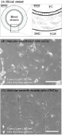
- staining is the most used assay to determine cell survival on a particular substrate and hence the cytotoxicity of a material is assessed [14][209]. Besides this live/dead cell investigation, biochemical proliferation assays, such as EdU or BrdU staining, are frequently applied to determine the cell



On the pathway of cellular uptake: new insight into the interaction between the cell membrane and very small nanoparticles
Beilstein J. Nanotechnol. 2016, 7, 1296–1311, doi:10.3762/bjnano.7.121
- : ATP depletion; calcium crystallization; cytotoxicity; endocytosis; HeLa cells; LDH; mesenchymal stem cells; morphology; necrosis; particle size; silica nanoparticles; TEM; Introduction Silicon dioxide nanoparticles (SiNPs) are used in a wide range of commercially available products to improve product
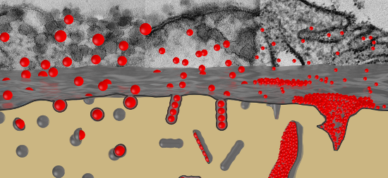
- have been reported in humans, general risk assessment and thereby the investigation of possible interactions of SiNPs with human cells and tissues is of crucial importance. A deeper understanding of SiNP uptake modes into cells may lead us one step further in grasping nanoparticle cytotoxicity and in
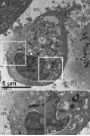
- indicative for the disintegration of the cell membrane and consequently for cytotoxicity. For quantitative estimation of the toxic potential HeLa cells were incubated with NPs for 2 h at different concentrations followed by determination of the LDH release (Figure 6). SiNP-22 induces only a moderate increase




Straightforward and robust synthesis of monodisperse surface-functionalized gold nanoclusters
Beilstein J. Nanotechnol. 2016, 7, 1278–1283, doi:10.3762/bjnano.7.118
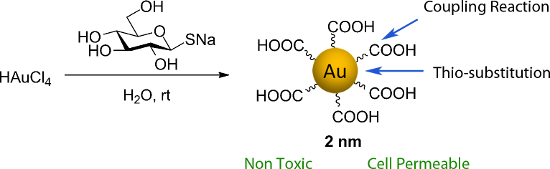
- ). Unfunctionalized Glc-NCs fail to aggregate since oxidized thio-glucose is not recognized by ConA (Figure 2C). Glc-NC@Man are monodisperse prior to aggregation by the addition of ConA as judged by TEM (Figure S12, Supporting Information File 1). Nanocluster cytotoxicity was assessed by incubating the nanoclusters
- for one day with the mouse cell line L929 for a proof-of-principle study. Cell viability was measured using the MTS [3-(4,5-dimethylthiazol-2-yl)-5-(3-carboxymethoxyphenyl)-2-(4-sulfophenyl)-2H-tetrazolium inner salt] assay [30]. The cytotoxicity of Glc-NCs, CTAB-NCs and THPC-NCs was compared. CTAB
- -NCs were toxic even at low concentrations (0.2–25 µM), whereas both THPC-NCs (Figure S13, Supporting Information File 1) and Glc-NCs did not show any toxicity at 500 µM (Figure 3A). To test whether the free stabilizers were affecting the cytotoxicity, both Glc-NCs and THPC-NCs were measured after


Reasons and remedies for the agglomeration of multilayered graphene and carbon nanotubes in polymers
Beilstein J. Nanotechnol. 2016, 7, 1174–1196, doi:10.3762/bjnano.7.109
- bones. However, some researchers reported that CNTs exhibit cytotoxicity to human dermal cells. Potential health hazards could also arise from inhalation. The discrepancy in such biocompatibility results can be attributed to the complicated physicochemical interactions between CNTs and biological cells


Multiwalled carbon nanotube hybrids as MRI contrast agents
Beilstein J. Nanotechnol. 2016, 7, 1086–1103, doi:10.3762/bjnano.7.102
- , e.g., stability of dispersion in buffers, lower cytotoxicity, visual contrast or selectivity for certain organs [33]. Finally, additional effects appear after introduction into a living organism, thus it is not surprising that further evolution of r2/r1 takes place there. It was found that the local
- the in vivo behavior of nanohybrids is essential in order to judge their applicability in MRI. Several types of studies were performed. The first group was to study cytotoxicity by determining cell viability upon incubation with the nanohybrids (Table 2). In some cases it was possible to indicate
- which component of the hybrid was responsible mainly for the cytotoxicity impact. The classical MTT assay is usually applied for this purpose. However, there have been reports of high measurement error in this method with MWCNT [40], thus Trypan Blue staining of the dead cells could be more reliable



Improved biocompatibility and efficient labeling of neural stem cells with poly(L-lysine)-coated maghemite nanoparticles
Beilstein J. Nanotechnol. 2016, 7, 926–936, doi:10.3762/bjnano.7.84
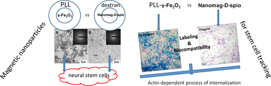
- their cellular uptake, the mechanism of internalization, cytotoxicity, viability and proliferation of neural stem cells, and compared them to the commercially available dextran-coated nanomag®-D-spio nanoparticles. Results: Light microscopy of Prussian blue staining revealed a concentration-dependent
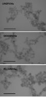
- efficiency, cellular viability, cytotoxicity, behavior after labeling, and the mechanism of internalization was determined and compared. Results Characterization of the nanoparticle morphology To compare the morphology of PLL-γ-Fe2O3 nanoparticles with commercially available nanomag®-D-spio particles
- effect on NSC, treated cells were assessed with regard to viability, proliferation and cytotoxicity. The MTT assay was applied to demonstrate NSC viability and proliferation. A constant amount of starting cells for culture was used and compared after 48 h of NSC proliferation in the culture. The non




Tight junction between endothelial cells: the interaction between nanoparticles and blood vessels
Beilstein J. Nanotechnol. 2016, 7, 675–684, doi:10.3762/bjnano.7.60
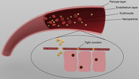
- for the adverse effects of NPs, e.g., cytotoxicity [8], unknown effects of its biological distribution [9] and genotoxicity [10]. At the same time, we realize that in most cases the pathway of how NPs enter the human body remains unknown. When we are exposed to NPs, they can enter our body though
- cytotoxicity in human umbilical vascular endothelial cells and these effects are related to the activation of potassium channels [49]. Iron oxide NPs also induce inflammation and malfunction in vascular endothelial systems [50]. In the following, we will present assumptions about how the NPs behave in blood
- vessels, in particular about (1) NPs pass through the endothelial layer of blood vessels and (2) NPs cause cytotoxicity in surrounding tissues under the endothelial layer. Endothelial cells and tight junction The endothelium provides a thin layer of cells that covers the internal surface of blood vessels

Comparison of the interactions of daunorubicin in a free form and attached to single-walled carbon nanotubes with model lipid membranes
Beilstein J. Nanotechnol. 2016, 7, 524–532, doi:10.3762/bjnano.7.46
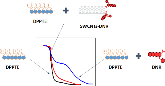
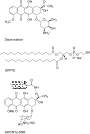
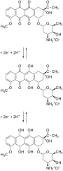
- conjugated to either polyethylene glycol (PEG) functionalized single-walled carbon nanotubes (SWCNTs) [20] or to aptamer-wrapped SWCNTs via π–π interactions. In both cases the cytotoxicity of the conjugates was verified on the selected cancer cell lines. In this study the influence of both free daunorubicin




Surface coating affects behavior of metallic nanoparticles in a biological environment
Beilstein J. Nanotechnol. 2016, 7, 246–262, doi:10.3762/bjnano.7.23
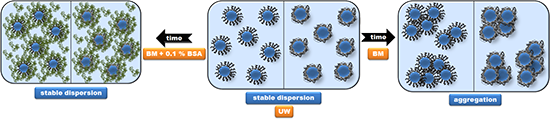
- uptake and toxicity of metallic NPs [41][42], whereas differences in surface coatings influence cytotoxicity and surface charge [43]. However, it is still unclear how different surface coatings affect the interaction of NPs with biological environments and the formation of the protein corona. Because